. 1998 Jul;18(7):4165-76.
doi: 10.1128/MCB.18.7.4165.

The capacity of polyomavirus enhancer binding protein 2alphaB (AML1/Cbfa2) to stimulate polyomavirus DNA replication is related to its affinity for the nuclear matrix

Affiliations

The capacity of polyomavirus enhancer binding protein 2alphaB (AML1/Cbfa2) to stimulate polyomavirus DNA replication is related to its affinity for the nuclear matrix

L F Chen et al. Mol Cell Biol. 1998 Jul.

Abstract

The nuclear matrix is thought to play an important role in the DNA replication of eukaryotic cells, although direct evidence for such a role is still lacking. A nuclear matrix-associated transcription factor, polyomavirus (Py) enhancer binding protein 2alphaB1 (PEBP2alphaB1) (AML1/Cbfa2), was found to stimulate Py replication through its cognate binding site. The minimal replication activation domain (RAD) was identified between amino acid (aa) 302 and aa 371 by using a fusion protein containing the GAL4 DNA binding domain (GAL4-RAD). In addition, the region showed affinity for the nuclear matrix and, on the basis of competition studies, binding activity for one or more proteins involved in the initiation of Py DNA replication. A leukemogenic chimeric protein, AML1/ETO(MTG8), which does not contain this region of PEBP2alphaB1/AML1, was also localized in the nuclear matrix fraction and competed for nuclear matrix association with PEBP2alphaB1 and GAL4-RAD. Moreover, AML1/ETO inhibited Py DNA replication stimulated by PEBP2alphaB1 and GAL4-RAD. The inhibition was specific for replication mediated by PEBP2alphaB1 and GAL4-RAD, and proportional to the degree of loss of these activators from the nuclear matrix, suggesting a requirement for nuclear matrix targeting in the stimulation of Py DNA replication by RAD. These results are the first to suggest a molecular link between the initiation of DNA replication and the nuclear matrix compartment.


Figures

FIG. 1
FIG. 1
Stimulation of Py DNA replication by PEBP2αB1. Replication assays were performed as described in Materials and Methods by using pPy(AE)4OICAT (lanes 1, 2, and 4 to 9) and pPy(AEM)6OICAT (lanes 10 to 15), which contain the wild-type and mutated PEBP2 binding site, respectively, as reporter plasmids. In lane 2, the Py TAg-expressing plasmid was omitted, and in lane 3, pPy(AE)4CAT, which has a deletion in the Py core origin, was used. Indicated amounts of PEBP2αB1 expression plasmids were included in lanes 4 to 15. The bands corresponding to the replicated reporter plasmid and the control plasmid are indicated.
FIG. 2
FIG. 2
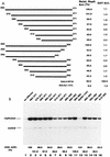
The C-terminal region of the PEBP2αB1 contains the replication activation domain. (A) Schematic representation of the N-terminal and C-terminal deletion mutants of PEBP2αB1 used in the experiments shown in panel B. RD, Runt domain. (B) Replication assays were performed by using pPy(AE)4OICAT as a reporter plasmid as described in Materials and Methods. A total of 0.5 μg of plasmid of the indicated deletion derivatives of PEBP2αB1 was cotransfected. The bands corresponding to the replicated reporter plasmid and the control plasmid are indicated. Assays without effector (lane 1) or without TAg (lane 2) were also included as controls. Replication activity relative to that of the wide type is indicated below each lane.
FIG. 3
FIG. 3
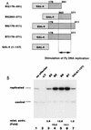
Stimulation of Py DNA replication by GAL4-PEBP2αB1 fusion proteins. (A) Schematic representation of the GAL4-PEBP2αB1 fusion constructs shown in panel B. (B) Replication assays for GAL4-PEBP2αB1 fusion constructs shown in panel A were performed by using pPyG5OICAT as a reporter plasmid. The bands corresponding to the replicated reporter and the control plasmid are indicated. Replication activity relative to that of the GAL4 DNA binding domain (1-147) is indicated below each lane.
FIG. 4
FIG. 4
Determination of the replication activation domain. (A) Schematic representation of the deletion mutants used in the experiments shown in panel B. Each mutant was fused to the GAL4 DNA binding domain. The relative replication activity of each deletion mutant measured in panel B and the level of transcription from the Py early promoter on pPyG5OICAT are indicated. The activities of GAL4-VP16 construct are also indicated for comparison. (B) The replication activity of each fusion protein shown in panel A was assayed as described in Materials and Methods by using pPyG5OICAT as a reporter plasmid. The bands corresponding to the replicated reporter and the control plasmid are indicated. Control experiments without effector (lane 1) or with an effector expressing the GAL4 DNA binding domain (1-147) are also indicated (lane 12).
FIG. 5
FIG. 5
AML1/ETO inhibited the nuclear matrix localization activity of PEBP2αB1 and GAL4-RAD but not that of VP16. (A) Schematic structure of PEBP2αB1 and two forms of the AML1(PEBP2αB1)/ETO fusion protein. (B) The plasmids expressing the indicated proteins were transfected into P19 cells. Twenty-four hours after transfection, soluble, chromatin, and nuclear matrix fractions were isolated as described in Materials and Methods. Proteins in each fraction were analyzed by Western blotting. (C) Increasing amounts of AML1/ETO expression plasmids were cotransfected with a constant amount of PEBP2αB1 expression plasmid. The nuclear matrix fraction was isolated and analyzed by Western blotting. (D) Increasing amounts of plasmids expressing AML1/ETO or CH15 were cotransfected with a constant amount of plasmid expressing GAL4-RAD (upper panel), and increasing amounts of plasmids expressing AML1/ETO were cotransfected with a constant amount of plasmid expressing GAL4-VP16 (lower panel). The nuclear matrix fraction was isolated and analyzed by Western blotting. (E) Nuclear skeleton. Five micrograms of plasmids expressing full-length PEBP2αB1 (lane 1), 1-371 (lane 2) and 1-291 (lane 3) was transfected into P19 cells. The nuclear skeleton fractions of the transfected cells were isolated as described in Materials and Methods and analyzed by Western blotting. The positions of three proteins are indicated, and the expected position of PEBP2αB1(1-291) is indicated by a dotted arrow.
FIG. 5
FIG. 5
AML1/ETO inhibited the nuclear matrix localization activity of PEBP2αB1 and GAL4-RAD but not that of VP16. (A) Schematic structure of PEBP2αB1 and two forms of the AML1(PEBP2αB1)/ETO fusion protein. (B) The plasmids expressing the indicated proteins were transfected into P19 cells. Twenty-four hours after transfection, soluble, chromatin, and nuclear matrix fractions were isolated as described in Materials and Methods. Proteins in each fraction were analyzed by Western blotting. (C) Increasing amounts of AML1/ETO expression plasmids were cotransfected with a constant amount of PEBP2αB1 expression plasmid. The nuclear matrix fraction was isolated and analyzed by Western blotting. (D) Increasing amounts of plasmids expressing AML1/ETO or CH15 were cotransfected with a constant amount of plasmid expressing GAL4-RAD (upper panel), and increasing amounts of plasmids expressing AML1/ETO were cotransfected with a constant amount of plasmid expressing GAL4-VP16 (lower panel). The nuclear matrix fraction was isolated and analyzed by Western blotting. (E) Nuclear skeleton. Five micrograms of plasmids expressing full-length PEBP2αB1 (lane 1), 1-371 (lane 2) and 1-291 (lane 3) was transfected into P19 cells. The nuclear skeleton fractions of the transfected cells were isolated as described in Materials and Methods and analyzed by Western blotting. The positions of three proteins are indicated, and the expected position of PEBP2αB1(1-291) is indicated by a dotted arrow.
FIG. 6
FIG. 6
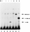
Effect of the AML1/ETO chimeric proteins on the replication stimulation activity of PEBP2αB1 and GAL4-RAD. (A) Binding of the in vitro-translated AML1/ETO (lanes 2 and 5), CH15 (lanes 3 and 6), and PEBP2αB1(1-185) (lanes 4 and 7) proteins to the PEBP2 site in the absence (lanes 2 to 4) or presence (lanes 5 to 7) of β2 (10 ng). (B) Replication assays for AML1/ETO chimeric proteins were performed with pPy(AE)4OICAT as a reporter plasmid. In lane 2, the TAg-expressing plasmid was omitted. The indicated amounts of plasmids expressing the long form (lanes 3 to 6) or the short form (lanes 7 to 10) of AML1/ETO, PEBP2αB1 lacking the region C-terminal to the runt domain (lanes 11 to 14), or wild-type PEBP2αB1 (lane 15) were cotransfected. (C) A constant amount (0.5 μg) of the plasmid expressing full-length PEBP2αB1 and the reporter plasmid, pPy(AE)4OICAT (0.2 μg), was cotransfected with increasing amounts of the plasmids expressing the long form of AML1/ETO (lanes 2 to 5), the short form of AML1/ETO (lanes 6 to 9), or truncated PEBP2αB1 that lacked the C-terminal region (1-185) (lanes 10 to 13). Replication of the reporter plasmid was measured as described in Materials and Methods. The bands corresponding to replicated reporter and the control plasmid are indicated. Activities relative to the wild-type domain are indicated below each lane. (D and E) Effect of the chimeric proteins and truncated protein on the replication stimulation activity of GAL4-RAD (D) or GAL4-VP16 (E) was measured as described for panel C. Activity relative to that of the wild-type domain is indicated below each lane.
FIG. 6
FIG. 6
Effect of the AML1/ETO chimeric proteins on the replication stimulation activity of PEBP2αB1 and GAL4-RAD. (A) Binding of the in vitro-translated AML1/ETO (lanes 2 and 5), CH15 (lanes 3 and 6), and PEBP2αB1(1-185) (lanes 4 and 7) proteins to the PEBP2 site in the absence (lanes 2 to 4) or presence (lanes 5 to 7) of β2 (10 ng). (B) Replication assays for AML1/ETO chimeric proteins were performed with pPy(AE)4OICAT as a reporter plasmid. In lane 2, the TAg-expressing plasmid was omitted. The indicated amounts of plasmids expressing the long form (lanes 3 to 6) or the short form (lanes 7 to 10) of AML1/ETO, PEBP2αB1 lacking the region C-terminal to the runt domain (lanes 11 to 14), or wild-type PEBP2αB1 (lane 15) were cotransfected. (C) A constant amount (0.5 μg) of the plasmid expressing full-length PEBP2αB1 and the reporter plasmid, pPy(AE)4OICAT (0.2 μg), was cotransfected with increasing amounts of the plasmids expressing the long form of AML1/ETO (lanes 2 to 5), the short form of AML1/ETO (lanes 6 to 9), or truncated PEBP2αB1 that lacked the C-terminal region (1-185) (lanes 10 to 13). Replication of the reporter plasmid was measured as described in Materials and Methods. The bands corresponding to replicated reporter and the control plasmid are indicated. Activities relative to the wild-type domain are indicated below each lane. (D and E) Effect of the chimeric proteins and truncated protein on the replication stimulation activity of GAL4-RAD (D) or GAL4-VP16 (E) was measured as described for panel C. Activity relative to that of the wild-type domain is indicated below each lane.
FIG. 7
FIG. 7
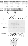
Effects of mutations in the replication activation domain on DNA replication. (A) Sequence comparison of PEBP2αB1, αA1, and αC1 in RAD. The amino acids conserved in this family of proteins are shown in bold, and the modified amino acids in each mutant (M1 to M6) are indicated below. (B) Replication assay for the mutants. Each mutant was fused to the GAL4 DNA binding domain and assayed for stimulation of Py DNA replication as described in Materials and Methods. The bands corresponding to the replicated reporter and the control plasmid are indicated. Activity relative to that of the wild-type RAD is indicated below each lane. (C) Binding of the M4 and M5 mutants to the nuclear matrix. Five micrograms each of the plasmids expressing GAL4-RAD (lane 1), M4 (lane 2), and M5 (lane 3) was transfected into P19 cells, and the nuclear matrix fraction was analyzed by Western blotting.
FIG. 8
FIG. 8
Demonstration of two types of competition. (A) Overexpression of GAL4-RAD inhibited the replication stimulation activity of PEBP2αB1. A total of 0.5 μg of the PEBP2αB1 expression plasmid and 0.2 μg of the reporter plasmid, pPy(AE)4OICAT, were cotransfected into P19 cells together with increasing amounts of the plasmids expressing GAL4-RAD (lanes 1 to 7) or M4 (lanes 8 to 14). In lanes 7 and 14, 4 μg of GAL4-RAD and M4 expression plasmids was transfected, respectively, without PEBP2αB1 expression plasmid. The replicated reporter and the control plasmid are indicated. Relative replication activity is indicated below each lane. (B) Overexpression of GAL4-RAD and M4 did not inhibit the nuclear matrix localization activity of PEBP2αB1. Increasing amounts of the GAL4-RAD (lanes 2 to 4) and M4 (lanes 5 to 7) expression plasmids were cotransfected with a constant amount of PEBP2αB1 expression plasmid into P19 cells. The nuclear matrix fraction was analyzed by Western blotting. Only the band representing PEBP2αB1 is shown. (C) Overexpression of GAL4-RAD and M4 inhibited the nuclear matrix binding activity of PEBP2αB1(1-371). Increasing amounts of GAL4-RAD (lanes 2 to 4) or M4 (lanes 5 to 7) expression plasmids were cotransfected with a constant amount of PEBP2αB1(1-371) expression plasmid into P19 cells. The nuclear matrix fraction was analyzed by Western blotting. The upper panel shows the blot probed with anti-GAL4 polyclonal antibody, while the lower panel shows the same blot probed with anti-PEBP2αB1 polyclonal antibody. (D) Effect of overexpression of GAL4-RAD and M4 on the replication stimulation activity of PEBP2αB1(1-371). A total of 0.5 μg of PEBP2αB1(1-371) expression plasmid and 0.2 μg of reporter plasmid, pPy(AE)4OICAT, were cotransfected into P19 cells together with increasing amounts of the plasmids expressing GAL4-RAD (lanes 2 to 4) or M4 (lanes 5 to 7). The replicated reporter and the control plasmid are indicated. Relative replication activity is indicated below each lane. (E) GAL4-VP16 did not compete with PEBP2αB1 for the replication stimulation activity. A total of 0.5 μg of the PEBP2αB1 expression plasmid and 0.2 μg of the reporter plasmid, pPy(AE)4OICAT, were cotransfected into P19 cells together with increasing amounts of the plasmids expressing GAL4-VP16 (lanes 2 to 4) or GAL4-RAD (lanes 5 to 7). The replicated reporter and the control plasmid are indicated. Relative replication activity is indicated below each lane.
FIG. 8
FIG. 8
Demonstration of two types of competition. (A) Overexpression of GAL4-RAD inhibited the replication stimulation activity of PEBP2αB1. A total of 0.5 μg of the PEBP2αB1 expression plasmid and 0.2 μg of the reporter plasmid, pPy(AE)4OICAT, were cotransfected into P19 cells together with increasing amounts of the plasmids expressing GAL4-RAD (lanes 1 to 7) or M4 (lanes 8 to 14). In lanes 7 and 14, 4 μg of GAL4-RAD and M4 expression plasmids was transfected, respectively, without PEBP2αB1 expression plasmid. The replicated reporter and the control plasmid are indicated. Relative replication activity is indicated below each lane. (B) Overexpression of GAL4-RAD and M4 did not inhibit the nuclear matrix localization activity of PEBP2αB1. Increasing amounts of the GAL4-RAD (lanes 2 to 4) and M4 (lanes 5 to 7) expression plasmids were cotransfected with a constant amount of PEBP2αB1 expression plasmid into P19 cells. The nuclear matrix fraction was analyzed by Western blotting. Only the band representing PEBP2αB1 is shown. (C) Overexpression of GAL4-RAD and M4 inhibited the nuclear matrix binding activity of PEBP2αB1(1-371). Increasing amounts of GAL4-RAD (lanes 2 to 4) or M4 (lanes 5 to 7) expression plasmids were cotransfected with a constant amount of PEBP2αB1(1-371) expression plasmid into P19 cells. The nuclear matrix fraction was analyzed by Western blotting. The upper panel shows the blot probed with anti-GAL4 polyclonal antibody, while the lower panel shows the same blot probed with anti-PEBP2αB1 polyclonal antibody. (D) Effect of overexpression of GAL4-RAD and M4 on the replication stimulation activity of PEBP2αB1(1-371). A total of 0.5 μg of PEBP2αB1(1-371) expression plasmid and 0.2 μg of reporter plasmid, pPy(AE)4OICAT, were cotransfected into P19 cells together with increasing amounts of the plasmids expressing GAL4-RAD (lanes 2 to 4) or M4 (lanes 5 to 7). The replicated reporter and the control plasmid are indicated. Relative replication activity is indicated below each lane. (E) GAL4-VP16 did not compete with PEBP2αB1 for the replication stimulation activity. A total of 0.5 μg of the PEBP2αB1 expression plasmid and 0.2 μg of the reporter plasmid, pPy(AE)4OICAT, were cotransfected into P19 cells together with increasing amounts of the plasmids expressing GAL4-VP16 (lanes 2 to 4) or GAL4-RAD (lanes 5 to 7). The replicated reporter and the control plasmid are indicated. Relative replication activity is indicated below each lane.

Similar articles

Cited by

References

    1. Amati B B, Gasser S M. Chromosomal ARS and CEN elements bind specifically to the yeast nuclear scaffold. Cell. 1988;54:967–978. - PubMed
    1. Amati B B, Gasser S M. Drosophila scaffold-attached regions bind nuclear scaffolds and can function as ARS elements in both budding and fission yeasts. Mol Cell Biol. 1990;10:5442–5454. - PMC - PubMed
    1. Bae S C, Ogawa E, Maruyama M, Oka H, Sataka M, Shigesada K, Jenkins N A, Gilbert D J, Copeland N G, Ito Y. PEBP2αB/mouse AML1 consists of multiple isoforms that possess differential transactivation potentials. Mol Cell Biol. 1994;14:3242–3252. - PMC - PubMed
    1. Bae S C, Takahashi E, Zhang Y W, Ogawa E, Shigesada K, Namba Y, Sataka M, Ito Y. Cloning, mapping and expression of PEBP2αC, a third gene encoding the mammalian Runt domain. Gene. 1995;159:245–248. - PubMed
    1. Baru M, Shlissel M, Manor H. The yeast GAL4 protein transactivates the polyomavirus origin of DNA replication in mouse cells. J Virol. 1991;65:3494–3503. - PMC - PubMed

Publication types

MeSH terms

LinkOut - more resources