. 1998 Jul;18(7):4272-81.
doi: 10.1128/MCB.18.7.4272.

Calcium and S100B regulation of p53-dependent cell growth arrest and apoptosis

Affiliations

Calcium and S100B regulation of p53-dependent cell growth arrest and apoptosis

C Scotto et al. Mol Cell Biol. 1998 Jul.

Abstract

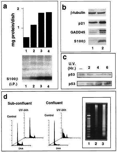
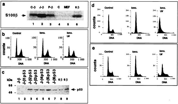
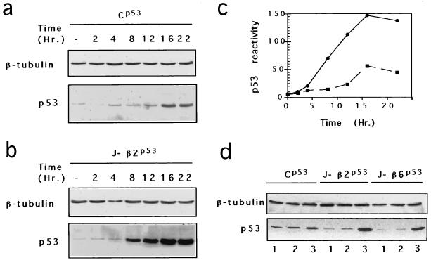
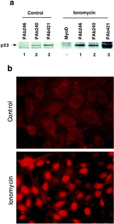
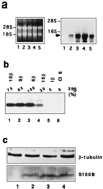
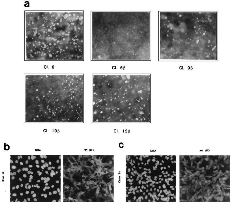
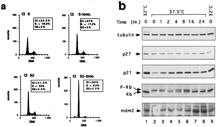
In glial C6 cells constitutively expressing wild-type p53, synthesis of the calcium-binding protein S100B is associated with cell density-dependent inhibition of growth and apoptosis in response to UV irradiation. A functional interaction between S100B and p53 was first demonstrated in p53-negative mouse embryo fibroblasts (MEF cells) by sequential transfection with the S100B and the temperature-sensitive p53Val135 genes. We show that in MEF cells expressing a low level of p53Val135, S100B cooperates with p53Val135 in triggering calcium-dependent cell growth arrest and cell death in response to UV irradiation at the nonpermissive temperature (37.5 degreesC). Calcium-dependent growth arrest of MEF cells expressing S100B correlates with specific nuclear accumulation of the wild-type p53Val135 conformational species. S100B modulation of wild-type p53Val135 nuclear translocation and functions was confirmed with the rat embryo fibroblast (REF) cell line clone 6, which is transformed by oncogenic Ha-ras and overexpression of p53Val135. Ectopic expression of S100B in clone 6 cells restores contact inhibition of growth at 37.5 degreesC, which also correlates with nuclear accumulation of the wild-type p53Val135 conformational species. Moreover, a calcium ionophore mediates a reversible G1 arrest in S100B-expressing REF (S100B-REF) cells at 37.5 degreesC that is phenotypically indistinguishable from p53-mediated G1 arrest at the permissive temperature (32 degreesC). S100B-REF cells proceeding from G1 underwent apoptosis in response to UV irradiation. Our data support a model in which calcium signaling and S100B cooperate with the p53 pathways of cell growth inhibition and apoptosis.


Figures

FIG. 1
FIG. 1
S100B synthesis in glial C6 cells is correlated with cell contact inhibition of growth and UV-induced apoptosis. (a) S100B synthesis in glial C6 cells is correlated with cell contact inhibition of growth. Glial C6 cells were seeded at 5 × 10⁵ cells/ml in 35-mm-diameter dishes. After 24 h (lane 1), 48 h (lane 2), 72 h (lane 3), and 82 h (lane 4), cells were metabolically labeled with [35S]Met-Cys mix for 6 h prior to harvesting. The upper panel shows the quantity of total protein per dish; the lower panel shows S100B protein immunoprecipitated (I.P.) from 1 mg of protein. (b) Western blot analysis for β-tubulin, p21WAF1, GADD45, and S100B in 50 μg of glial C6 cell extracts from exponentially growing cultures (lane 1) or from those at the end of logarithmic growth (lane 2). (c) Time course of p53 induction in subconfluent (upper panel) or confluent (lower panel) glial C6 cells after UV irradiation (20 J/m2). (d) Cell density-dependent inhibition of growth of glial C6 cells is correlated with sensitivity to UV-induced apoptosis. Exponentially growing cultures (Sub-confluent) or those at the end of logarithmic growth (Confluent) were left untreated (Control) or UV irradiated (30 J/m2) (UV-24 h). Cell DNA was analyzed after 24 h by flow cytometry analysis or on an agarose gel. (Inset) Lane 1, DNA markers; lane 2, exponentially growing cells; lane 3, cells at the end of logarithmic growth.
FIG. 2
FIG. 2
Ectopic expression of S100B and p53Val135 in p53-negative MEF cells activates a calcium-dependent G1 checkpoint at the nonpermissive temperature. (a) Comparison of S100B synthesis in hygromycin-resistant MEF cell clones C-β (lane 1), J-β (lane 2), and P-β (lane 3), hygromycin-resistant MEF cell clone C (lane 4), parental MEF cells (lane 5), and REF clone 6β cells (lane 6). Cells were metabolically labeled with [35S]Met-Cys mix and lysed in RIPA buffer. Extracts, corresponding to 1 mg of protein, were incubated with affinity-purified rabbit polyclonal anti-S100B. (b) Flow cytometry analysis of the DNA content of S100B-MEF J-β cells grown at 37.5°C not stimulated (Control), stimulated with ionomycin (Iono.) for 24 h, or stimulated for 24 h with ionomycin and UV irradiated (Iono. UV). (c) Western blot analysis of the p53Val135 content in total cell extracts of S100B-MEF J-β cells infected with pLXSNp53val135 recombinant retrovirus. S100B-MEF J-β cells (lane 1) and six derived clones (J-β1p53 to J-β6p53) were selected by limiting dilution (lanes 2 to 7). Fifty micrograms of protein was loaded in each lane. In lanes 8 and 9, 0.05 and 0.1 μg of total protein from clone 6β cell extract were loaded for comparison. (d and e) Flow cytometry analysis of the DNA content of clone J-β2p53 cells (d) and clone Cp53 cells (e) grown at 37.5°C not stimulated (Control), stimulated with ionomycin (Iono.) for 36 h, or stimulated with ionomycin for 24 h and UV irradiated (Iono. UV). Clone Cp53 was generated by transfection of clone C (panel a, lane 4) with p53Val135 recombinant retrovirus and limiting dilution. In panels b, d, and e, ionomycin was used at 1 μM (final concentration). The irradiation dose was 10 J/m2, and cells were analyzed 24 h postirradiation.
FIG. 3
FIG. 3
Ionomycin stimulates p53Val135 accumulation in S100B-MEF cells. (a to c) Time course of p53Val135 protein induction in clone Cp53 (a) and clone J-β2p53 (b) cells after ionomycin stimulation. (c) p53Val135 immunoreactivity quantified for clone Cp53 (▪) and clone J-β2p53 (•). (d) Comparison of p53Val135 contents in clone Cp53, clones J-β2p53, and clone J-β6p53 grown at 37.5°C (lanes 1) or 32°C (lanes 2) or stimulated with ionomycin for 18 h at 37.5°C (lanes 3).
FIG. 4
FIG. 4
Ionomycin promotes nuclear accumulation of the wild-type p53Val135 species in clone J-β2p53 cells. (a) Clone J-β2p53 cells grown at 37.5°C were not stimulated (Control) or stimulated for 18 h with ionomycin (Ionomycin) prior to labeling with [35S]Met-Cys mix (100 μCi/ml) for 3 h. Nuclear extracts were prepared and 35S-labeled p53Val135 was immunoprecipitated with the wild-type-specific PAb246 (lanes 1), the mutant-specific PAb240 (lanes 2), or the pan-specific PAb421 (lanes 3). Anti-MyoD immunoglobulin G was used as a control. (b) Immunofluorescence analysis of PAb246 immunoreactivities in subconfluent clone J-β2p53 cells grown at 37.5°C not stimulated (Control) or stimulated with 1 μM ionomycin (Ionomycin) for 20 h.
FIG. 5
FIG. 5
Expression of S100B in clone 6 cells. (a) Northern blot analysis for S100B mRNA in clone 6 cells (lane 1) and transfected clone 6β (lane 4), 9β (lane 3), 10β (lane 5), and 15β (lane 2) cells. In the left panel is ethidium bromide staining of electrophoresed RNAs; the right panel shows the autoradiograph of the hybridized membrane. Positions of the 18S and 28S RNAs are shown. The arrow points to S100B mRNA. (b) Immunoprecipitation analysis of 35S-labeled S100B in clone 6 cells (lane 6) and transfected clone 1β (lane 5), 15β (lane 4), 6β (lane 3), 9β (lane 2), and 10β (lane 1) cells. Cells were lysed in RIPA buffer. Extracts, corresponding to 1 mg of protein, were incubated with affinity-purified rabbit polyclonal anti-S100B. The relative percentage of 35S incorporated into the S100B protein as quantified with a phosphorimager is shown above each lane. (c) Western blot analysis of β-tubulin and S100B protein in total protein extracts (50 μg) from exponentially growing (lanes 1 and 3) and confluent (lanes 2 and 4) glial C6 cells (lanes 1 and 2) and clone 6β cells (lanes 3 and 4).
FIG. 6
FIG. 6
S100B expression promotes cell contact inhibition of growth of clone 6 cells. (a) S100B expression inhibits anchorage-independent growth of clone 6 cells. Clone (Cl.) 6, clone 6β, clone 9β, clone 10β and clone 15β cells were tested for anchorage-independent growth in soft agar. Colonies were photographed 15 days after plating. The results are representative of two different experiments carried out in duplicate. (b and c) p53Val135 is located in the nuclei of confluent clone 9β cells. Clone 6 (b) and clone 9β (c) cells were grown to confluence, and p53Val135 was immunostained with purified wild-type (wt)-specific monoclonal antibody PAb246 (right panels). Left panels show DNA staining with Hoechst. Magnification, ×36.
FIG. 7
FIG. 7
S100B cooperates with ionomycin to rescue a G1-phase growth arrest in REF clone 6 cells at the nonpermissive temperature (37.5°C). (a) Flow cytometry analysis of the DNA content of clone 6 and clone 9β cells stimulated with 1 μM ionomycin (−Iono) for 24 h as indicated. Insets show the cell cycle parameters of clone 6 and clone 9β cells unstimulated or stimulated for 20 h with ionomycin as determined by pulsing cells with BrdU. (b) Effects of temperature shift and ionomycin on cell cycle regulatory proteins in clone 6β cells. Immunoblots show cellular lysates corresponding to clone 6β cells growth arrested by shifting the temperature to 32°C for 24 h (lanes 1 and 9) or grown at 37.5°C (lane 2) and stimulated with ionomycin for 1 h (lane 3), 2 h (lane 4), 4 h (lane 5), 8 h (lane 6), 16 h (lane 7), and 24 h (lane 8) prior lysis in SDS sample buffer. Cl, clone.
FIG. 8
FIG. 8
S100B cooperates with ionomycin to promote UV-dependent apoptosis of clone 6 cells at the nonpermissive temperature. (a) Clone 6 cells stimulated for 24 h with 1 μM ionomycin (Iono.) and UV irradiated. (b) Unstimulated clone 9β cells UV irradiated. (c and d) Clone 9β and 6β cells, respectively, stimulated for 24 h with 1 μM ionomycin and UV irradiated. Cells were collected after 4, 17, and 26 h and analyzed for DNA content by FACS analysis. (e) Agarose gel analysis of DNA fragmentation in apoptotic clone 9β cells 4 h (lane 1), 17 h (lane 2), and 26 h (lane 3) after UV irradiation. In all experiments, after irradiation (10 J/m2), cells were grown in medium without ionomycin.

Similar articles

Cited by

References

    1. Allore R, O’Hanlon D, Price R, Neilson K, Willard H F, Cox D R, Marks A, Dunn R J. Gene encoding the β-subunit of S100 protein is on chromosome 21: implication for Down syndrome. Science. 1988;239:1311–1313. - PubMed
    1. Aloni-Grinstein R, Schwartz D, Rotter V. Accumulation of wild-type p53 protein upon γ-irradiation induces a G2 arrest-dependent immunoglobulin κ light chain gene expression. EMBO J. 1995;14:1392–1401. - PMC - PubMed
    1. Artuso M, Esteve A, Brésil H, Vuillaume M, Hall J. The role of the Ataxia telangiectasia gene in the p53, WAF1/CIP1 (p21) and GADD45-mediated response to DNA damage produced by ionising radiation. Oncogene. 1995;11:1427–1435. - PubMed
    1. Asai A, Miyagi Y, Sugiyama A, Gamanuma M, Hong S I, Takamoto S, Nomura K, Matsutani M, Takakura K, Kuchino Y. Negative effects of wild-type p53 and s-Myc on cellular growth and tumorigenicity of glioma cells. J Neuro-Oncol. 1994;19:259–268. - PubMed
    1. Barak Y, Juven T, Haffner R, Oren M. mdm2 expression is induced by wild type p53 activity. EMBO J. 1993;12:461–468. - PMC - PubMed

Publication types

MeSH terms