. 1998 Jul;18(7):4282-90.
doi: 10.1128/MCB.18.7.4282.

Involvement of prolonged ras activation in thrombopoietin-induced megakaryocytic differentiation of a human factor-dependent hematopoietic cell line

Affiliations

Involvement of prolonged ras activation in thrombopoietin-induced megakaryocytic differentiation of a human factor-dependent hematopoietic cell line

I Matsumura et al. Mol Cell Biol. 1998 Jul.

Abstract

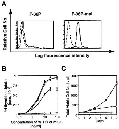
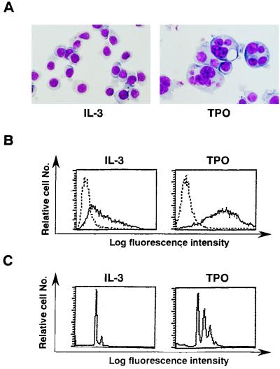
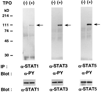
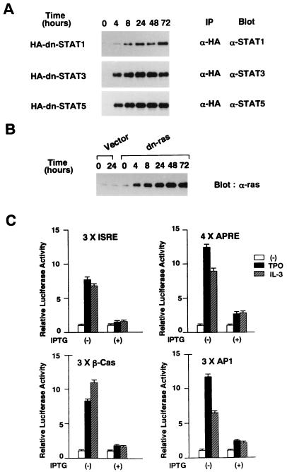
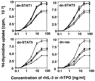
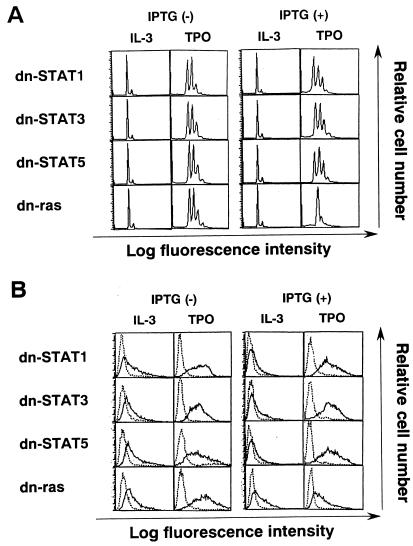
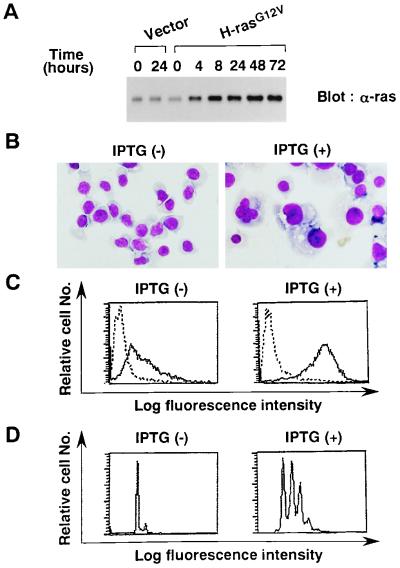
Thrombopoietin (TPO) is a hematopoietic growth factor that plays fundamental roles is both megakaryopoiesis and thrombopoiesis through binding to its receptor, c-mpl. Although TPO has been shown to activate various types of intracellular signaling molecules, such as the Janus family of protein tyrosine kinases, signal transducers and activators of transcription (STATs), and ras, the precise mechanisms underlying TPO-induced proliferation and differentiation remain unknown. In an effort to clarify the mechanisms of TPO-induced proliferation and differentiation, c-mpl was introduced into F-36P, a human interleukin-3 (IL-3)-dependent erythroleukemia cell line, and the effects of TPO on the c-mpl-transfected F-36P (F-36P-mpl) cells were investigated. F-36P-mpl cells were found to proliferate and differentiate at a high rate into mature megakaryocytes in response to TPO. Dominant-negative (dn) forms of STAT1, STAT3, STAT5, and ras were inducibly expressed in F-36P-mpl cells, and their effects on TPO-induced proliferation and megakaryocytic differentiation were analyzed. Among these dn molecules, both dn ras and dn STAT5 reduced TPO- or IL-3-induced proliferation of F-36P-mpl cells by approximately 30%, and only dn ras could inhibit TPO-induced megakaryocytic differentiation. In accord with this result, overexpression of activated ras (H-rasG12V) for 5 days led to megakaryocytic differentiation of F-36P-mpl cells. In a time course analysis on H-rasG12V-induced differentiation, activation of the ras pathway for 24 to 28 h was required and sufficient to induce megakaryocytic differentiation. Consistent with this result, the treatment of F-36P-mpl cells with TPO was able to induce prolonged activation of ras for more than 24 h, whereas IL-3 had only a transient effect. These results suggest that prolonged ras activation may be involved in TPO-induced megakaryocytic differentiation.


Figures

FIG. 1
FIG. 1
(A) Flow cytometric analysis of F-36P and F-36P-mpl cells. The expression of c-Mpl was examined by staining with rabbit anti-c-Mpl serum (——) or preimmune rabbit serum (–––). (B) Dose response of F-36P and F-36P-mpl to rhIL-3 and rhTPO. Triplicate aliquots of cells were cultured in serum-free medium with various concentration of rhIL-3 or rhTPO for 48 h, and then cell proliferation was measured by a [3H]thymidine incorporation assay (F-36P; □, IL-3; ○, TPO; F-36P-mpl; ▪, IL-3; •, TPO). (C) Changes in the total viable cell number during culture with or without rhIL-3 or rhTPO. F-36P-mpl cells were resuspended in 10% fetal calf serum–RPMI containing 10-ng/ml rhIL-3 or 30-ng/ml rhTPO at a cell density of 100/μl, and the total viable cell number was determined by the trypan blue dye exclusion method at the time indicated (□, IL-3; ○, TPO; ▵, free of growth factor). The results shown are the means ± the standard deviations of triplicate cultures.
FIG. 2
FIG. 2
(A) Light micrograph of F-36P-mpl cells after a 5-day culture with rhIL-3 or rhTPO. A cytocentrifugation preparation from each culture was stained with May-Grunwald-Giemsa (original magnification, ×100). (B and C) Flow cytometric analyses of F-36P-mpl cells. Expression of GPIIb/IIIa was examined by staining with MAb AP2 (——) and a control Ab of the same isotype (–––) (B). The DNA content of F-36P-mpl cells was examined by staining with PI solution and analysis on a FACSort apparatus (C).
FIG. 3
FIG. 3
TPO-induced tyrosine phosphorylation of STAT proteins in F-36P-mpl cells. F-36P-mpl cells were serum and factor starved for 12 h and then either left unstimulated or stimulated with rhTPO for 15 min. Total cell lysates were immunoprecipitated with an anti-STAT1, an anti-STAT3, or an anti-STAT5b Ab. The blots were probed with an antiphosphotyrosine (α-PY) MAb. The filters were then stripped and reprobed with anti-STAT1, anti-STAT3, and anti-STAT5b Abs, respectively.
FIG. 4
FIG. 4
(A and B) Inducible expression of dn STATs and dn ras. Each clone was treated with 0.5 mM IPTG for the times indicated. HA-immunoprecipitated proteins (A) or total cell lysates (B) were subjected to SDS-PAGE. The blots were probed with anti-STAT1, anti-STAT3, anti-STAT5b, and anti-ras Abs. (C) Effects of dn mutants on each signaling pathway. Four types of reporter plasmids, each containing 3× ISRE, 4× APRE, 3× β-Cas, and 3× AP1, were used as reporter genes. F-36P-mpl cells were electroporated with 30 μg of the reporter gene together with 30 μg of pRL-CMV-Rluc. The cells were serum and IL-3 starved for 12 h and then stimulated with rhTPO (30 ng/ml) or rhIL-3 (100 ng/ml) for 5 h. To examine the effects of dn mutants, the cells were pretreated with 0.5 mM IPTG for 24 h before electroporation and cultured with IPTG during the assay. Relative firefly luciferase activities were calculated by normalizing transfection efficiency to renilla luciferase activities. The results shown are means ± the standard deviations of triplicate experiments. Open bars, unstimulated.
FIG. 5
FIG. 5
Effects of dn STATs and dn ras on rhTPO- and rhIL-3-induced proliferation. Cells of each clone were IL-3 starved for 24 h and then cultured for 48 h with rhTPO or rhIL-3 at the concentrations indicated. The cells were left untreated or treated with 0.5 mM IPTG during the 24-h starvation period and the 48-h culture period. Cell proliferation was quantitated with a [3H]thymidine incorporation assay. The results shown are means ± the standard deviations of triplicate experiments. □, IL-3 without IPTG; ▪, IL-3 with IPTG; ○, TPO without IPTG; •, TPO with IPTG.
FIG. 6
FIG. 6
Light micrograph of F-36P-mpl cells. Clones were cultured for 5 days under the conditions indicated. To examine the effects of dn mutants, the cells were pretreated with 0.5 mM IPTG for 24 h and then cultured in the presence of 0.5 mM IPTG for 5 days. A cytocentrifugation preparation from each culture was stained with May-Grunwald-Giemsa (original magnification, ×100).
FIG. 7
FIG. 7
Flow cytometric analyses of dn mutant clones after a 5-day culture (as described in the legend to Fig. 6) with rhTPO. DNA content was examined by staining with PI solution and analysis on a FACSort apparatus (A). Expression of GPIIb/IIIa was examined by staining with MAb AP2 (——) and a control Ab of the same isotype (–––) (B).
FIG. 8
FIG. 8
Effects of H-rasG12V on megakaryocytic differentiation. (A) Inducible expression of H-rasG12V. F-36P-H-rasG12V and F-36P-Vector (transfected with an empty vector) cells were treated with 0.5 mM IPTG for the times indicated, and expression of H-rasG12V was examined by Western blot analysis. (B) Light micrograph of F-36P-H-rasG12V after a 5-day culture with or without IPTG (original magnification, ×100). (C and D) Flow cytometric analyses of GPIIb/IIIa expression (C) and DNA content (D) after a 5-day culture with or without IPTG.
FIG. 9
FIG. 9
(A) Northern blot analysis of luciferase mRNA. F-36P-AP1-Lu cells were serum and rhIL-3 starved for 24 h and then stimulated with rhTPO (30 ng/ml) or rhIL-3 (10 ng/ml) for the times indicated. Expression of luciferase mRNA was examined by Northern blot analysis. (B) Detection of an active, GTP-bound form of ras during treatment with rhTPO or rhIL-3. F-36P-mpl cells were serum and rhIL-3 starved for 24 h and then stimulated with rhTPO (30 ng/ml) or rhIL-3 (10 ng/ml) for the times indicated. GTP-bound ras was pulled from total cell lysates by incubation with a GST-RBD fusion protein precoupled with glutathione-Sepharose beads. The beads were collected and subjected to SDS-PAGE. Immunoblotting was performed with a murine anti-ras MAb (17). The total amount of ras was estimated by immunoblotting with an anti-ras MAb using total cell lysates.
FIG. 10
FIG. 10
(A) Changes in H-rasG12V expression after IPTG deprivation. F-36P-H-rasG12V cells were treated with IPTG for 24 h, washed, and then cultured in IPTG-free medium for up to 12 h. Changes in H-rasG12V expression were examined by Western blot analysis. (B) Time-dependent effects of H-rasG12V on megakaryocytic differentiation. F-36P-H-rasG12V cells were treated with IPTG for the times indicated, washed, and then resuspended in IPTG-free culture medium. At 120 h after the addition of IPTG, GPIIb/IIIa expression and DNA content were determined by flow cytometric analyses.

Similar articles

Cited by

References

    1. Alexander W S, Maurer A B, Novak U, Harrison-Smith M. Tyrosine-599 of the c-Mpl receptor is required for Shc phosphorylation and the induction of cellular differentiation. EMBO J. 1996;15:6531–6540. - PMC - PubMed
    1. Chiba S, Takaku F, Tange T, Shibuya K, Misawa C, Sasaki K, Miyagawa K, Yazaki Y, Hirai H. Establishment and erythroid differentiation of a cytokine-dependent human leukemia cell line F-36P: a parental line requiring granulocyte-macrophage colony-stimulating factor or interleukin-3, and a subline requiring erythropoietin. Blood. 1991;78:2261–2268. - PubMed
    1. Chretien S, Varlet P, Verdier F, Gobert S, Cartron J-P, Gisselbrecht S, Mayeux P, Lacombe C. Erythropoietin-induced erythroid differentiation of the human erythroleukemia cell line TF-1 correlates with impaired STAT5 activation. EMBO J. 1996;15:4174–4181. - PMC - PubMed
    1. de Rooij J, Bos J L. Minimal ras-binding domain of raf1 can be used as an activation-specific probe for ras. Oncogene. 1997;14:623–625. - PubMed
    1. de Sauvage F J, Carver-Moore K, Luoh S M, Ryan A, Dowd M, Eaton D L, Moore M W. Physiological regulation of early and late stages of megakaryopoiesis by thrombopoietin. J Exp Med. 1996;183:651–656. - PMC - PubMed

Publication types

MeSH terms