. 1998 Jul;18(7):4301-14.
doi: 10.1128/MCB.18.7.4301.

CCAAT/enhancer binding protein alpha is a regulatory switch sufficient for induction of granulocytic development from bipotential myeloid progenitors

Affiliations

CCAAT/enhancer binding protein alpha is a regulatory switch sufficient for induction of granulocytic development from bipotential myeloid progenitors

H S Radomska et al. Mol Cell Biol. 1998 Jul.

Abstract

The transcription factor CCAAT/enhancer binding protein alpha (C/EBPalpha) regulates a number of myeloid cell-specific genes. To delineate the role of C/EBPalpha in human granulopoiesis, we studied its expression and function in human primary cells and bipotential (granulocytic/monocytic) myeloid cell lines. We show that the expression of C/EBPalpha initiates with the commitment of multipotential precursors to the myeloid lineage, is specifically upregulated during granulocytic differentiation, and is rapidly downregulated during the alternative monocytic pathway. Conditional expression of C/EBPalpha alone in stably transfected bipotential cells triggers neutrophilic differentiation, concomitant with upregulation of the granulocyte-specific granulocyte colony-stimulating factor receptor and secondary granule protein genes. Moreover, induced expression of C/EBPalpha in bipotential precursors blocks their monocytic differentiation program. These results indicate that C/EBPalpha serves as a myeloid differentiation switch acting on bipotential precursors and directing them to mature to granulocytes.


Figures

FIG. 1
FIG. 1
Human C/EBPα mRNA and protein increase with granulocytic and shut off with monocytic differentiation. (a) Human C/EBPα mRNA is expressed specifically in cells of the myeloid lineage, as determined by Northern blotting of total RNA isolated from nonhematopoietic HeLa cells (lane 1), Jurkat T cells (lane 2), BJA-B (lane 3) and Raji (lane 4) B cells, promyelocytic Mono Mac 6 (lane 5), U937 (lane 6), and HL-60 (lane 7) cells, and K562 erythroleukemia cells (lane 8). The same blot was hybridized sequentially with a probe for human C/EBPα (hC/EBPα) and for 18S rRNA, as indicated to the right. Positions of migration of 28S and 18S rRNAs are marked on the left. Exposure was for 5 days with an intensifying screen. (b) The C/EBPα gene is activated at the stage of hematopoietic stem cell commitment to the myeloid lineage. Single human bone marrow cells were isolated as described in Materials and Methods and analyzed by Southern blotting of PCR-amplified cDNA. Shown are hybridization of the blot to a human C/EBPα-specific probe (top) and ethidium bromide staining of the gel before blotting (bottom). Each panel (bracketed on top) contains cDNA from five individual cells representing various stages of differentiation (marked above each bracket). Exposure was for 42 h with an intensifying screen. (c) C/EBPα is preferentially expressed in primary granulocytic cells, as determined by Northern blotting of total RNA purified from normal human peripheral blood monocytes and neutrophils. HeLa and HL-60 RNAs were included as negative and positive controls, respectively. The top panel shows results from hybridization to the human C/EBPα probe. Exposure was for 3 days with an intensifying screen. The same blot was stripped and rehybridized to an IL-8 receptor B (IL8RB; neutrophil-specific) probe (middle panel; 2-day exposure) and then to a 28S oligonucleotide as an internal control (bottom panel). (d) Expression of C/EBPα mRNA increases during granulocytic induction of myeloid cell lines and shuts off with monocytic differentiation. K562, U937, HL-60, and NB4 cells were stimulated to erythroid differentiation with DMSO, to monocytic differentiation with TPA, or to granulocytic differentiation with retinoic acid (RA); cell aliquots were withdrawn at the time points indicated above the blot, and RNA was analyzed by Northern blotting. Top, expression of human C/EBPα; middle, expression of the myeloid maturation marker CD18; bottom, 18S RNA as a control for RNA loading and quantitation of C/EBPα expression. Exposure was for 3 days (left) and 4 days (right) for C/EBPα and 4 days (left) and 18 h (right) for CD18, using an intensifying screen. (e) C/EBPα protein increases with retinoic acid-induced granulocytic induction of myeloid cell lines and decreases with TPA-induced monocytic differentiation. Shown is a Western blot of whole-cell extracts from uninduced U937 (lanes 1 and 11) and HL-60 (lanes 6 and 14) cells and retinoic acid (lanes 2 to 5 and 7 to 10)- or TPA (lanes 12, 13, 15, and 16)-induced cells. The cell lines and the inducers, as well as the times of induction in days, are indicated above the lanes. In the top panel, the blot was stained with anti-C/EBPα antibody; in the bottom panel, the same blot was stripped and subsequently stained with anti-β-tubulin antibody. Molecular size standards are shown in kilodaltons to the left. Arrows on the right indicate full-length C/EBPα and β-tubulin, and the asterisk indicates truncated C/EBPα protein (45, 53).
FIG. 2
FIG. 2
Conditional expression of C/EBPα is sufficient for induction of granulocytic differentiation of bipotential U937 cells. Two individual stable clones of U937 cells with an empty expression vector, U937(vect)#1 (A) and U937(vect)#3 (B), were grown in zinc-containing medium for 17 days. Three individual clones of U937 cells were transfected with an inducible C/EBPα expression vector and grown in the absence of zinc (C [U937α#2]) or the presence of zinc for 17 days (D [U937α#2], E [U937α#3], and F [U937α#5]). Cell aliquots were centrifuged onto glass slides and stained with Wright-Giemsa stain. N, representative cell with the appearance of a polymorphonuclear granulocyte.
FIG. 3
FIG. 3
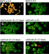
Induced expression of C/EBPα does not lead to apoptosis. IL-3-dependent BaF3 cells cultured in medium supplemented with IL-3 (B; negative control), or in conditions such that IL-3 was withdrawn for 24 h (A; positive control, showing apoptosis of nearly all cells as demonstrated by typical orange-yellow staining of the cell nuclei), as well as U937 cells stably transfected with the empty expression vector [U937(vect)#1] and cultured for 17 days in the presence of zinc (C), or transfected with the rat C/EBPα construct (U937α#2) and grown in the absence (D) or presence of zinc (E and F), were tested by TUNEL assay for apoptosis. Panels E and F show different fields from the same slide. The red arrow in panel F indicates an apoptotic cell.
FIG. 4
FIG. 4
Induced expression of C/EBPα causes upregulation of granulocytic maturation marker genes. (a) Northern blot with total RNA isolated from the U937 cell line stably transfected with the inducible rat C/EBPα expression gene (clone α#2). Prior to RNA extraction cells were cultured for 13 days in zinc-deficient (−) or zinc-supplemented (+) medium, as indicated at the top. The blot was sequentially hybridized to probes indicated below each panel. Hybridization to 18S rRNA served as an internal control for loading and RNA transfer. The migration of 28S and 18S rRNAs is indicated to the left. Similar results were obtained with other stable clones of U937 and HL-60 cells. (b) Western blot of protein extracts from parental untransfected lines (U937 [lane 2] and HL-60 [lane 9]) and their C/EBPα transfectants (U937α#2, U937α#3, and HL60α#5). C/EBPα-negative K562 extract served as a control of antibody specificity (lane 1). Cells were grown in the absence of zinc (−Zn) or in the presence of zinc (+Zn) for 1 day (d1), 2 days (d2), or 3 days (d3), as indicated. Positions of molecular size markers are shown in kilodaltons on the left, and the arrow on the right marks the migration of the 42-kDa C/EBPα protein detected by the anti-rat C/EBPα antibody, which also detects the endogenous human C/EBPα protein. All lanes except lane 9 contained 20 μg of protein extract; in lane 9, 40 μg of total protein was loaded.
FIG. 5
FIG. 5
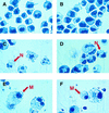
Induced expression of C/EBPα prevents monocytic differentiation. Shown are Wright-Giemsa-stained cytospin preparations of U937α#2 cells. (A) Cells grown in zinc-deficient medium for 9 days. (B) Cells grown in zinc-containing medium for 9 days. (C and D) Cells cultured in the presence of zinc for 7 days, with TPA then added for 2 days, in the continued presence of zinc (total of 9 days of zinc treatment). N, representative cell with the appearance of a polymorphonuclear granulocyte. (E and F) Cells treated with TPA for 2 days in the absence of zinc. M, representative cell with the morphology of a macrophage.
FIG. 6
FIG. 6
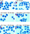
Short-term induction of C/EBPα expression in U937 cells is sufficient for morphological granulocytic maturation. C/EBPα-transfected U937 cells (clone U937α#2) were cultured in the absence (A) or continued presence (B) of zinc for 16 days. The same cell line was also initially seeded in medium containing zinc, subsequently washed after 1 day (E and F), 2 days (D), or 3 days (C), and maintained in zinc-deficient medium for an additional 20 days (C), 21 days (D), 22 days (E), or 23 days (F).
FIG. 7
FIG. 7
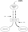
Model of bipotential myeloid lineage differentiation. The expression of increased levels of C/EBPα in a bipotential myeloid precursor induces genetic changes over the course of a week that lead to irreversible granulocytic differentiation. Prior to this switch point, monocytic differentiation agents such as TPA can induce macrophage development, but subsequent to this point the same agents can accelerate granulocytic differentiation. After the switch point, C/EBPα silencing is required for the monocytic pathway to proceed, as forced expression of C/EBPα prevents monocytic differentiation. At present, the factors which direct monocytic maturation of the bipotential myeloid precursor cell are not clearly defined, but candidate genes include the egr-1 family (48) and/or PU.1 (30).

Similar articles

Cited by

References

    1. Ahuja S K, Shetty A, Tiffany H L, Murphy P M. Comparison of the genomic organization and promoter function for human interleukin-8 receptors A and B. J Biol Chem. 1994;269:26381–26389. - PubMed
    1. Andrews R G, Bryant E M, Bartelmez S H, Muirhead D Y, Knitter G H, Bensinger W, Strong D M, Bernstein I D. CD34+ marrow cells, devoid of T and B lymphocytes, reconstitute stable lymphopoiesis and myelopoiesis in lethally irradiated allogeneic baboons. Blood. 1992;80:1693–1701. - PubMed
    1. Antonson P, Stellan B, Yamanaka R, Xanthopoulos K G. A novel human CCAAT/enhancer binding protein gene, C/EBPɛ, is expressed in cells of lymphoid and myeloid lineages and is localized on chromosome 14q11.2 close to the T-cell receptor α/δ locus. Genomics. 1996;35:30–38. - PubMed
    1. Antonson P, Xanthopoulos K G. Molecular cloning, sequence, and expression patterns of the human gene encoding CCAAT/enhancer binding protein α (C/EBPα) Biochem Biophys Res Commun. 1995;215:106–113. - PubMed
    1. Barbu V, Dautry F. Northern blot normalization with a 28S rRNA oligonucleotide probe. Nucleic Acids Res. 1989;17:7115. - PMC - PubMed

Publication types