. 1998 Jul;18(7):4315-23.
doi: 10.1128/MCB.18.7.4315.

Interactions among Drosophila nuclear envelope proteins lamin, otefin, and YA

Affiliations

Interactions among Drosophila nuclear envelope proteins lamin, otefin, and YA

M Goldberg et al. Mol Cell Biol. 1998 Jul.

Abstract

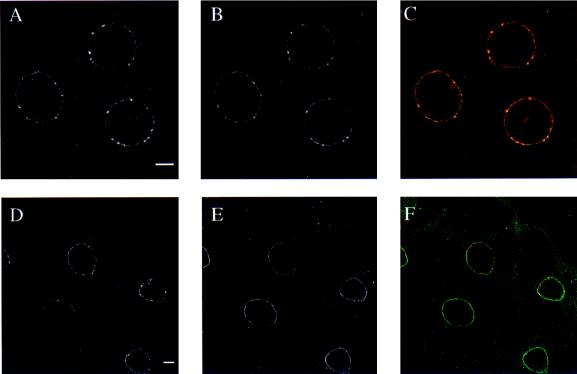
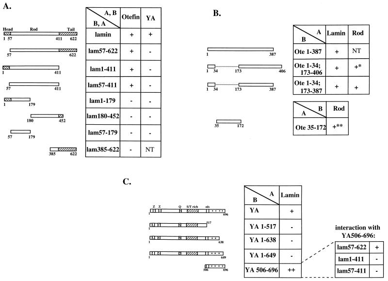
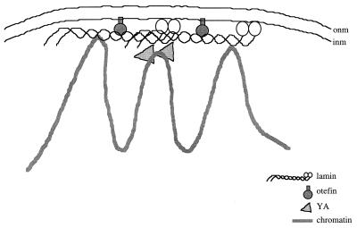
The nuclear envelope plays many roles, including organizing nuclear structure and regulating nuclear events. Molecular associations of nuclear envelope proteins may contribute to the implementation of these functions. Lamin, otefin, and YA are the three Drosophila nuclear envelope proteins known in early embryos. We used the yeast two-hybrid system to explore the interactions between pairs of these proteins. The ubiquitous major lamina protein, lamin Dm, interacts with both otefin, a peripheral protein of the inner nuclear membrane, and YA, an essential, developmentally regulated protein of the nuclear lamina. In agreement with this interaction, lamin and otefin can be coimmunoprecipitated from the vesicle fraction of Drosophila embryos and colocalize in nuclear envelopes of Drosophila larval salivary gland nuclei. The two-hybrid system was further used to map the domains of interaction among lamin, otefin, and YA. Lamin's rod domain interacts with the complete otefin protein, with otefin's hydrophilic NH2-terminal domain, and with two different fragments derived from this domain. Analogous probing of the interaction between lamin and YA showed that the lamin rod and tail plus part of its head domain are needed for interaction with full-length YA in the two-hybrid system. YA's COOH-terminal region is necessary and sufficient for interaction with lamin. Our results suggest that interactions with lamin might mediate or stabilize the localization of otefin and YA in the nuclear lamina. They also suggest that the need for both otefin and lamin in mediating association of vesicles with chromatin might reflect the function of a protein complex that includes these two proteins.


Figures

FIG. 1
FIG. 1
Coimmunoprecipitation of lamin and otefin from extracts of membrane vesicles. Proteins were extracted with buffer containing 0.5 M NaCl from membrane vesicles prepared from 0- to 6-h-old Drosophila embryos. Immunoprecipitation of proteins from the extract was with affinity-purified polyclonal antiotefin antibodies (left panel) or with affinity-purified antilamin antibodies (right panel). Lanes 1, total nuclear extracts from 0- to 6-h-old embryos (lane 1 in the right panel contains 3.5 times more extract than lane 1 in the left panel; lanes 2, proteins immunoprecipitated with affinity-purified antibodies as noted above; lanes 3, proteins immunoprecipitated with total Ig. The proteins were separated by SDS-10% PAGE and subjected to protein blot analysis with MAb 611A3A6 antilamin (left panel) and MAb 618A2O7 antiotefin (right panel). The positions of lamin isoforms Dm1 (76 kDa), Dmmit (75 kDa), and Dm2 (74 kDa) and the position of otefin (53 kDa) are indicated.
FIG. 2
FIG. 2
Lamin and otefin colocalize in salivary gland nuclei. Salivary glands were dissected from third-instar larvae, fixed, and probed with affinity-purified polyclonal antibodies specific for otefin (A and D), monoclonal antilamin antibody 611A3A6 (B), and monoclonal antibody RL1 against O-linked sugar-containing glycoproteins (E). FITC-conjugated goat-anti-mouse IgG and Cy3-conjugated goat-anti-rabbit IgG were used as the secondary antibodies for the monoclonal and polyclonal antibodies, respectively. To assess colocalization visually in double-labeled salivary gland cells (panels A and B, and panels D and E), the images from the Cy3 channel and the FITC channel were combined into a 24-bit RGB image as the red and green components, respectively (C and F). The resulting color image was yellow where the red and green features overlapped. Bar, 10 μm.
FIG. 3
FIG. 3
Characterization of the interaction domains in lamin, otefin, and YA. A and B, vectors containing the GAL4 activation domain and binding domain, respectively. The otefin and lamin constructs were cloned in frame in either A or B as indicated; YA was fused in frame to B. All constructs were tested for induction of β-Gal, and all except YA506-696 and lam385-622 were tested for histidine-independent growth as described in Materials and Methods. +, detection of lacZ and (when tested) his reporter gene expression with levels as shown in Table 1; −, lack of detectable signals (again as shown in Table 1); +*, histidine-independent growth of transformed HF7c, but no detectable activity of lacZ in transformed SFY526; +**, weak his reporter gene expression, as indicated by reduced, but still detectable, ability of transformed HF7c to grow on plates lacking histidine, as compared to a positive control; NT, not tested. All fusion constructs in either vector are not able to induce β-Gal activity or permit histidine-independent growth ability when cotransformed with the other empty vector, and all fusion constructs (except for lam385-622 and YA506-696, which were not tested) resulted in the production of fusion proteins of the correct size and antigenicity (data not shown). (A) To define the region of lamin that interacts with otefin, combinations of full-length otefin constructs in vector A were cotransformed into yeast hosts SFY526 and HF7c with lamin deletion constructs (diagrammed on the left; lamin’s head, rod, and tail are marked) in vector B. The same results were obtained with the reciprocal construct-vector pairs, except that interaction was not detected between otefin fused to the binding domain and lam 57-411 or lam 1-411 fused to the activation domain. As described in Materials and Methods, in the case of such conflicts, we consider the positive results significant, since changes in the conformation of the fusion protein could have abolished interaction in the negative cases (11). To test lamin regions needed for interaction with YA, full-length YA in vector B was cotransformed with the lamin deletion constructs in vector A. (B) To define the region of otefin that interacts with lamin, two-hybrid fusions of otefin deletions (diagrammed on the left) containing aa 1 to 387, aa 1 to 34 and 173 to 406, or 1 to 34 and 173 to 387 were introduced into the yeast strains along with full-length lamin fusions. Results with the otefin deletions in vector B and lamin in vector A are shown. In the reciprocal vector-insert pairs, the doubly deleted otefin (ote1-34; 173-387) interacted with full-length lamin, although the singly deleted otefins (ote1-387 or ote1-34; 173-406) did not. In the conflicting cases, we consider the positive results significant, as discussed in Materials and Methods. We tested for interaction between ote35-172 and the lamin rod domain using only the vector-insert combination shown in the figure. (C) To determine whether YA’s COOH terminus is needed for interaction with lamin, we tested the YA deletions or fragments diagrammed on the left, in vector B, for interaction with full-length lamin and, for YA506-696, with the lamin deletions shown, cloned in vector A. For this panel only, relative signal strengths are indicated (by the number of plus signs) to indicate why we could detect an interaction between the lamin deletions and the YA C terminus but not between those deletions and full-length YA (in panel A). The diagrams show YA’s two potential zinc fingers (Z), glutamine-rich (Q) and serine/threonine-rich (S/T rich) regions, its two putative nuclear localization signals (nls), and its polar COOH terminus (∗∗∗) (34, 37).
FIG. 4
FIG. 4
YA interactor candidates hybridize to 32P-labeled lamin probes on Southern blots. Plasmids retrieved from candidate yeast colonies were digested with EcoRI to release their cDNA inserts. Examples of two representative isolates are shown in lanes 1 and 2 (100 ng/lane). 32P-labeled lamin Dm0 probes were used to probe the inserts (see Materials and Methods). As a positive control, we loaded the same amount of plasmid pGBTT-lam57-411 (lamin rod domain; insert size, 1.3 kb) digested with EcoRI (lane 3).
FIG. 5
FIG. 5
A model for the organization of lamin, otefin, and YA in the nuclear envelope. Otefin interacts with both lamin (this study) and the inner nuclear membrane (1, 2) and hence is shown on the membrane-facing side of the nuclear lamina. The contacts of otefin with lamin require only lamin’s rod domain. YA associates with lamin (this study) and chromatin (40) and hence is placed at the nucleoplasmic side of the nuclear lamina and in contact with chromatin; YA is also shown as self-associating as reported previously (38). Lamin is shown associating with itself, with chromatin and, via its C terminus, with the inner nuclear membrane as reported elsewhere (4, 7, 57, 58, 64). onm, outer nuclear membrane; inm, inner nuclear membrane.

Similar articles

Cited by

References

    1. Ashery-Padan R, Ulitzur N, Arbel A, Goldberg M, Weiss A M, Maus N, Fisher P A, Gruenbaum Y. Localization and posttranslational modification of otefin, a protein required for vesicle attachment to chromatin, during Drosophila melanogaster development. Mol Cell Biol. 1997;17:4114–4123. - PMC - PubMed
    1. Ashery-Padan R, Weiss A M, Feinstein N, Gruenbaum Y. Distinct regions specify the targeting of otefin to the nucleoplasmic side of the nuclear envelope. J Biol Chem. 1997;272:2493–2499. - PubMed
    1. Ashery-Padan, R., and Y. Gruenbaum. Unpublished observations.
    1. Ausubel F M, Brent R, Kingston R E, Moore D D, Seidman J G, Smith J A, Struhl K. Current protocols in molecular biology. New York, N.Y: John Wiley & Sons, Inc.; 1994.
    1. Baricheva E A, Berrios M, Bogachev S S, Borisevich I V, Lapik E R, Sharakhov I V, Stuurman N, Fisher P A. DNA from Drosophila melanogaster beta-heterochromatin binds specifically to nuclear lamins in vitro and the nuclear envelope in situ. Gene. 1996;171:171–176. - PubMed

Publication types