. 1998 Jul;18(7):4324-36.
doi: 10.1128/MCB.18.7.4324.

PEX12, the pathogenic gene of group III Zellweger syndrome: cDNA cloning by functional complementation on a CHO cell mutant, patient analysis, and characterization of PEX12p

Affiliations

PEX12, the pathogenic gene of group III Zellweger syndrome: cDNA cloning by functional complementation on a CHO cell mutant, patient analysis, and characterization of PEX12p

K Okumoto et al. Mol Cell Biol. 1998 Jul.

Abstract

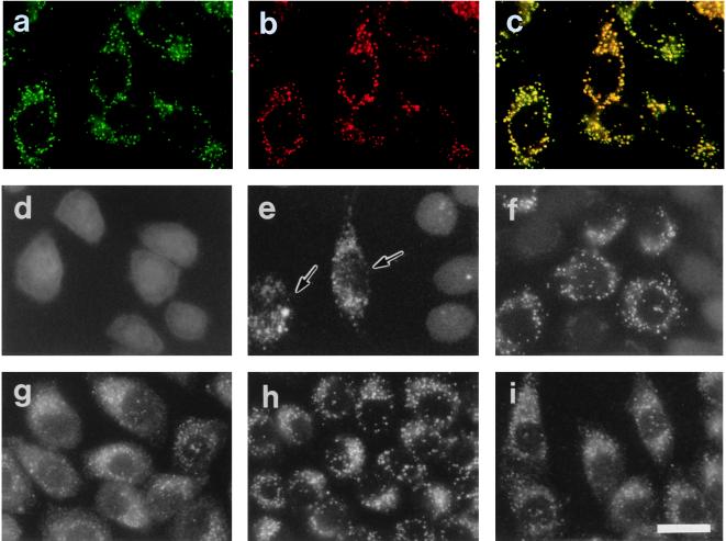
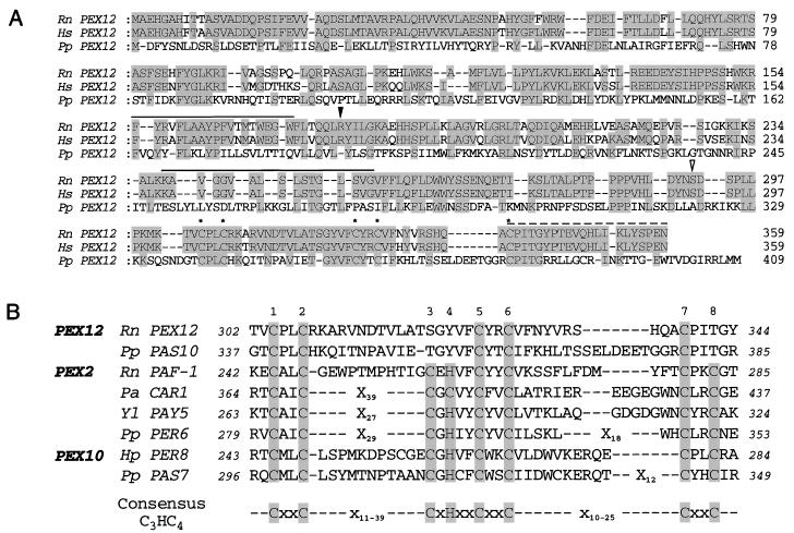
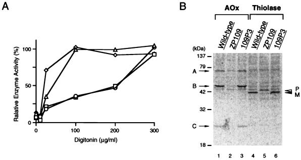
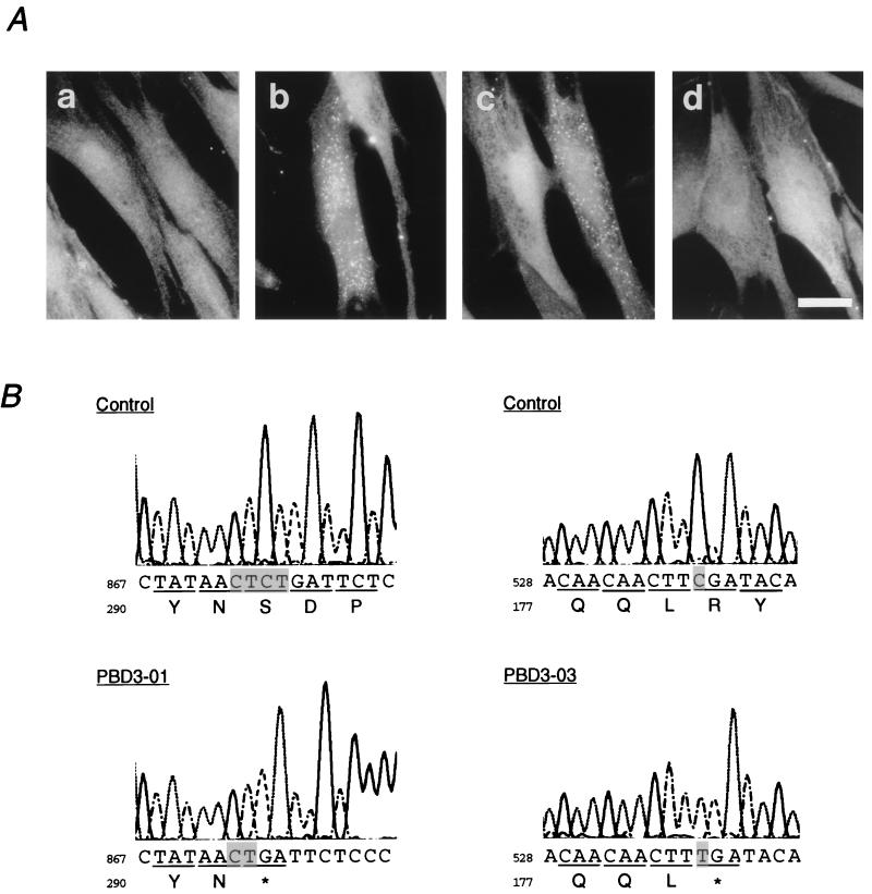
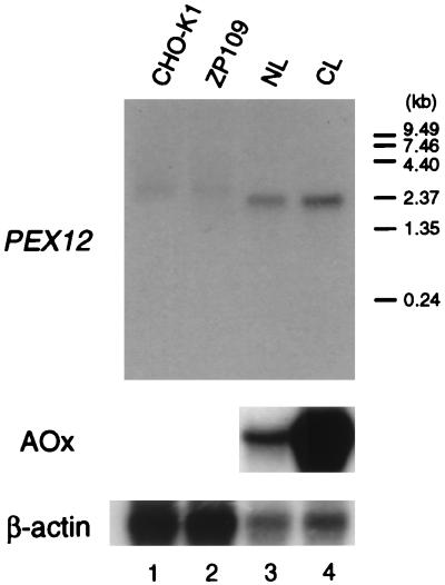
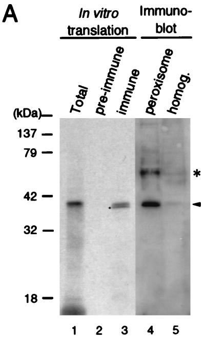
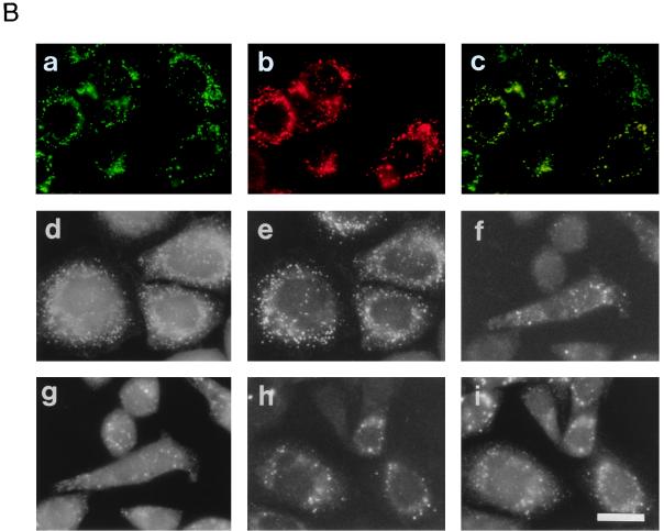
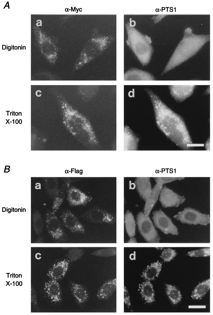
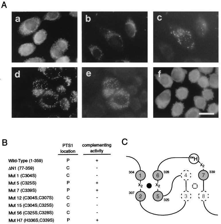
Rat PEX12 cDNA was isolated by functional complementation of peroxisome deficiency of a mutant CHO cell line, ZP109 (K. Okumoto, A. Bogaki, K. Tateishi, T. Tsukamoto, T. Osumi, N. Shimozawa, Y. Suzuki, T. Orii, and Y. Fujiki, Exp. Cell Res. 233:11-20, 1997), using a transient transfection assay and an ectopic, readily visible marker, green fluorescent protein. This cDNA encodes a 359-amino-acid membrane protein of peroxisomes with two transmembrane segments and a cysteine-rich zinc finger, the RING motif. A stable transformant of ZP109 with the PEX12 was morphologically and biochemically restored for peroxisome biogenesis. Pex12p was shown by expression of bona fide as well as epitope-tagged Pex12p to expose both N- and C-terminal regions to the cytosol. Fibroblasts derived from patients with the peroxisome deficiency Zellweger syndrome of complementation group III (CG-III) were also complemented for peroxisome biogenesis with PEX12. Two unrelated patients of this group manifesting peroxisome deficiency disorders possessed homozygous, inactivating PEX12 mutations: in one, Arg180Thr by one point mutation, and in the other, deletion of two nucleotides in codons for 291Asn and 292Ser, creating an apparently unchanged codon for Asn and a codon 292 for termination. These results indicate that the gene encoding peroxisome assembly factor Pex12p is a pathogenic gene of CG-III peroxisome deficiency. Moreover, truncation and site mutation studies, including patient PEX12 analysis, demonstrated that the cytoplasmically oriented N- and C-terminal parts of Pex12p are essential for biological function.


Figures

FIG. 1
FIG. 1
Restoration of peroxisomes in CHO mutant cells. (a and d to f) Fluorescence microscopy of peroxisomes. Intracellular location of GFP in CHO cells stably expressing GFP tagged with tripeptide PTS1 (AKL) was monitored on unfixed cells, except for panel a, under fluorescence microscopy. (b and g to i) Immunofluorescent staining of peroxisomes. (a) Wild-type CHOG1 cells; (b) CHOG1 stained with goat anti-rat catalase antibody; (c) merged view of panels a and b; (d) peroxisome-deficient mutant ZP109G1 cells; (e) peroxisome-restored ZP109G1, after lipofection with a combined pool (B4) of rat cDNA library. Arrows indicate the complemented cells. Cytosolic appearance of GFP-AKL was apparent in the other cells. (f) ZP109G1 transfected with plasmid pUcD2Hyg · RnPEX12. (g to i) 109P3 cells (stable transformants of ZP109 cells obtained with pUcD2Hyg · RnPEX12) were stained with antisera to catalase, PTS1 peptide, and PMP70, respectively. Magnification, ×570; bar, 20 μm.
FIG. 2
FIG. 2
Deduced amino acid sequence of rat (R. norvegicus [Rn]) PEX12. (A) Alignment with human (Homo sapiens [Hs]) and P. pastoris (Pp) Pex12p. Dashes represent spaces. The putative membrane-spanning segment is overlined; the dashed line indicates the sequence used for chemical synthesis of the Pex12p peptide. Identical amino acids between two or more species are shaded. Conserved cysteine residues of the RING finger are designated by asterisks. Open and solid arrowheads indicate the positions of mutations in CG-III patients PBD3-01 and PBD3-03, respectively. (B) Conserved RING finger in the C-terminal regions of the peroxins required for peroxisome assembly. RING fingers were found in Pex2p of mammals (44, 54, 56), P. pastoris (59), Yarrowia lipolytica (Yl) (8), and Podospora anserina (Pa) (1), Pex10p of P. pastoris (19) and Hansenula polymorpha (Hp) (49), and Pex12p of P. pastoris (18). Italic numbers designate positions in respective deduced amino acid sequence; Xn, numbers of amino acid residues. Consensus cysteine and histidine residues in the RING are shaded and numbered (see text).
FIG. 3
FIG. 3
Complementation of biogenesis of peroxisomal enzymes. (A) Latency of catalase activity in CHO-K1, ZP109, and 109P3 cells. Circles, CHO-K1; triangles, ZP109; squares, 109P3, stable rat PEX12 transformant of ZP109; diamond, lactate dehydrogenase in CHO-K1. Relative free enzyme activity is expressed as a percentage of the total activity measured in the presence of 1% Triton X-100 (57). The results represent means of duplicate assays. (B) Biogenesis of peroxisomal proteins. Cells were labeled for 24 h with [35S]methionine and [35S]cysteine. Cell types are indicated at the top. Immunoprecipitation was done with rabbit anti-rat acyl-CoA oxidase (AOx) and 3-ketoacyl-CoA thiolase (Thiolase) antibodies. Immunoprecipitates were analyzed by SDS-PAGE (12% polyacrylamide gel). Radioactive polypeptide bands were detected using a FujiX BAS1000 Bio-Imaging Analyzer (Fuji Photo Film, Tokyo, Japan). Exposure time was 14 h. Arrows show the positions of AOx components A, B, and C; open and solid arrowheads indicate a larger precursor (P) and mature protein (M) of 3-ketoacyl-CoA thiolase, respectively.
FIG. 4
FIG. 4
Mutation analysis of PEX12 from CG-III patients. (A) Complementation of peroxisome assembly in CG-III fibroblasts. Transfection to fibroblasts from patient PBD3-01 of complementation CG-III (a) was done with rat and human PEX12 (b and c) or a PEX12 isolated from the PBD3-01 fibroblasts (d). Immunofluorescence microscopic analysis was done as for Fig. 1, using anti-human catalase antibody. Numerous peroxisomes were present in the cytoplasm of the transfected cells in panels b and c but not in panel d. Magnification, ×630; bar, 30 μm. (B) Nucleotide sequence analysis of PEX12 isolated from CG-III patients and a control. Partial sequence and deduced amino acid sequence are shown. A two-base deletion in the nucleotide sequence at residues 873 to 876 created a codon 292 (open arrowhead in Fig. 2A) for termination in patient PBD3-01 (left); a point mutation at nucleotide residue 538 changes a codon 180 for Arg (solid arrowhead in Fig. 2A) to a termination codon in a patient (PBD3-03) with Zellweger syndrome (right).
FIG. 5
FIG. 5
Northern blot analysis of PEX12 mRNA. RNA was separated, transferred to a Zeta-Probe GT membrane (Bio-Rad), and hybridized with 32P-labeled cDNA probes for rat PEX12 (top) and rat acyl-CoA oxidase (AOx; middle), respectively. Human β-actin (bottom) cDNA was used as a control probe to check the amount of RNA loaded. Washing was done twice with 0.15 M NaCl–10 mM sodium phosphate (pH 7.4)–1 mM EDTA–0.5% SDS at 60°C. Lanes: 1 and 2, poly(A)+ RNA (5 μg) from wild-type CHO-K1 and ZP109 cells, respectively; 3 and 4, total RNA (20 μg) from the livers of a normal (NL) and a clofibrate-treated (CL) rat, respectively. Exposure times were 90 h (top), 28 h (middle), and 24 h (bottom).
FIG. 6
FIG. 6
Intracellular localization of Pex12p. (A) Size comparison of rat liver Pex12p and in vitro transcription-translation product of Pex12p cDNA. The [35S]methionine-labeled in vitro transcription-translation product of rat PEX12 and rat liver fractions were subjected to SDS-PAGE and transferred to a polyvinylidene difluoride membrane. The autoradiograph (lanes 1 to 3) was exposed for 3 days; immunodetection (lanes 4 and 5) was done with rabbit anti-Pex12p peptide antibody. Lanes: 1, in vitro transcription-translation product (1 μl) of PEX12 cDNA; 2 and 3, immunoprecipitation of 35S-Pex12p (1 μl) with preimmune and anti-Pex12p immune sera, respectively; 4, peroxisomes (20 μg); 5, liver homogenates (50 μg). The dot indicates an apparently cleaved product of 35S-Pex12p during the immunoprecipitation; an unidentified band is shown by an asterisk. (B) Morphological analysis. Myc-tagged rat Pex12p was expressed in CHO-K1, Z65, and ZP92 cells. ZP109 was transfected with rat PEX12. (a and b) CHO-K1 cells stained with anti-Myc antibody and anti-PTS1 peptide antibody, respectively; (c) merged view of panels a and b; (d and e) ZP109 cells stained with antisera against Pex12p and catalase, respectively; (f and g) Z65 cells stained with antibodies to Myc and PMP70, respectively; (h and i) ZP92 cells stained with antibodies to Myc and PMP70, respectively. Magnification, ×500; bar, 20 μm.
FIG. 6
FIG. 6
Intracellular localization of Pex12p. (A) Size comparison of rat liver Pex12p and in vitro transcription-translation product of Pex12p cDNA. The [35S]methionine-labeled in vitro transcription-translation product of rat PEX12 and rat liver fractions were subjected to SDS-PAGE and transferred to a polyvinylidene difluoride membrane. The autoradiograph (lanes 1 to 3) was exposed for 3 days; immunodetection (lanes 4 and 5) was done with rabbit anti-Pex12p peptide antibody. Lanes: 1, in vitro transcription-translation product (1 μl) of PEX12 cDNA; 2 and 3, immunoprecipitation of 35S-Pex12p (1 μl) with preimmune and anti-Pex12p immune sera, respectively; 4, peroxisomes (20 μg); 5, liver homogenates (50 μg). The dot indicates an apparently cleaved product of 35S-Pex12p during the immunoprecipitation; an unidentified band is shown by an asterisk. (B) Morphological analysis. Myc-tagged rat Pex12p was expressed in CHO-K1, Z65, and ZP92 cells. ZP109 was transfected with rat PEX12. (a and b) CHO-K1 cells stained with anti-Myc antibody and anti-PTS1 peptide antibody, respectively; (c) merged view of panels a and b; (d and e) ZP109 cells stained with antisera against Pex12p and catalase, respectively; (f and g) Z65 cells stained with antibodies to Myc and PMP70, respectively; (h and i) ZP92 cells stained with antibodies to Myc and PMP70, respectively. Magnification, ×500; bar, 20 μm.
FIG. 7
FIG. 7
Topology of Pex12p. (A, a and b), CHO-K1 cells transfected with rat PEX12-myc were treated with 25 μg of digitonin per ml, under which the plasma membrane was permeabilized (28). (c and d) PEX12-myc-transfected CHO-K1 cells were treated with 1% Triton X-100. Cells were stained with anti-Myc antibody (a and c) and anti-PTS1 peptide antibody (b and d). Note that Pex12p-Myc was detected after both types of treatments. Magnification, ×630; bar, 20 μm. (B) CHO-K1 cells transfected with N-terminally flag-tagged rat PEX12 were treated with 25 μg of digitonin per ml (a and b) or with 1% Triton X-100 (c and d). Cells were stained with rabbit anti-flag antibody (a and c) and anti-PTS1 antibody (b and d). Magnification, ×630; bar, 20 μm.
FIG. 8
FIG. 8
N-terminal deletion and RING finger mutagenesis of Pex12p. (A) ΔN1, an N-terminally flag-tagged Pex12p variant lacking amino acid residues from the N terminus to position 76. Conserved cysteine residues of the RING finger were mutated to serine: C304S (mut1), C339S (mut7), and C325S/C328S (mut56). mut1 and mut7 were epitope tagged with flag and Myc at the N and C termini, respectively; mut56 was C-terminally tagged with Myc. ZP109 cells were transfected by PEX12 mutant ΔN1 (a), mut1 (b and c), mut7 (d and e), or mut56 (f). Cells were stained with antibodies to PTS1 (a, b, d, and f) and flag (c and e). Magnification, ×630; bar, 20 μm. (B) Complementing activity of Pex12p mutants. ZP109 was complemented (+) or not complemented (−), as assessed by PTS1 protein import. (C) Schematic view of the RING finger. One zinc finger is formed in Pex12p by four cysteine residues, C1 (at 304C), C2 (307C), C5 (325C), and C6 (328C), corresponding to those in authentic RING, C3HC4 (Fig. 2B). Solid circle, a divalent zinc ion; X2, two amino acids.

Similar articles

Cited by

References

    1. Berteaux-Lecellier V, Picard M, Thompson-Coffe C, Zickler D, Panvier-Adoutte A, Simonet J-M. A nonmammalian homolog of the PAF1 gene (Zellweger syndrome) discovered as a gene involved in caryogamy in the fungus Podospora anserina. Cell. 1995;81:1043–1051. - PubMed
    1. Braverman N, Steel G, Obie C, Moser A, Moser H, Gould S J, Valle D. Human PEX7 encodes the peroxisomal PTS2 receptor and is responsible for rhizomelic chondrodysplasia punctata. Nat Genet. 1997;15:369–376. - PubMed
    1. Chalfie M, Tu Y, Euskirchen G, Ward W W, Prasher D C. Green fluorescent protein as a marker for gene expression. Science (Washington, DC) 1994;263:802–805. - PubMed
    1. Chang C-C, Lee W-H, Moser H, Valle D, Gould S J. Isolation of the human PEX12 gene, mutated in group 3 of the peroxisome biogenesis disorders. Nat Genet. 1997;15:385–388. - PubMed
    1. Chomczynski P, Sacchi N. Single-step method of RNA isolation by acid guanidinium thiocyanate-PhOH-chloroform extraction. Anal Biochem. 1987;162:156–159. - PubMed

MeSH terms

Associated data