. 1998 Jul;18(7):4358-67.
doi: 10.1128/MCB.18.7.4358.

Targeting to transcriptionally active loci by the hydrophilic N-terminal domain of Drosophila DNA topoisomerase I

Affiliations

Targeting to transcriptionally active loci by the hydrophilic N-terminal domain of Drosophila DNA topoisomerase I

W L Shaiu et al. Mol Cell Biol. 1998 Jul.

Abstract

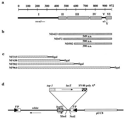
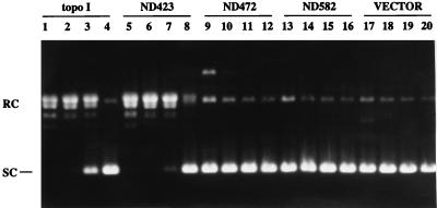
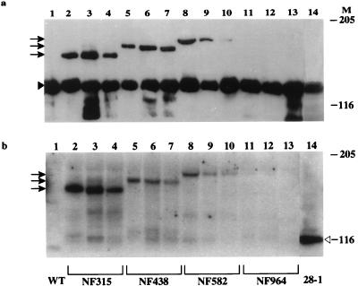
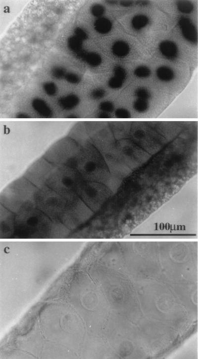
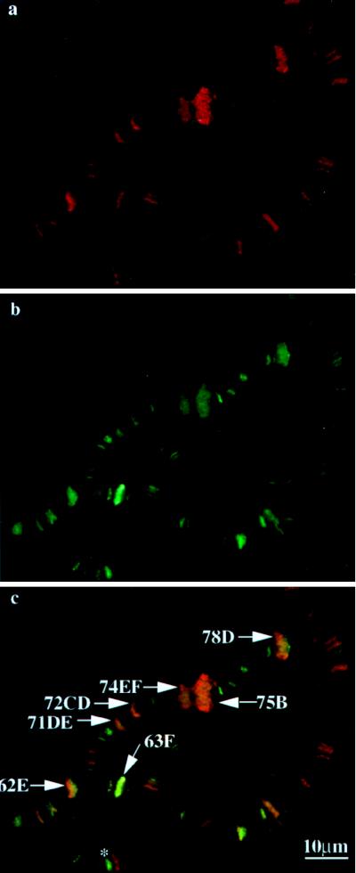
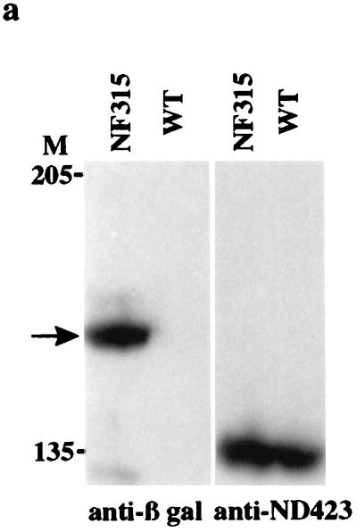
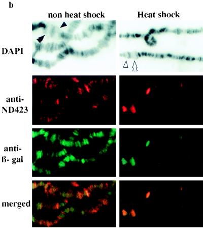
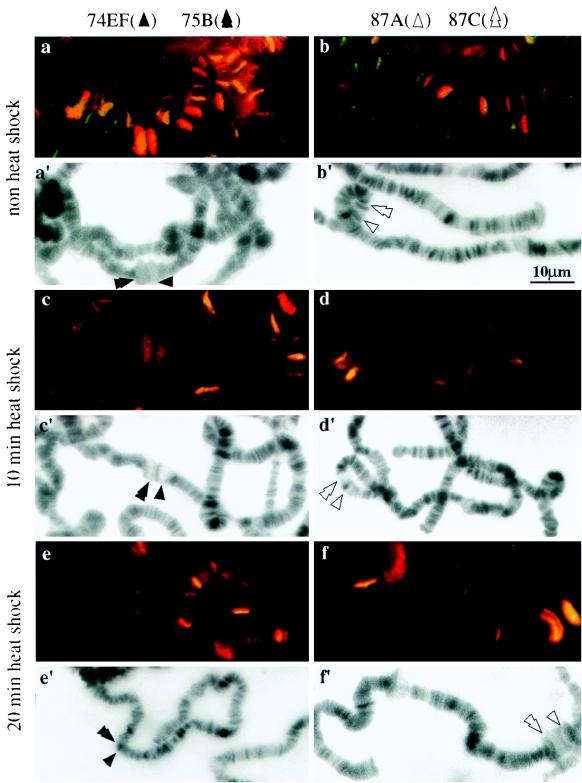
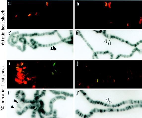
DNA topoisomerase I (topo I) from Drosophila melanogaster contains a nonconserved, hydrophilic N-terminal domain of about 430 residues upstream of the conserved core domains. Deletion of this N terminus did not affect the catalytic activity of topo I, while further removal of sequences into the conserved regions inactivated its enzymatic activity. We have investigated the cellular function of the Drosophila topo I N-terminal domain with top1-lacZ transgenes. There was at least one putative nuclear localization signal within the first 315 residues of the N-terminal domain that allows efficient import of the large chimeric proteins into Drosophila nuclei. The top1-lacZ fusion proteins colocalized with RNA polymerase II (pol II) at developmental puffs on the polytene chromosomes. Either topo I or the top1-lacZ fusion protein was colocalized with RNA pol II in some but not all of the nonpuff, interband loci. However, the fusion proteins as well as RNA pol II were recruited to heat shock puffs during heat treatment, and they returned to the developmental puffs after recovery from heat shock. By immunoprecipitation, we showed that two of the largest subunits of RNA pol II coprecipitated with the N-terminal 315-residue fusion protein by using antibodies against beta-galactosidase. These data suggest that the topo I fusion protein can be localized to the transcriptional complex on chromatin and that the N-terminal 315 residues were sufficient to respond to cellular processes, especially during the reprogramming of gene expression.


Figures

FIG. 1
FIG. 1
Drosophila topo I N-terminal deletion and top1-lacZ fusion constructs. (a) Schematic diagram of the structural features of the enzyme. The top of the panel indicates the number of amino acid residues. In the lower part of the panel, the hatched boxes (II, III, IV, and VI) represent regions of amino acid sequence homology among topo I proteins from humans, Drosophila melanogaster, Arabidopsis thaliana, and budding and fission yeasts (26). The active-site tyrosine is shown as Y, and +/− and +++/−−− refer to charged and hydrophilic residues. (b) ND423, ND472, and ND582 are three proteins with N-terminal deletions; they have codons for amino acid residues from 1 to 423, 472, and 582, respectively, removed from the full-length top1 cDNA. (c) Fusion constructs of N-terminal segments of topo I and β-gal. NF stands for N-terminal fusion, and the suffix numbers are the numbers of amino acid residues of topo I remaining in the fusion constructs. (d) Linear representation of pCaSpeRhsp83 plasmid with the top1-lacZ insert (not drawn to scale). The solid arrowheads indicate the 5′ and 3′ terminal repeats of the P element, and the thin arrows show the direction of transcription.
FIG. 2
FIG. 2
Supercoil relaxation activities of bacterial extracts expressing topo I and truncation mutants. Supercoiled plasmid DNA substrate was incubated with extracts prepared from bacteria expressing the full-length protein (lanes 1 to 4), ND423 (lanes 5 to 8), ND472 (lanes 9 to 12), ND582 (lanes 13 to 16), and vector pET3b (lanes 17 to 20). The DNA products were analyzed by agarose gel electrophoresis, and the positions of supercoiled (SC) as well as relaxed-circular (RC) DNA are indicated on the left side. Four successive 10-fold dilutions were carried out in each set of the activity assays. Lanes 1, 5, 9, 13, and 17 show the assays of undiluted extracts, while lanes 4, 8, 12, 16, and 20 show the assays with 1,000-fold-diluted extracts. Expression plasmid DNAs, present in higher concentrations in the reaction mixtures, were visible in lanes 9 and 17.
FIG. 3
FIG. 3
Expression of top1-lacZ fusion proteins in transgenic flies. Protein extracts from adult flies were prepared as described previously (30), and about 50 μg of total protein from each transgenic sample was loaded on an SDS–5% PAGE gel. Western blots were probed with either affinity purified rabbit anti-topo I antibody (a) or rabbit anti-β-gal antibody (b). Lane 1 contains a protein extract from wild-type flies, and lane 14 contains a protein extract from 28-1 flies (a transgenic strain transformed with a control construct of β-gal). Lanes 2 to 13 contain adult fly extracts from the following independent transformants: NF315 (lanes 2, 3, and 4), NF438 (lanes 5, 6, and 7), NF582 (lanes 8, 9, and 10), and NF964 (lanes 11, 12, and 13). Arrows indicate the migration positions of different fusion products. The solid and open arrowheads label the positions of endogenous topo I and β-gal, respectively. Molecular masses for size markers in kilodaltons are indicated under M.
FIG. 4
FIG. 4
Nuclear localization of the top1-lacZ fusion protein in salivary glands detected by X-Gal staining for β-gal activity. Salivary glands from third-instar larvae of NF315 (a), 28-1 (b), and wild-type (c) flies were prepared as described in Materials and Methods. The β-gal activity is readily detected in the nuclei of NF315 transgenic animals and is shown as the dark staining. Note that the smaller nuclei of adjacent tissue also exhibit the activity (a). Scale bar (b), 100 μm.
FIG. 5
FIG. 5
top1-lacZ fusion products colocalize with RNA pol IIo on developmental puffs. Preparations of chromosome squashes from transgenic flies carrying NF438 were reacted with goat anti-IIo and rabbit anti-β-gal, followed by species-specific, fluorescence-tagged secondary antibody. Red fluorescence for RNA pol IIo (a) and green fluorescence for β-gal (b) are shown. Panel c is a superimposed fluorescence image of panels a and b, showing blended colors of green and red at the indicated developmental puffs. The asterisk in panel c marks cytological position 66B, which is enriched in topo I fusion protein but not RNA pol IIo.
FIG. 6
FIG. 6
top1-lacZ fusion products are recruited to chromosome puffs as the endogenous topo I. (a) Extracts from wild-type (WT) and NF315 adult flies were separated on an SDS–7% PAGE gel. Western blots were probed with either mouse anti-β-gal or affinity-purified rabbit anti-ND423 antibodies. The arrow indicates the top1-lacZ fusion protein. The size markers are marked on the left of the Western blot. (b) Immunofluorescence images of both untreated and heat-shocked polytene chromosomes double stained with affinity-purified rabbit anti-ND423 and mouse anti-β-gal. DAPI-stained fluorescence images were used to identify chromosome bands and puffs. The symbols ▴ and ▴▴ identify developmental puffs 74EF and 75B, respectively; the symbols ▵ and ▵▵ indicate heat shock puffs 87A and 87C, respectively.
FIG. 6
FIG. 6
top1-lacZ fusion products are recruited to chromosome puffs as the endogenous topo I. (a) Extracts from wild-type (WT) and NF315 adult flies were separated on an SDS–7% PAGE gel. Western blots were probed with either mouse anti-β-gal or affinity-purified rabbit anti-ND423 antibodies. The arrow indicates the top1-lacZ fusion protein. The size markers are marked on the left of the Western blot. (b) Immunofluorescence images of both untreated and heat-shocked polytene chromosomes double stained with affinity-purified rabbit anti-ND423 and mouse anti-β-gal. DAPI-stained fluorescence images were used to identify chromosome bands and puffs. The symbols ▴ and ▴▴ identify developmental puffs 74EF and 75B, respectively; the symbols ▵ and ▵▵ indicate heat shock puffs 87A and 87C, respectively.
FIG. 7
FIG. 7
Localization of top1-lacZ fusion products to the developmental and heat shock-induced puffs during heat treatment. Polytene chromosome squashes were prepared from NF315 flies with no heat shock (a and b); heat treatment for 10 (c and d), 20 (e and f), or 60 (g and h) min; or recovery at 25°C for 60 min after heat shock for 60 min (i and j). They were then immunostained with anti-β-gal and anti-IIo antibodies. Merged images are shown here for comparing the colocalizations of topo I fusion protein and RNA pol IIo. Panels a′ to j′ are identical to panels a to j except that DAPI fluorescence micrographs are presented for identifying chromosome puffs.
FIG. 7
FIG. 7
Localization of top1-lacZ fusion products to the developmental and heat shock-induced puffs during heat treatment. Polytene chromosome squashes were prepared from NF315 flies with no heat shock (a and b); heat treatment for 10 (c and d), 20 (e and f), or 60 (g and h) min; or recovery at 25°C for 60 min after heat shock for 60 min (i and j). They were then immunostained with anti-β-gal and anti-IIo antibodies. Merged images are shown here for comparing the colocalizations of topo I fusion protein and RNA pol IIo. Panels a′ to j′ are identical to panels a to j except that DAPI fluorescence micrographs are presented for identifying chromosome puffs.
FIG. 8
FIG. 8
Coimmunoprecipitation of RNA pol II with the top1-lacZ fusion product. Samples from immunoprecipitation with either rabbit preimmune serum (pre-immu) or rabbit anti-β-gal (IP) were separated on an SDS–7% PAGE gel. Western blots were probed with either affinity-purified goat anti-E2 (exon 2 of RNA pol IIa; left panel), anti-IIc (middle panel), or mouse anti-β-gal (right panel). The open arrowhead indicates RNA pol IIa, while the solid arrowhead designates IIc, the second-largest subunit of RNA pol II. The arrow indicates the top1-lacZ fusion product. Degradation products from the top1-lacZ protein were also detected in an anti-β-gal blot.

Similar articles

Cited by

References

    1. Alsner J, Svejstrup J Q, Kjeldsen E, Sorensen B S, Westergaard O. Identification of an N-terminal domain of eukaryotic DNA. J Biol Chem. 1992;267:12408–12411. - PubMed
    1. Amero S A, Matunis M J, Matunis E L, Hockensmith J W, Raychaudhuri G, Beyer A L. A unique ribonucleoprotein complex assembles preferentially on ecdysone-responsive sites in Drosophila melanogaster. Mol Cell Biol. 1993;13:5323–5330. - PMC - PubMed
    1. Ashburner M, Bonner J J. The induction of gene activity in Drosophila by heat shock. Cell. 1979;17:241–254. - PubMed
    1. Bharti A K, Olson M O J, Kufe D M, Rubin E H. Identification of a nucleolin binding site in human topoisomerase I. J Biol Chem. 1996;271:1993–1997. - PubMed
    1. Bjornsti M A, Wang J C. Expression of yeast DNA topoisomerase I can complement a conditional-lethal DNA topoisomerase I mutation in Escherichia coli. Proc Natl Acad Sci USA. 1987;84:8971–8975. - PMC - PubMed

Publication types

Substances

LinkOut - more resources