Comparative Study
. 1998 Jul 1;18(13):4815-24.
doi: 10.1523/JNEUROSCI.18-13-04815.1998.

Identification of the amino terminus of neuronal Ca2+ channel alpha1 subunits alpha1B and alpha1E as an essential determinant of G-protein modulation

Affiliations
Comparative Study

Identification of the amino terminus of neuronal Ca2+ channel alpha1 subunits alpha1B and alpha1E as an essential determinant of G-protein modulation

K M Page et al. J Neurosci. .

Abstract

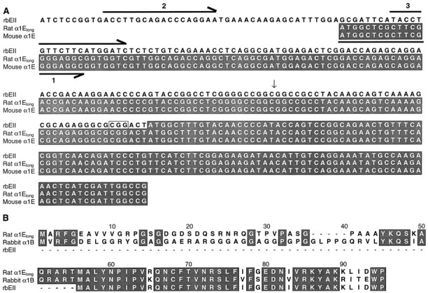
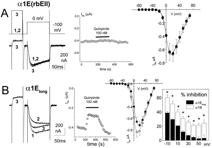
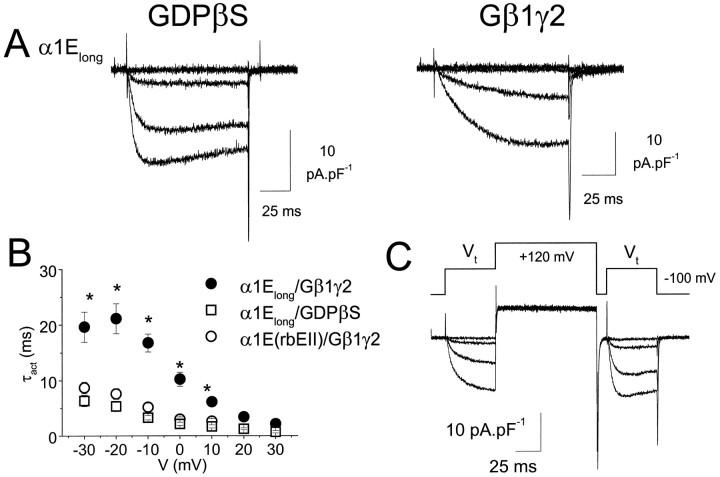
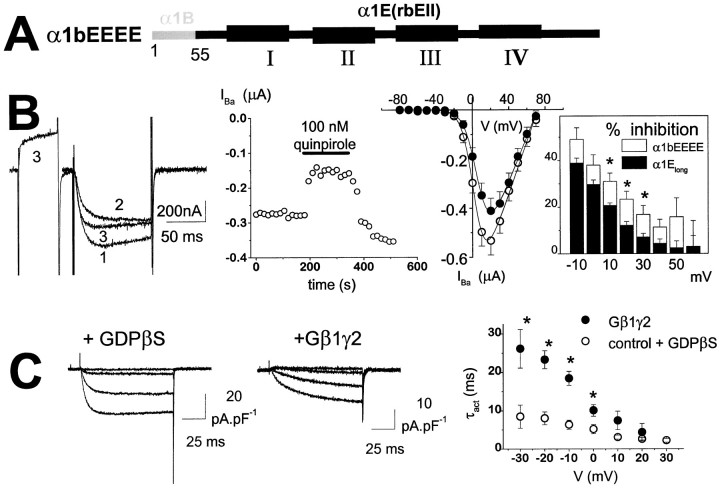
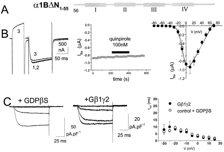
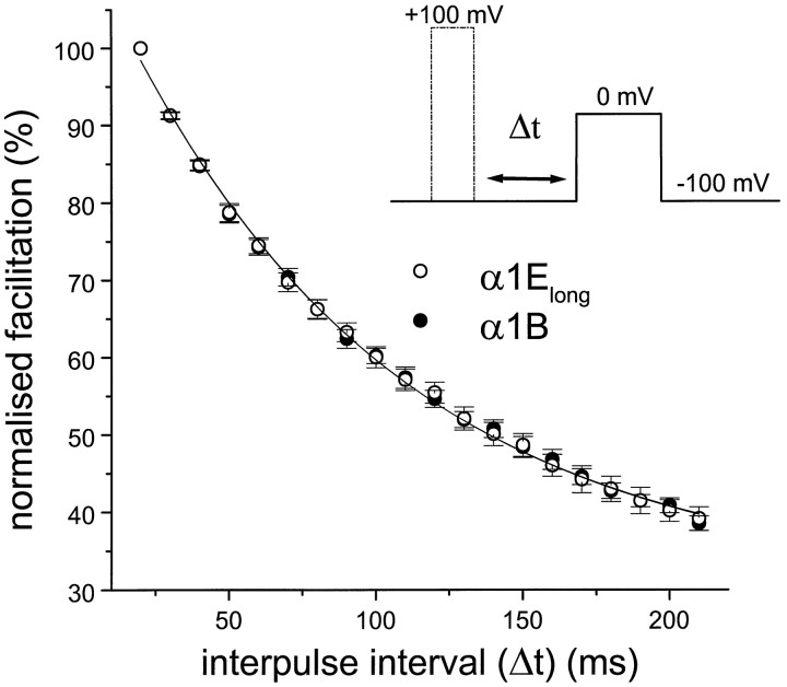
We have examined the basis for G-protein modulation of the neuronal voltage-dependent calcium channels (VDCCs) alpha1E and alpha1B. A novel PCR product of alpha1E was isolated from rat brain. This contained an extended 5' DNA sequence and was subcloned onto the previously cloned isoform rbEII, giving rise to alpha1Elong whose N terminus was extended by 50 amino acids. VDCC alpha1 subunit constructs were co-expressed with the accessory alpha2-delta and beta2a subunits in Xenopus oocytes and mammalian (COS-7) cells. The alpha1Elong showed biophysical properties similar to those of rbEII; however, when G-protein modulation of expressed alpha1 subunits was induced by activation of co-expressed dopamine (D2) receptors with quinpirole (100 nM) in oocytes, or by co-transfection of Gbeta1gamma2 subunits in COS-7 cells, alpha1Elong, unlike alpha1E(rbEII), was found to be G-protein-modulated, in terms of both a slowing of activation kinetics and a reduction in current amplitude. However, alpha1Elong showed less modulation than alpha1B, and substitution of the alpha1E1-50 with the corresponding region of alpha1B1-55 produced a chimera alpha1bEEEE, with G-protein modulation intermediate between alpha1Elong and alpha1B. Furthermore, deletion of the N-terminal 1-55 sequence from alpha1B produced alpha1BDeltaN1-55, which could not be modulated, thus identifying the N-terminal domain as essential for G-protein modulation. Taken together with previous studies, these results indicate that the intracellular N terminus of alpha1E1-50 and alpha1B1-55 is likely to contribute to a multicomponent site, together with the intracellular I-II loop and/or the C-terminal tail, which are involved in Gbetagamma binding and/or in subsequent modulation of channel gating.


Figures

Fig. 1.
Fig. 1.
Sequence of α1Elong used in this study. A, DNA alignment of the 5′ sequences of α1E(rbEII) (L15453), rat α1Elong (AF057029), and mouse α1E (L29346). Shaded areas show translated sequences. The vertical arrow shows the position of the restriction site NotI, which was used to subclone the extended 5′ sequence onto α1E(rbEII). The boxed CGG nucleotides before the ATG start site in the α1E(rbEII) were found to be present in the rbEII clone but are absent from the L15453sequence in the database. This triplet is also present in the published mouse, human, and rabbit α1E sequences. The forward primers used (see Materials and Methods) are shown as horizontal arrows, below (primer 1) or above (primers 2 and3) the corresponding sequence. Note that the extended N-terminal sequence of α1Elong shows a high degree of homology with part of the reported 5′ untranslated sequence of the rbEII cDNA. B, Amino acid alignment for the N termini of rat α1Elong, rabbit α1B (published sequence), and rat α1E(rbEII, published sequence). Conserved residues areshaded. The rat α1Elong N-terminal amino acid sequence was also identical to that of the published mouse α1E sequence (L29346).
Fig. 2.
Fig. 2.
Properties and G-protein modulation of α1Elong: comparison with α1E(rbEII). Ashows the lack of modulation of α1E(rbEII) in the absence of co-transfected VDCC β subunits. α1E(rbEII) was expressed with α2-δ but without β2a subunits in Xenopus oocytes (together with D2 dopamine receptors). Left panel, Example currents, control (1), plus quinpirole (2), and after a depolarizing prepulse to +100 mV in the presence of quinpirole (3). The voltage protocol is shown above the current traces. Middle panel, Time course of IBa amplitude during quinpirole application. Right panel,IV plot before (○) and during (•) quinpirole application (n = 6). TheIV data were fitted with a modified Boltzmann equation as described previously (Page et al., 1997).B shows the modulation of α1Elong in the presence of co-transfected VDCC β subunits. α1Elong was expressed with both α2-δ and β2a subunits inXenopus oocytes (together with D2 dopamine receptors). Activation of dopamine D2 receptors by quinpirole (100 nm) in oocytes caused a reversible inhibition ofIBa. Left panel, Example currents, control (1), plus quinpirole (2), and after a depolarizing prepulse in the presence of quinpirole (3). Middle panel, Time course of inhibition by quinpirole. Right panel, IV plot before (○) and during (•) quinpirole application (n = 9). TheIV data were fitted as described inA. The boxed inset shows the voltage-dependence of the inhibition by quinpirole from theIV data of α1Elong(solid bars, n = 9). Data for α1B (open bars, n = 8) are plotted for comparison; * p < 0.01 (Student’st test).
Fig. 3.
Fig. 3.
G-protein modulation of α1Elongexpressed in COS-7 cells. α1Elong was expressed with accessory VDCC α2-δ and β2a subunits in the presence or absence of co-expressed Gβ1γ2. A, Examples of current density–voltage profiles for α1Elong in a control cell in the presence of GDPβS to limit any tonic G-protein modulation (left), and a cell co-expressing Gβ1γ2 (right) (Vt = −40 to −10 mV, in 10 mV steps). B, Voltage-dependence of τact for α1Elong with co-expressed Gβ1γ2 (•, n = 10), α1Elong in the presence of GDPβS (□, n = 7), and α1E(rbEII) with co-expressed Gβ1γ2 (○, n = 5), * p < 0.01 compared with respective control. C, Example of facilitation of α1ElongIBa in the presence of co-expressed Gβ1γ2 by a depolarizing prepulse to +120 mV, 10 msec before and immediately after equivalent test pulses P1 and P2, to test potentials (Vt) between −40 and −10 mV in 10 mV intervals. The voltage protocol is shown above the current traces. Facilitation was then determined as the P2/P1 ratio of the current amplitudes in P1 and P2 (Table 1).
Fig. 4.
Fig. 4.
G-protein modulation of an α1E construct containing the N terminus of α1B. A, The α1 subunit construct in which the α1B1–55 sequence was added to α1E(rbEII) to form α1bEEEE was expressed with accessory VDCC α2-δ and β2a subunits in Xenopus oocytes (together with D2 receptors) or in COS-7 cells (together with Gβ1γ2 subunits). B, α1bEEEE currents expressed in oocytes.Left panel, Example currents, control (1), plus quinpirole (2), and after a depolarizing prepulse in the presence of quinpirole (3). The voltage protocol is the same as shown in Figure 2A. Middle panel, Time course of inhibition by quinpirole. Right panel,IV plot before (○) and during (•) quinpirole application (n = 9). TheIV data were fitted according to the legend to Figure 2. The boxed inset shows the voltage-dependence of the inhibition by quinpirole from theIV data (open bars,n = 9). Data for α1Elong(solid bars, n = 9) are plotted for comparison; * p < 0.05 (Student’st test). C, α1bEEEE currents expressed in COS-7 cells. Left panel, Example current density–voltage profiles for control α1bEEEEIBa in the presence of 2 mmGDPβS. Middle panel, α1bEEEEIBa in the presence of Gβ1γ2 (Vt = −40 to −10 mV in 10 mV steps).Right panel, Voltage-dependence of τactfor α1bEEEE in the presence (•, n = 5) or absence (○, n = 3) of co-expressed Gβ1γ2; *p < 0.01 compared with respective control.
Fig. 5.
Fig. 5.
Lack of G-protein modulation of an N-terminally truncated α1B construct. A, The α1 construct in which the α1B1–55 sequence was deleted from α1B to form α1BΔN1–55 was expressed with accessory VDCC α2-δ and β2a subunits in Xenopus oocytes (together with D2 receptors) or in COS-7 cells (together with Gβ1γ2 subunits). B, α1BΔN1–55 currents expressed in oocytes. Left panel, Example currents, control (1), plus quinpirole (2), and after a depolarizing prepulse in the presence of quinpirole (3). The voltage protocol is the same as shown in Figure 2A. Middle panel, Time course of IBa amplitude during quinpirole application. Right panel,IV plot before (○) and during (•) quinpirole application (n = 7). TheIV data were fitted according to the legend to Figure 2. C, α1BΔN1–55currents expressed in COS-7 cells. Left panel, Example current density–voltage profiles in the absence or presence of Gβ1γ2 (Vt = −40 to −10 mV in 10 mV steps). Right panel, Voltage-dependence of τact in the presence (•, n = 10) or absence (○, n = 7) of co-expressed Gβ1γ2.
Fig. 6.
Fig. 6.
Reinhibition kinetics of α1Elong and α1B. Prepulses of 50 msec duration to +100 mV were applied, and the time between prepulse and test pulse to 0 mV (interpulse interval Δt at −100 mV) was increased, in 10 msec steps, up to 220 msec. There was no difference between the τreinhibition for α1Elong (○,n = 9) and α1B (•, n = 9)IBa.

References

    1. Bourinet E, Soong TW, Stea A, Snutch TP. Determinants of the G protein-dependent opioid modulation of neuronal calcium channels. Proc Natl Acad Sci USA. 1996;93:1486–1491. - PMC - PubMed
    1. Brice NL, Berrow NS, Campbell V, Page KM, Brickley K, Tedder I, Dolphin AC. Importance of the different β subunits in the membrane expression of the α1A and α2 calcium channel subunits: studies using a depolarisation-sensitive α1A antibody. Eur J Neurosci. 1997;9:749–759. - PubMed
    1. Campbell V, Berrow N, Brickley K, Page K, Wade R, Dolphin AC. Voltage-dependent calcium channel β-subunits in combination with α1 subunits have a GTPase activating effect to promote hydrolysis of GTP by Gαo in rat frontal cortex. FEBS Lett. 1995a;370:135–140. - PubMed
    1. Campbell V, Berrow NS, Fitzgerald EM, Brickley K, Dolphin AC. Inhibition of the interaction of G protein Go with calcium channels by the calcium channel β-subunit in rat neurones. J Physiol (Lond) 1995b;485:365–372. - PMC - PubMed
    1. Currie KPM, Fox AP. Comparison of N and P/Q type voltage-gated calcium channel current inhibition. J Neurosci. 1997;17:4570–4579. - PMC - PubMed

Publication types

MeSH terms

LinkOut - more resources