Comparative Study
. 1998 Jul 1;18(13):4891-900.
doi: 10.1523/JNEUROSCI.18-13-04891.1998.

Upregulation of the enzyme chain hydrolyzing extracellular ATP after transient forebrain ischemia in the rat

Affiliations
Comparative Study

Upregulation of the enzyme chain hydrolyzing extracellular ATP after transient forebrain ischemia in the rat

N Braun et al. J Neurosci. .

Abstract

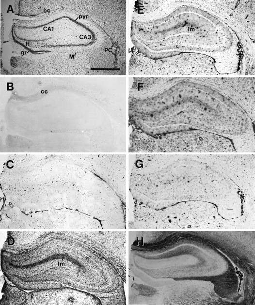
A short ischemic period induced by the transient occlusion of major brain arteries induces neuronal damage in selectively vulnerable regions of the hippocampus. Adenosine is considered to be one of the major neuroprotective substances produced in the ischemic brain. It can be released from damaged cells, but it also could be generated extracellularly from released ATP via a surface-located enzyme chain. Using the rat model of global forebrain ischemia, we applied a short (10 min) transient interruption of blood flow and studied the distribution of ectonucleotidase activities in the hippocampus. Northern hybridization of mRNA isolated from hippocampi of sham-operated and ischemic animals revealed an upregulation of ectoapyrase (capable of hydrolyzing nucleoside 5'-tri- and diphosphates) and ecto-5'-nucleotidase (capable of hydrolyzing nucleoside 5'-monophosphates). A histochemical analysis that used ATP, UTP, ADP, or AMP as substrates revealed a strong and selective increase in enzyme activity in the injured areas of the hippocampus. Enhanced staining could be observed first at 2 d. Staining increased within the next days and persisted at 28 d after ischemia. The spatiotemporal development of catalytic activities was identical for all substrates. It was most pronounced in the CA1 subfield and also could be detected in the dentate hilus and to a marginal extent in CA3. The histochemical staining corresponded closely to the development of markers for reactive glia, in particular of microglia. The upregulation of ectonucleotidase activities implies increased nucleotide release from the damaged tissue and could play a role in the postischemic control of nucleotide-mediated cellular responses.


Figures

Fig. 1.
Fig. 1.
Northern blot analysis of ectonucleotidases. Equal amounts (0.5 μg) of polyadenylated RNA isolated from hippocampi 7 d after sham operation (a) or 7 d after 10 min of global ischemia (b) were subjected to electrophoresis, blotted, and hybridized with rat ecto-5′-nucleotidase (e-5′-nucl.), ectoapyrase (e-apyrase), and ecto-ATPase (e-ATPase) antisense riboprobes. A significant postischemic enhancement of the hybridization signals is detectable for ecto-5′-nucleotidase and ectoapyrase. The riboprobe for ectoapyrase hybridizes with two major bands at 4.9 and 5.4 kb. A riboprobe for mouse β-actinwas used as an internal control.
Fig. 2.
Fig. 2.
Corresponding hippocampal sections 7 d after sham operation. Comparison of Nissl staining (A), immunocytochemistry (B–D), and nucleotidase histochemistry (E–H). A, Nissl staining revealed no neuronal damage in the hippocampus. B, OX42 immunostaining is significant only in fiber tracts, as for example in the corpus callosum. C, Vimentin is detectable in capillaries. D, GFAP immunoreaction is strongest in the stratum lacunosum moleculare. E–G, ATPase (E), UTPase (F), and ADPase (G) reaction product in the hippocampus is restricted mainly to capillaries. Staining is enhanced slightly in the stratum lacunosum. H, 5′-Nucleotidase activity is highest in CA1 and in the stratum lacunosum moleculare of CA1 and CA3. The pyramidal cell layers and the dentate granule cell layer are free of reaction product. CA1, CA3, Subfields of the hippocampus; cc, corpus callosum;gr, granular cell layer of the dentate gyrus;H, hilus of the dentate gyrus; lm, stratum lacunosum moleculare; pyr, stratum pyramidale;PC, plexus choroideus; M, meninx. Scale bar, 1 μm.
Fig. 3.
Fig. 3.
Corresponding hippocampal sections 3 d after global ischemia. Comparison of Nissl staining (A), immunocytochemistry (B–D), and nucleotidase histochemistry (E–H). A, Reduced Nissl staining in CA1 (arrowheads) indicates damaged pyramidal neurons. Immunostaining for OX42 (B), vimentin (C), and GFAP (D) is upregulated most significantly in CA1 and the dentate hilus.E–H, ATPase (E), UTPase (F), ADPase (G), and 5′-nucleotidase activity (H) is enhanced in the pyramidal cell layer of CA1(arrows). Abbreviations as in Figure 2. Scale bar, 1 μm.
Fig. 4.
Fig. 4.
Corresponding hippocampal sections 7 d after global ischemia. Comparison of Nissl staining (A), immunocytochemistry (B–D), and nucleotidase histochemistry (E–H). A, Reduced Nissl staining in the CA1 subfield (arrowheads) indicates damaged pyramidal cells. Immunolabeling for OX42 (B), vimentin (C), and GFAP (D) is strongly enhanced in CA1 and the dentate hilus. In contrast to GFAP immunoreactivity, OX42 and vimentin immunostaining is very distinct in the layer of damaged pyramidal cells.E–H, Strong upregulation of ATPase (E), UTPase (F), ADPase (G), and 5′-nucleotidase (H) activity in all layers of CA1 and the dentate hilus. The reaction product is most intense in the layer of damaged pyramidal cells and the stratum lacunosum moleculare. Abbreviations as in Figure 2. Scale bar, 1 μm.
Fig. 5.
Fig. 5.
Immunocytochemical detection of the postischemic glial reaction in CA1. A–C, OX42 immunoreactivity 7 d after sham operation (A) and 3 d (B) and 7 d (C) after ischemia. Arrows depict activated microglial cells that are apparent at 3 and 7 d. At 7 d the accumulation of the OX42 immunoreaction is strongest in the pyramidal cell layer.D–F, Vimentin immunoreactivity 7 d after sham operation (D) and 3 d (E) and 7 d (F) after ischemia. Capillaries are strongly labeled for vimentin (arrowheads). Vimentin-expressing cells with radiate ramifications (arrows) presumably represent reactive astrocytes, whereas rod-shaped cells presumably represent microglia.G–I, GFAP immunoreactivity 7 d after sham operation (G) and 3 d (H) and 7 d (I) after ischemia. Reactive and swollen astrocytes are clearly detectable at 3 and 7 d. Note that astrocytes do not accumulate in the pyramidal cell layer after the disappearance of neurons. ori, Stratum oriens;pyr, pyramidal cell layer; rad, stratum radiatum. Scale bar, 100 μm.

References

    1. Abbracchio MP. P1 and P2 receptors in cell growth and differentiation. Drug Dev Res. 1996;39:393–406.
    1. Boarder MR, Weisman GA, Turner JT, Wilkinson GF. G-protein-coupled P2 purinoceptors: from molecular biology to functional responses. Trends Pharmacol Sci. 1995;16:133–139. - PubMed
    1. Bolego C, Ceruti S, Brambilla R, Puglisi L, Cattabeni F, Burnstock G, Abbracchio MP. Characterization of the signalling pathways involved in ATP and basic fibroblast growth factor-induced astrogliosis. Br J Pharmacol. 1997;121:1692–1699. - PMC - PubMed
    1. Braun N, Lenz C, Gillardon F, Zimmermann M, Zimmermann H. Focal cerebral ischemia enhances glial expression of 5′-nucleotidase. Brain Res. 1997;766:213–226. - PubMed
    1. Buell G, Collo G, Rassendren F. P2X receptors: an emerging channel family. Eur J Neurosci. 1996;8:2221–2228. - PubMed

Publication types

MeSH terms