. 1998 Jul 1;18(13):5008-18.
doi: 10.1523/JNEUROSCI.18-13-05008.1998.

Differential withdrawal of retinal axons induced by a secreted factor

Affiliations

Differential withdrawal of retinal axons induced by a secreted factor

H Ichijo et al. J Neurosci. .

Abstract

To understand the development of the topographic map in the chick retinotectal projection, we studied the long-term interactions between retinal axons and tectal cell processes using a novel coculture system, the ryomen chamber. Both nasal and temporal retinal axons initially grew equally well on a substrate consisting of posterior tectal cell processes; however, subsequently most temporal axons withdrew from this surface, whereas most nasal axons did not. Experiments using conditioned media indicate that posterior tectal cells induced withdrawal of the temporal axons by secreting a soluble factor. This withdrawal seems to be distinct from the immediate repulsive effect of ephrin-A2 (ELF-1) and ephrin-A5 (RAGS) seen in the stripe assay because (1) the withdrawal-inducing factor was diffusible, whereas ephrin-A2 and -A5 are membrane-bound, and (2) the withdrawal-inducing factor appeared later in development than ephrin-A2 and -A5. Furthermore, sensitivity to the withdrawal-inducing factor decreased continuously from the temporal to nasal retina. These results suggest that target cell-induced axonal withdrawal may be involved during a late stage of the development of the retinotectal map.


Figures

Fig. 1.
Fig. 1.
Schematic drawings of the experimental design.A, The ryomen chamber consists of a pair of stainless steel rings and a Nuclepore filter.B, RGC axons grow on tectal cell processes that have penetrated the filter. C, D, Scanning electronmicrographs of the substrates on the retinal side of the filter at (C) low and (D) high magnifications. In D, round processes (arrow) and filter pores (arrowhead) are shown. Immunofluorescent staining of the substrates (5 d in vitro) by anti-MAP2 (E) and anti-vimentin (F) antibodies. Most of the round processes (arrowheads in F) and some flat processes with thin spikes (arrows inF) are labeled by the anti-vimentin antibody. Scale bars: C, 30 μm; D, 10 μm;E, F, 25 μm.
Fig. 2.
Fig. 2.
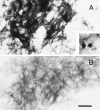
Expression of EphA3 ligands with RAP in situ histochemistry. EphA3 ligands are expressed by the posterior tectal cells on the tectal side of the filter intensely after 4 d (A) and weakly after 8 d (B) in vitro. A round process on the retinal side (arrow) is labeled by EphA3-AP (inset in A). Scale bar, 25 μm.
Fig. 3.
Fig. 3.
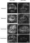
Growth of retinal axons in the standard overnight preculture schedule. AD, Views of the axons from the retinal side of the filter after 3 d. The axons grow well on both the anterior and posterior substrates. Black areas in the middle of the pictures are the nitrocellulose filters mechanically supporting the retinal tissues. The nasal axons on the anterior (A) and posterior (B) substrates. The temporal axons on the anterior (C) and posterior (D) substrates.EH, Views of the axons after 7 d. The temporal axons remain on the anterior substrate (G), but most of them do not stay on the posterior substrate (H). The nasal axons remain on the anterior (E) and posterior (F) substrates. Axon–substrate combinations are also shown at the left side of pictures. Scale bar, 1 mm.
Fig. 4.
Fig. 4.
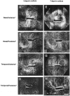
Growth of retinal axons in the long preculture schedule for 4 d (see Fig. 5A).AD, The axons after 3 d. The temporal axons grow on the anterior substrate (C) but do not grow on the posterior substrate (D). The nasal axons grow on both the anterior (A) and posterior (B) substrates.EH, The axons after 7 d. The temporal axons recover to grow on the posterior substrate (H), and they remain on the anterior substrate (G). The nasal axons remain on both the anterior (E) and posterior (F) substrates. Axon–substrate combinations are also shown at the left side of pictures.Anterior* and Posterior* indicate the anterior and posterior substrates precultured for 4 d, respectively. Scale bar, 1 mm.
Fig. 5.
Fig. 5.
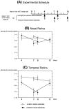
The experimental schedule is shown inA. Retinal explants are added at day 1 (the standard overnight preculture schedule) or day 4 (the long preculture schedule)in vitro of the tectal cells. The explants are incubated for an additional 3 or 7 d. Maximum densities of nasal (B) and temporal (C) axonal bundles are shown as a function of days in vitro for the tectal cells. The densities of the temporal bundles are reduced on the posterior substrates at the seventh and eighth day. Openand closed circles indicate the densities of axonal bundles on the anterior and posterior substrates, respectively. SEs are shown. n = 10 for each condition.
Fig. 6.
Fig. 6.
Growth of retinal axons in the conditioned media. Nasal and temporal explants are indicated as n andt, respectively. Temporal (A) and nasal (B) explants are cultured in the medium conditioned for 8 d by the E7 posterior tectal cells.C, A temporal explant is cultured in heat-treated medium conditioned for 8 d by the E7 posterior tectal cells. Scale bar, 200 μm. D, Axonal outgrowth from the nasal and temporal explants in the conditioned media or in the presence of the soluble ephrins. Light gray and dark gray bars indicate amounts of the nasal and temporal outgrowth (the total areas covered with the axonal bundles from an explant), respectively. 4A and 8A indicate the axonal outgrowth in media conditioned by the E7 anterior tectal cells for 4 and 8 d, respectively. 4P and8P indicate the axonal outgrowth in media conditioned by the E7 posterior tectal cells for 4 and 8 d, respectively.Heated 8P indicates the axonal outgrowth in heat-treated medium that was conditioned by the E7 posterior tectal cells for 8 d. n is between 12 and 22 for each condition.ephrin A2 and A5 indicate the axonal outgrowth in the media conditioned by the E7 anterior tectal cells for 8 d with 20 nm ephrin-A2-AP and ephrin-A5-AP, respectively. control indicates the axonal outgrowth in the F12 minimum medium. n is between 5 and 10 for each condition. The difference between the nasal and temporal outgrowth is significant in 8P (p = 0.0033, indicated by *) but not in 4P(p > 0.05).
Fig. 7.
Fig. 7.
Mapping nasotemporal transitions in retinae with the stripe assay and the ryomen chamber assays.A, Original positions of the retinal explants are shown.B, Average preferences of the retinal axons in the stripe assay as white bars (the left ordinate), maximum densities in the ryomen chamber assay (the right ordinate) on the substrates made from the posterior third of the tecta (closed circles) and on the substrates made from the posterior sixth of the tecta (open squares). The explants are represented at their original positions in the retinae (the abscissa). A steep slope of transition from the center to the periphery in the nasal retina is seen on the substrate made from the posterior third of the tecta, and a gradual slope on the (Figure legend continues). substrates made from the posterior sixth of the tecta. In contrast, a step transition is shown between the temporal and nasal retinae in the stripe assay. SEs are shown. n is between 5 and 8 for each condition. Serial retinal explants are placed (C) on the carpets containing alternating membrane stripes (scale bar, 200 μm) or (D) on the substrates made from the posterior third of the tecta (scale bar, 1 mm).

References

    1. Bonhoeffer F, Huf J. In vitro experiments on axon guidance demonstrating an anterior-posterior gradient on the tectum. EMBO J. 1982;1:427–431. - PMC - PubMed
    1. Carri NG, Ebendal T. Target-field specificity in the induction of retinal neurite outgrowth. Brain Res. 1987;428:83–90. - PubMed
    1. Cheng H-J, Flanagan JG. Identification and cloning of ELF-1, a developmentally expressed ligand for the Mek4 and Sek receptor tyrosine kinases. Cell. 1994;79:157–168. - PubMed
    1. Cheng H-J, Nakamoto M, Bergemann AD, Flanagan JG. Complementary gradients in expression and binding of ELF-1 and Mek4 in development of the topographic retinotectal projection map. Cell. 1995;82:371–381. - PubMed
    1. Constantine-Paton M, Blum AS, Mendez Otero R, Barnstable CJ. A cell surface molecule distributed in a dorsoventral gradient in the perinatal rat retina. Nature. 1986;324:459–462. - PubMed

Publication types

LinkOut - more resources