Striatal extracellular dopamine levels in rats with haloperidol-induced depolarization block of substantia nigra dopamine neurons
- PMID: 9634572
- PMCID: PMC6792547
- DOI: 10.1523/JNEUROSCI.18-13-05068.1998
Striatal extracellular dopamine levels in rats with haloperidol-induced depolarization block of substantia nigra dopamine neurons
Abstract
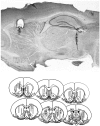
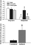
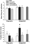
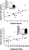
Correlations between substantia nigra (SN) dopamine (DA) cell activity and striatal extracellular DA were examined using simultaneous extracellular single-unit recordings and in vivo microdialysis performed in drug-naive rats and in rats treated repeatedly with haloperidol (HAL). Intact rats treated with HAL for 21-28 d exhibited significantly fewer active DA cells, indicating the presence of depolarization block (DB) in these cells. However, in rats that received surgical implantation of the microdialysis probe followed by a 24 hr recovery period, HAL-induced DA cell DB was reversed, as evidenced by a number of active DA neurons that was significantly higher than that in HAL-treated intact rats and similar to that of drug-naive rats. In contrast, using a modified probe implantation procedure that did not reverse SN DA neuron DB, we found striatal DA efflux to be significantly lower than in controls and significantly correlated with the reduction in DA neuron spike activity. Furthermore, although basal striatal DA efflux was independent of SN DA cell burst-firing activity in control rats, these variables were significantly correlated in rats with HAL-induced DA cell DB. Therefore, HAL-induced DB of SN DA neurons is disrupted by implantation of a microdialysis probe into the striatum using standard procedures. However, a modified microdialysis method that allowed reinstatement of DA neuron DB revealed that the HAL-induced inactivation of SN DA neurons was associated with significantly lower extracellular DA levels in the striatum. Moreover, the residual extracellular DA maintained in the presence of DB may, in part, depend on the burst-firing pattern of the noninactivated DA neurons in the SN.
Figures


Similar articles
-
Neurochemical and electrophysiological evidence that 5-HT4 receptors exert a state-dependent facilitatory control in vivo on nigrostriatal, but not mesoaccumbal, dopaminergic function.Eur J Neurosci. 2001 Mar;13(5):889-98. doi: 10.1046/j.0953-816x.2000.01453.x. Eur J Neurosci. 2001. PMID: 11264661
-
Differential regulation of dopamine release by N-methyl-D-aspartate receptors in rat striatum after partial and extreme lesions of the nigro-striatal pathway.Brain Res. 1998 Jun 29;797(2):255-66. doi: 10.1016/s0006-8993(98)00381-3. Brain Res. 1998. PMID: 9666143
-
Glucose-regulated dopamine release from substantia nigra neurons.Brain Res. 2000 Aug 25;874(2):158-64. doi: 10.1016/s0006-8993(00)02573-7. Brain Res. 2000. PMID: 10960600
-
Assessment of striatal extracellular dopamine and dopamine metabolites by microdialysis in haloperidol-treated rats exhibiting oral dyskinesia.Neuropsychopharmacology. 1993 Sep;9(2):101-9. doi: 10.1038/npp.1993.48. Neuropsychopharmacology. 1993. PMID: 8216693
-
Tyrosine augments acute clozapine- but not haloperidol-induced dopamine release in the medial prefrontal cortex of the rat: an in vivo microdialysis study.Neuropsychopharmacology. 2001 Jul;25(1):149-56. doi: 10.1016/S0893-133X(01)00220-2. Neuropsychopharmacology. 2001. PMID: 11377928
Cited by
-
Limited access to ethanol increases the number of spontaneously active dopamine neurons in the posterior ventral tegmental area of nondependent P rats.Alcohol. 2010 May;44(3):257-64. doi: 10.1016/j.alcohol.2010.02.009. Alcohol. 2010. PMID: 20682193 Free PMC article.
-
Activation of ventral tegmental area cells by the bed nucleus of the stria terminalis: a novel excitatory amino acid input to midbrain dopamine neurons.J Neurosci. 2002 Jun 15;22(12):5173-87. doi: 10.1523/JNEUROSCI.22-12-05173.2002. J Neurosci. 2002. PMID: 12077212 Free PMC article.
-
Insights on current and novel antipsychotic mechanisms from the MAM model of schizophrenia.Neuropharmacology. 2020 Feb;163:107632. doi: 10.1016/j.neuropharm.2019.05.009. Epub 2019 May 8. Neuropharmacology. 2020. PMID: 31077730 Free PMC article. Review.
-
How antipsychotics work-from receptors to reality.NeuroRx. 2006 Jan;3(1):10-21. doi: 10.1016/j.nurx.2005.12.003. NeuroRx. 2006. PMID: 16490410 Free PMC article. Review.
-
Glutamatergic dysfunction in schizophrenia: from basic neuroscience to clinical psychopharmacology.Eur Neuropsychopharmacol. 2008 Nov;18(11):773-86. doi: 10.1016/j.euroneuro.2008.06.005. Epub 2008 Jul 23. Eur Neuropsychopharmacol. 2008. PMID: 18650071 Free PMC article. Review.
References
-
- Albe-Fessard D, Sanderson P. A demonstration of tonic inhibitory and facilitatory striatal actions on substantia nigra neurons. Adv Behav Biol. 1987;32:321–326.
-
- Arnt J, Skarsfeldt T. Do novel antipsychotics have similar pharmacological characteristics? A review of evidence. Neuropsychopharmacology. 1998;18:63–101. - PubMed
-
- Asin KE, Wirtshafter D. Evidence for dopamine involvement in reinforcement obtained using a latent extinction paradigm. Pharmacol Biochem Behav. 1990;36:417–420. - PubMed
-
- Benveniste H. Brain microdialysis. J Neurochem. 1989;52:1667–1679. - PubMed
-
- Benveniste H, Drejer J, Schousboe A, Diemer NH. Regional cerebral glucose phosphorylation and blood flow after insertion of a microdialysis fiber through the dorsal hippocampus of the rat. J Neurochem. 1987;49:729–734. - PubMed
Publication types
MeSH terms
Substances
Grants and funding
LinkOut - more resources
Full Text Sources
Miscellaneous
