. 1998 Jun 23;95(13):7433-8.
doi: 10.1073/pnas.95.13.7433.

Resistance gene N-mediated de novo synthesis and activation of a tobacco mitogen-activated protein kinase by tobacco mosaic virus infection

Affiliations

Resistance gene N-mediated de novo synthesis and activation of a tobacco mitogen-activated protein kinase by tobacco mosaic virus infection

S Zhang et al. Proc Natl Acad Sci U S A. .

Abstract

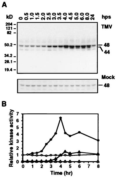
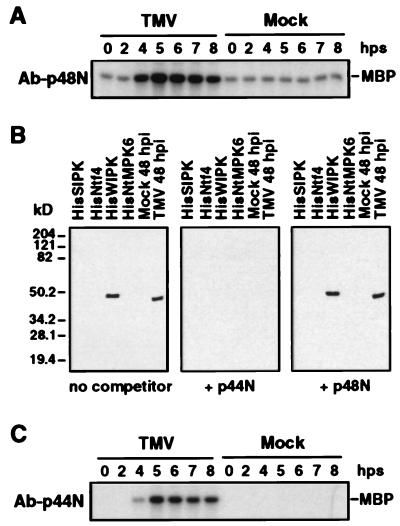
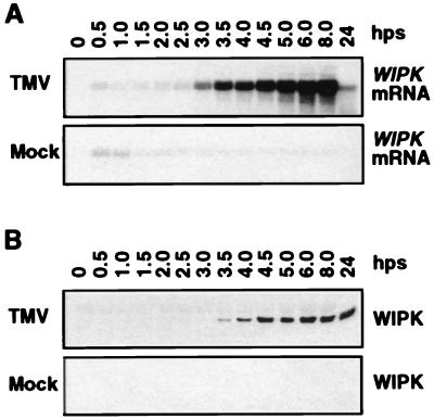
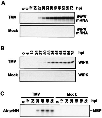
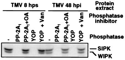
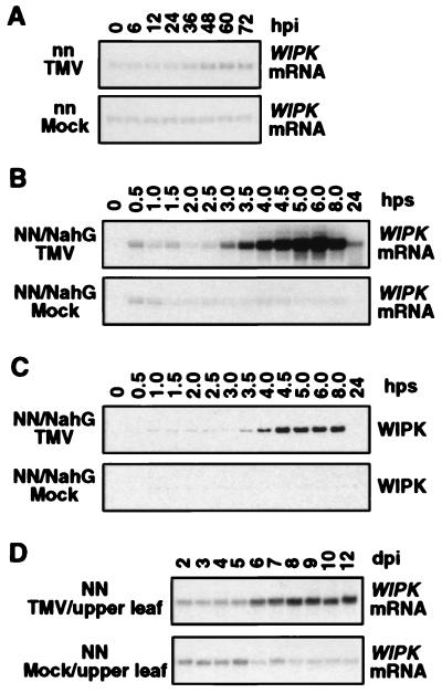
Salicylic acid-induced protein kinase (SIPK) and wounding-induced protein kinase (WIPK), two distinct members of the mitogen-activated protein (MAP) kinase family, are activated in tobacco resisting infection by tobacco mosaic virus (TMV). WIPK activation by TMV depends on the disease-resistance gene N because infection of susceptible tobacco not carrying the N gene failed to activate WIPK. Activation of WIPK required not only posttranslational phosphorylation but also a preceding rise in its mRNA and de novo synthesis of WIPK protein. The induction by TMV of WIPK mRNA and protein also occurred systemically. Its activation at the mRNA, protein, and enzyme levels was independent of salicylic acid. The regulation of WIPK at multiple levels by an N gene-mediated signal(s) suggests that this MAP kinase may be an important component upstream of salicylic acid in the signal-transduction pathway(s) leading to local and systemic resistance to TMV.


Figures

Figure 1
Figure 1
Activation of 48-kDa and 44-kDa kinases in TMV-infected tobacco. Tobacco plants carrying resistance gene N [N. tabacum cv. Xanthi nc (NN)] were inoculated with either TMV (U1 strain, 1 μg/ml in 50 mM phosphate buffer, pH 7.0) or buffer only (mock). After infection, plants were maintained at 32°C for 48 hr. Discs from the infected leaves were collected at various time after the plants were shifted back to 22°C (hps, hr postshift), and protein extracts were prepared. (A) In-gel kinase activity assay. Extracts containing 15 μg protein were electrophoresed in 10% SDS-polyacrylamide gels imbedded with 0.25 mg/ml of MBP in the separating gel. After protein renaturation, the kinase reaction was carried out as described in Materials and Methods. The sizes of activated kinases are given in kilodaltons. (B) The activities of 48-kDa kinase [in TMV-inoculated (•) and mock-inoculated (○) leaves] and 44-kDa kinase [in TMV-inoculated (▴) and mock-inoculated (▵) leaves] were quantitated by using a PhosphorImager, and the relative activities were plotted against time. Kinase activities were normalized to the level present at the zero time point for the 48-kDa kinase, which was given a value of 1.
Figure 2
Figure 2
Immune-complex kinase assays using sequence-specific antibodies against SIPK and WIPK. (A) Immune-complex kinase assay of TMV-activated kinase using SIPK-specific antibody, Ab-p48N. Protein extracts (50 μg) from TMV- or mock-inoculated leaf tissue were reacted with Ab-p48N (2.5 μg). The resultant antigen–antibody complex was precipitated with protein A-agarose beads and washed extensively before addition to a kinase assay mixture with [γ-32P]ATP and MBP as substrates. The reaction mixture, including the phosphorylated MBP, was then fractionated by SDS/PAGE. (B) An antibody raised against a peptide (p44N) corresponding to the unique N terminus of WIPK, Ab-p44N, specifically recognized the WIPK protein. Two nanograms each of recombinant HisSIPK, HisNtf4, HisWIPK, and HisNtMPK6 or 20 μg of protein extracts from 48-hr mock- or TMV-inoculated tobacco leaves (maintained throughout infection at 22°C) were subjected to immunoblot analysis with Ab-p44N in the absence or presence of 0.2 μg/ml competitor peptides p44N or p48N. (C) Immune-complex kinase assay of TMV-activated kinase using WIPK-specific antibody, Ab-p44N. Protein extracts (50 μg) from TMV- or mock-inoculated leaves were immunoprecipitated with Ab-p44N (2.5 μg), and the kinase activity of the immune-complex was determined as above. Times in A and C are given in hps from 32°C to 22°C.
Figure 3
Figure 3
Activation of WIPK gene expression by TMV in tobacco plants [cv. Xanthi nc (NN)] after temperature shift. (A) Increase in steady-state levels of WIPK mRNA in TMV-infected plants. Duplicates of leaf discs used in Fig. 1 were extracted for total RNA, thus facilitating direct comparison of the induction kinetics of mRNA and enzymatic activity. Twenty micrograms of total RNA per lane was separated on 1.2% formaldehyde/agarose gels and transferred to Zeta-probe membranes. Blots were hybridized with random primer-labeled inserts consisting of either a full-length cDNA of WIPK (data shown) or its 3′ untranslated region (data not shown). Equal loading of RNA was confirmed by ethidium bromide staining of the rRNA (data not shown). (B) Increase of WIPK protein in TMV-infected tobacco after temperature shift. Samples containing 20 μg of protein from the leaf extracts used for Fig. 1A were separated on 10% SDS-polyacrylamide gels. After blotting to nitrocellulose, the WIPK protein was detected with Ab-p44N.
Figure 4
Figure 4
Activation of WIPK by TMV in tobacco plants [cv. Xanthi nc (NN)] maintained at 22°C throughout infection. (A) Increase in steady-state levels of WIPK mRNA in TMV-infected tobacco plants. Tobacco plants were inoculated with TMV or buffer (mock) as in Fig. 1 except a higher concentration of TMV was used (5 μg/ml). Leaf discs were taken at the indicated times in hr postinoculation (hpi). Total RNA was prepared and analyzed for WIPK mRNA as described in Fig. 3. (B) Increase of WIPK protein in TMV-infected tobacco maintained at 22°C. Protein extracts were prepared from duplicate leaf discs to those used in A. Twenty micrograms of protein was analyzed by immunoblotting by using Ab-p44N as described in Fig. 3. (C) Induction of WIPK enzymatic activity in TMV-infected tobacco maintained at 22°C. Selected protein extracts from B were analyzed by immune-complex kinase assay by using WIPK-specific Ab-p44N as described in Fig. 2.
Figure 5
Figure 5
Active 44-kDa WIPK required both threonine and tyrosine phosphorylation. Protein extracts were prepared in the absence of phosphatase inhibitors from TMV-infected leaves at 8 hr after shifting plants from 32°C to 22°C or 48 hr after infection at 22°C. Samples containing 20 μg of protein were treated with either the serine/threonine-specific phosphatase, PP-2A1 (0.25 unit in 30 μl), or the tyrosine-specific protein phosphatase, YOP (2 units in 30 μl), for 20 min at 30°C in the presence or absence of a phosphatase inhibitor. The PP-2A1 inhibitor, okadaic acid (OA), and YOP inhibitor, Na3VO4 (Van), were used at a concentration of 1 μM and 1 mM, respectively. After phosphatase treatment, kinase activity was detected by the in-gel kinase activity assay.
Figure 6
Figure 6
TMV activation of WIPK transcription in tobacco is N gene-dependent, SA-independent, and systemic. (A) WIPK mRNA induction in tobacco by TMV infection is N gene-dependent. TMV-susceptible tobacco plants [N. tabacum cv. Xanthi (nn), which lacks N resistance gene] were infected and WIPK mRNA was detected in the inoculated leaves by RNA blot analysis. (B) Induction of WIPK mRNA by TMV infection is SA-independent. Transgenic tobacco [cv. Xanthi nc (NN)] plants expressing the NahG gene were infected and WIPK mRNA was determined in the inoculated leaves by RNA gel blot analysis. (C) Increase of WIPK protein in TMV-infected NahG transgenic tobacco after temperature shift. Protein extracts were prepared from duplicate leaf discs to those used in B. Immunoblot analysis was performed as in Fig. 3. (D) Systemic induction of WIPK mRNA after TMV infection. Three leaves from each tobacco plant [cv. Xanthi nc (NN)] were either inoculated with TMV or buffer only (mock) and maintained at 22°C. At indicated days postinoculation (dpi), leaf discs were taken from the upper uninoculated leaves. Total RNA was isolated and WIPK mRNA levels were determined.

References

    1. Hammond-Kosack K E, Jones J D G. Plant Cell. 1996;8:1773–1791. - PMC - PubMed
    1. Yang Y, Shah J, Klessig D F. Genes Dev. 1997;11:1621–1639. - PubMed
    1. Ryals J A, Neuenschwander U H, Willits M G, Molina A, Steiner H-Y, Hunt M D. Plant Cell. 1996;8:1809–1819. - PMC - PubMed
    1. Bent A F. Plant Cell. 1996;8:1757–1771. - PMC - PubMed
    1. Baker B, Zambryski P, Staskawicz B, Dinesh-Kumar S P. Science. 1997;276:726–733. - PubMed

Publication types

MeSH terms

LinkOut - more resources