. 1998 Jun 23;95(13):7445-50.
doi: 10.1073/pnas.95.13.7445.

Protein kinase mutants of human ATR increase sensitivity to UV and ionizing radiation and abrogate cell cycle checkpoint control

Affiliations

Protein kinase mutants of human ATR increase sensitivity to UV and ionizing radiation and abrogate cell cycle checkpoint control

J A Wright et al. Proc Natl Acad Sci U S A. .

Abstract

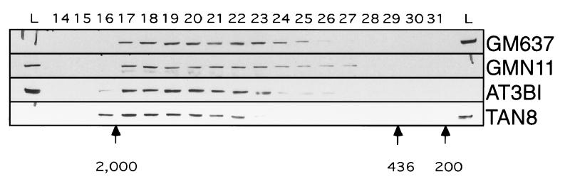
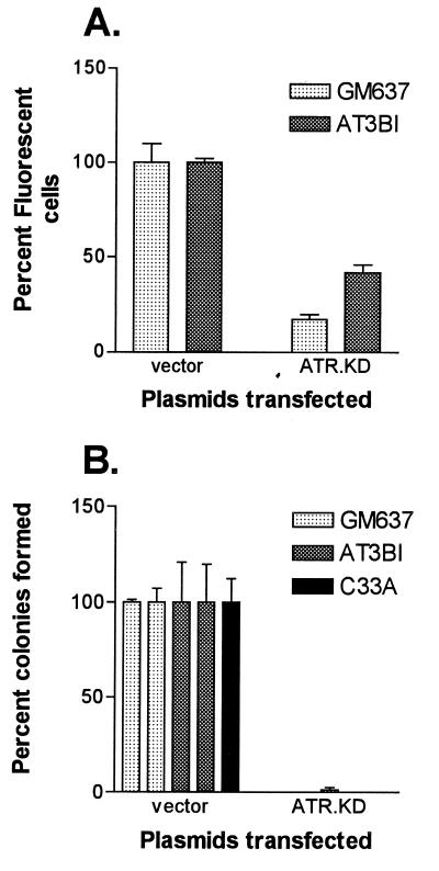
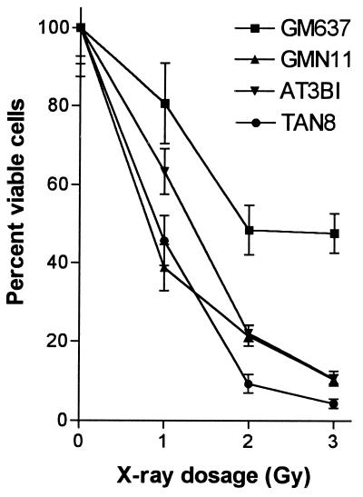
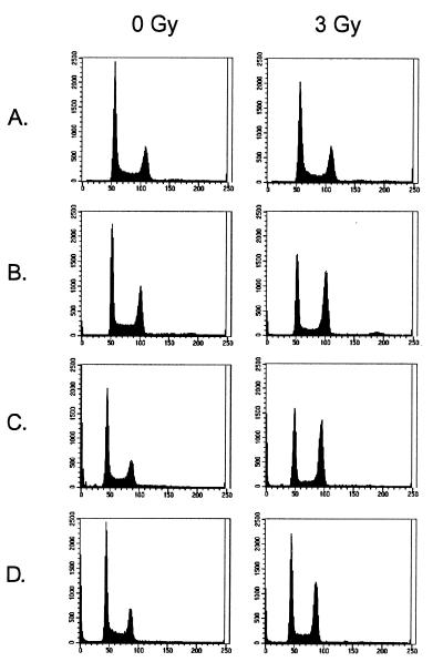
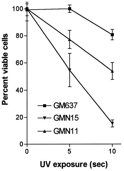
In fission yeast, the rad3 gene product plays a critical role in sensing DNA structure defects and activating damage response pathways. A structural homologue of rad3 in humans (ATR) has been identified based on sequence similarity in the protein kinase domain. General information regarding ATR expression, protein kinase activity, and cellular localization is known, but its function in human cells remains undetermined. In the current study, the ATR protein was examined by gel filtration of protein extracts and was found to exist predominantly as part of a large protein complex. A kinase-inactivated form of the ATR gene was prepared by site-directed mutagenesis and was used in transfection experiments to probe the function of this complex. Introduction of this kinase-dead ATR into a normal fibroblast cell line, an ATM-deficient fibroblast line derived from a patient with ataxia-telangiectasia, or a p53 mutant cell line all resulted in significant losses in cell viability. Clones expressing the kinase-dead ATR displayed increased sensitivity to x-rays and UV and a loss of checkpoint control. We conclude that ATR functions as a critical part of a protein complex that mediates responses to ionizing and UV radiation in human cells. These responses include effects on cell viability and cell cycle checkpoint control.


Figures

Figure 1
Figure 1
Sizing of ATR-containing complexes by gel filtration. Protein extracts prepared as described in Materials and Methods were fractionated by Superose 6 chromatography. Immunoblot analysis was used to determine the elution profile of ATR. Native ATR is ≈300 kDa in size. Elution peaks of molecular size standards are indicated below.
Figure 2
Figure 2
Effect of transfection of ATR.KD expression plasmid on cell viability. (A) GM637 and AT3BI cells were transfected transiently in triplicate with a 3:1 molar excess of the indicated constructs relative to a plasmid expressing green fluorescent protein and quantitated by FACS. For each cell line, 10,000 events were collected. Transfection efficiency was 15–25% with vector alone, and results were normalized to these values. (B) Colonies arising from independent transfections with the indicated plasmids were counted after 10–14 days of selection in G418. Average numbers of colonies with vector alone were ≈1,000 for GM637, ≈30 for AT3BI, and ≈65,000 for C-33A. Results were normalized to vector controls.
Figure 3
Figure 3
Effect of ionizing radiation on colony-forming ability of ATR.KD-transfected cell lines. Cells were exposed to the indicated doses of x-rays and plated in quadruplicate. Colonies were counted after 10–14 days. Colony counts were normalized to those for unirradiated controls.
Figure 4
Figure 4
Effect of ionizing radiation on cell cycle distribution of ATR.KD-transfected cell lines. Cell lines, unirradiated and irradiated with 3 Gy, were stained with propidium iodide and analyzed for DNA content by FACS. (A) GM637. (B) GMN11. (C) AT3BI. (D) TAN8.
Figure 5
Figure 5
Effect of UV radiation on colony-forming ability of ATR.KD-transfected cell lines. Cells were exposed to 310 nM UV for indicated period of time and plated in quadruplicate. Colonies were counted after 10–14 days. Colony counts were normalized to those for unirradiated controls.

References

    1. Hartwell L H, Weinert T A. Science. 1989;246:629–634. - PubMed
    1. Carr A M, Hoekstra M F. Trends Cell Biol. 1995;5:32–40. - PubMed
    1. Carr A M. Curr Opin Genet Dev. 1997;7:93–98. - PubMed
    1. Jiminez G, Yucel J, Rowley R, Subramani S. Proc Natl Acad Sci USA. 1992;87:4952–4956. - PMC - PubMed
    1. Seaton B L, Yucel J, Sunnerhagen P, Subramani S. Gene. 1992;119:83–89. - PubMed

Publication types

Substances