. 1998 Jun 23;95(13):7475-9.
doi: 10.1073/pnas.95.13.7475.

Mitogenic and oncogenic properties of the small G protein Rap1b

Affiliations

Mitogenic and oncogenic properties of the small G protein Rap1b

D L Altschuler et al. Proc Natl Acad Sci U S A. .

Abstract

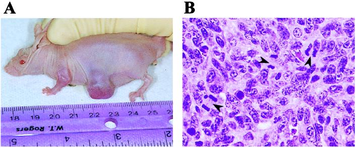
It has been widely reported that the small GTP-binding protein Rap1 has an anti-Ras and anti-mitogenic activity. Thus, it is generally accepted that a normal physiological role of Rap1 proteins is to antagonize Ras mitogenic signals, presumably by forming nonproductive complexes with proteins that are typically effectors or modulators of Ras. Rap1 is activated by signals that raise intracellular levels of cAMP, a molecule that has long been known to exert both inhibitory and stimulatory effects on cell growth. We have now tested the intriguing hypothesis that Rap1 could have mitogenic effects in systems in which cAMP stimulates cell proliferation. The result of experiments addressing this possibility revealed that Rap1 has full oncogenic potential. Expression of Rap1 in these cells results in a decreased doubling time, an increased saturation density, and an unusual anchorage-dependent morphological transformation. Most significantly, however, Rap1-expressing cells formed tumors when injected into nude mice. Thus, we propose that the view that holds Rap1 as an antimitogenic protein should be restricted and conclude that Rap1 is a conditional oncoprotein.


Figures

Figure 1
Figure 1
Establishment of wild-type Rap1b-expressing Swiss 3T3 cell lines. (A) Expression of HA-tagged Rap1b as evaluated by immunoprecipitation-coupled blotting techniques by using a monoclonal anti-HA antibody (HA.11, Babco, Richmond, CA). Samples were analyzed on nonreducing SDS/PAGE with lysates from control (pL7-Hy) and two independent Rap1b-expressing clones (pL7-3-24 and pL7-3-50). HC and IgG indicate the position of heavy chain and whole Ig, respectively. (B) pL7-3-50 cells were analyzed by indirect immunofluorescence with the HA.11 antibody (dilution, 1/150). HA-tagged Rap1b shows a perinuclear localization, as reported for endogenous Rap proteins. (C) Morphological changes induced by wild-type Rap1b. Cells plated on six-well dishes were photographed under low magnification 1 day after reaching confluency. Typical fields with foci-like structures are shown here for Rap1b-expressing cells, as compared with a single monolayer observed in control cells.
Figure 2
Figure 2
Growth properties of Rap1b-expressing Swiss 3T3 cells. (A) Growth curves of control (pL7-Hy, ▪) and Rap1b-expressing (pL-3-24, ◊; and pL-7-3-50, ▴) cells in serum-containing medium. Cells were plated on six-well dishes (50,000 per well, per triplicate) and counted daily after trypsinization. Results are expressed as average of triplicates (variation <5%). (B) Cell culture inserts (Falcon; 0.45-μm pore size) containing 50,000 cells of each cell line were placed on top of wells containing 50,000 pL7-Hy cells. Plates were treated as before and bottom wells (control cells) were counted daily to test for potential autocrine activity.
Figure 3
Figure 3
Rap1b-expressing cells show an increased ability to traverse S phase. (A) [3H]Thymidine incorporation assays indicate that Rap1b- expressing cells (pL7-3-24, ▪ and pL7-3-50, ▴) show an increase in the maximal response, as compared with control (pL7-Hy, •) cells. Dose-responses were performed for fetal bovine serum (FBS), platelet-derived growth factor (PDGF) and forskolin (FK). Forskolin dose-response was performed in the presence of a constant amount of insulin (Ins, 1 μg/ml) and 3-isobutyl-1-methylxanthine (100 μM). (B) Nuclear incorporation of BrdUrd was assayed by indirect immunofluorescence (32). Cells were left untreated (−), or stimulated with insulin (INS, 1 μg/ml) or insulin plus forskolin (INS+FK, 1 μg/ml and 10 μM, respectively) in the presence of 100 μM 3-isobutyl-1-methylxanthine. Total nuclei was visualized by 4′,6-diamidino-2-phenylindole staining, and the results expressed as % BrdUrd/4′,6-diamidino-2-phenylindole. Experiments were done in duplicates and three or four independent fields per sample analyzed and expressed as averages (variation <10%).
Figure 4
Figure 4
Rap1b-expressing cells induce tumor formation in nude mice, (A) pL7-3-50 cells were injected into nude mice, and a tumor scored 2 months after injection is shown here. (B) A typical hematoxylin/eosin staining showing several mitotic figures is shown here (arrowheads).

References

    1. Noda M. Biochem Biophys Acta. 1993;1155:97–109. - PubMed
    1. Boguski M S, McCormick F. Nature (London) 1993;366:643–654. - PubMed
    1. Nagata K, Itoh H, Katada T, Takenaka K, Ui M, Kaziro Y, Nozawa Y. J Biol Chem. 1989;264:17000–17005. - PubMed
    1. Pizon V, Lerosey I, Chardin P, Tavitian A. Nucleic Acids Res. 1988;16:7719. - PMC - PubMed
    1. Kitayama H, Sugimoto Y, Matsuzaki T, Ikawa Y, Noda M. Cell. 1989;56:77–84. - PubMed

Publication types

LinkOut - more resources