. 1998 Jun 23;95(13):7603-7.
doi: 10.1073/pnas.95.13.7603.

Incomplete embryonic lethality and fatal neonatal hemorrhage caused by prothrombin deficiency in mice

Affiliations

Incomplete embryonic lethality and fatal neonatal hemorrhage caused by prothrombin deficiency in mice

J Xue et al. Proc Natl Acad Sci U S A. .

Abstract

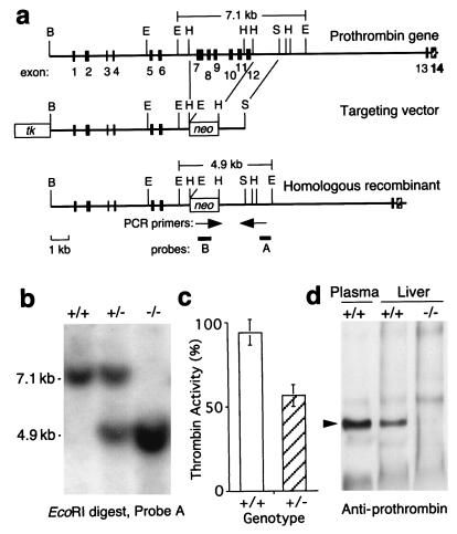
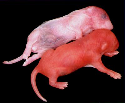
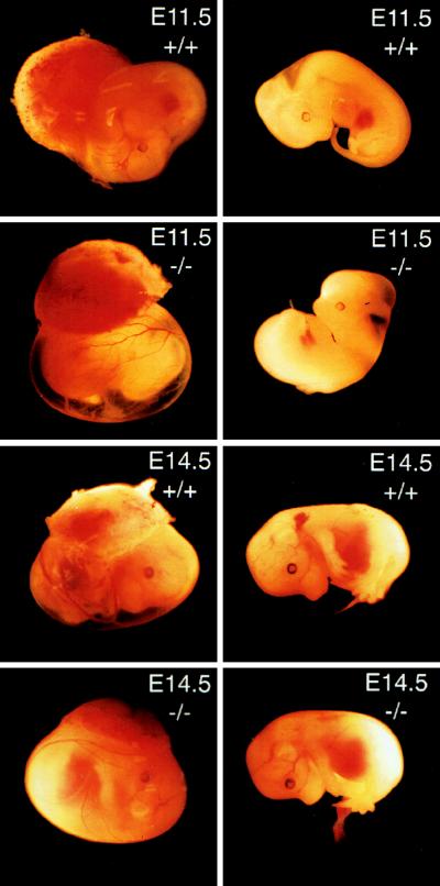
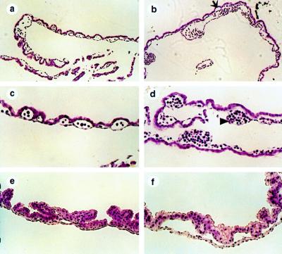
Deficiency of blood coagulation factor V or tissue factor causes the death of mouse embryos by 10.5 days of gestation, suggesting that part of the blood coagulation system is necessary for development. This function is proposed to require either generation of the serine protease thrombin and cell signaling through protease-activated receptors or an activity of tissue factor that is distinct from blood clotting. We find that murine deficiency of prothrombin clotting factor 2 (Cf2) was associated with the death of approximately 50% of Cf2(-/-) embryos by embryonic day 10.5 (E10.5), and surviving embryos had characteristic defects in yolk sac vasculature. Most of the remaining Cf2(-/-) embryos died by E15.5, but those surviving to E18.5 appeared normal. The rare Cf2(-/-) neonates died of hemorrhage on the first postnatal day. These studies suggest that a part of the blood coagulation system is adapted to perform a developmental function. Other mouse models show that the absence of platelets or of fibrinogen does not cause fetal wastage. Therefore, the role of thrombin in development may be independent of its effects on blood coagulation and instead may involve signal transduction on cells other than platelets.


Figures

Figure 1
Figure 1
Targeting of the prothrombin gene by homologous recombination. (a) Structure of mouse prothrombin gene and targeting vector. The cloned genomic DNA fragment extended from the first BamHI site to the last shown EcoRI site. Solid rectangles represent exons 1–12. Additional exons 13 and 14 (hatched rectangles) are indicated based on the structure of the human prothrombin gene. Cleavage sites are shown for BamHI (B), EcoRI (E), HindIII (H), and SalI (S). The targeting vector contains a pgk-neo cassette in place of exons 7–12 and a pgk-tk cassette. The product of homologous recombination is shown at the bottom. Positions of PCR primers (arrows) and hybridization probes (A and B) used to detect successful gene targeting are indicated. The EcoRI digest will generate a 7.1 kb fragment from the untargeted allele and a 4.9 kb fragment from the targeted allele, both of which are recognized by probe A. (b) Southern blot of EcoRI-digested tail DNA from Cf2+/+, Cf2+/−, and Cf2−/− littermates hybridized with probe A. (c) Prothrombin levels in mouse plasma sampled from 10 mice of each genotype (mean ± SD). (d) Western blot of prothrombin in mouse liver extracts. Arrowhead indicates the position of 72 kDa prothrombin.
Figure 2
Figure 2
Newborn mice. Both wild-type (Lower) and homozygous prothrombin deficient mice (Upper) were able to breath and suckle. Compared with wild type, the Cf2−/− mouse is pale, with extensive bruising over the swollen abdomen and head. Dissection of the Cf2−/− mouse showed massive intraperitoneal hemorrhage with no clotted blood.
Figure 3
Figure 3
Prothrombin-deficient embryos. Embryos from a single pregnancy at either E11.5 or E14.5 are shown with their placentas and extraembryonic membranes (Left) and without (dissected) (Right). For all genotypes, the yolk sac vasculature and embryos appear grossly normal.
Figure 4
Figure 4
Histological analysis of the yolk sac. Sections are shown for normal Cf2+/+ embryos (a, c, and e) and null Cf2−/− embryos (b, d, and f). At E9.5 (ad), 100× magnification of a wild-type yolk sac (a) shows a normal outer layer of visceral endoderm and inner layer of small capillaries surrounded by endothelium, whereas the prothrombin-deficient yolk sac (b) shows greatly dilated, enlarged vascular spaces. In some places the visceral endoderm and mesothelial layers appear to be separated (arrow). Under higher (200×) magnification, the normal small yolk sac capillaries (c) contain erythrocytes and hematopoietic cells; the yolk sac of the Cf2−/− embryo has normal appearing blood islands (triangle) adjacent to dilated vascular spaces. The difference in appearance of yolk sac capillaries is evident also at E11.5 (200× magnification (e and f). Capillary channels in the normal yolk sac (e) are small, whereas those in Cf2−/− yolk sac are greatly enlarged (f).

References

    1. Mann K G. In: Hemostasis and Thrombosis: Basic Principles and Clinical Practice. Colman R W, Hirsch J, Marder V J, Salzman E W, editors. Philadelphia: Lippincott; 1994. pp. 184–199.
    1. Coughlin S R, Vu T K, Hung D T, Wheaton V I. J Clin Invest. 1992;89:351–355. - PMC - PubMed
    1. Weinstein J R, Gold S J, Cunningham D D, Gall C M. J Neurosci. 1995;15:2906–2919. - PMC - PubMed
    1. Cavanaugh K P, Gurwitz D, Cunningham D D, Bradshaw R A. J Neurochem. 1990;54:1735–1743. - PubMed
    1. Gurwitz D, Cunningham D D. Proc Natl Acad Sci USA. 1988;85:3440–3444. - PMC - PubMed

Publication types

LinkOut - more resources