

Human carbonic anhydrase XII: cDNA cloning, expression, and chromosomal localization of a carbonic anhydrase gene that is overexpressed in some renal cell cancers
- PMID: 9636197
- PMCID: PMC22698
- DOI: 10.1073/pnas.95.13.7608
Human carbonic anhydrase XII: cDNA cloning, expression, and chromosomal localization of a carbonic anhydrase gene that is overexpressed in some renal cell cancers
Abstract
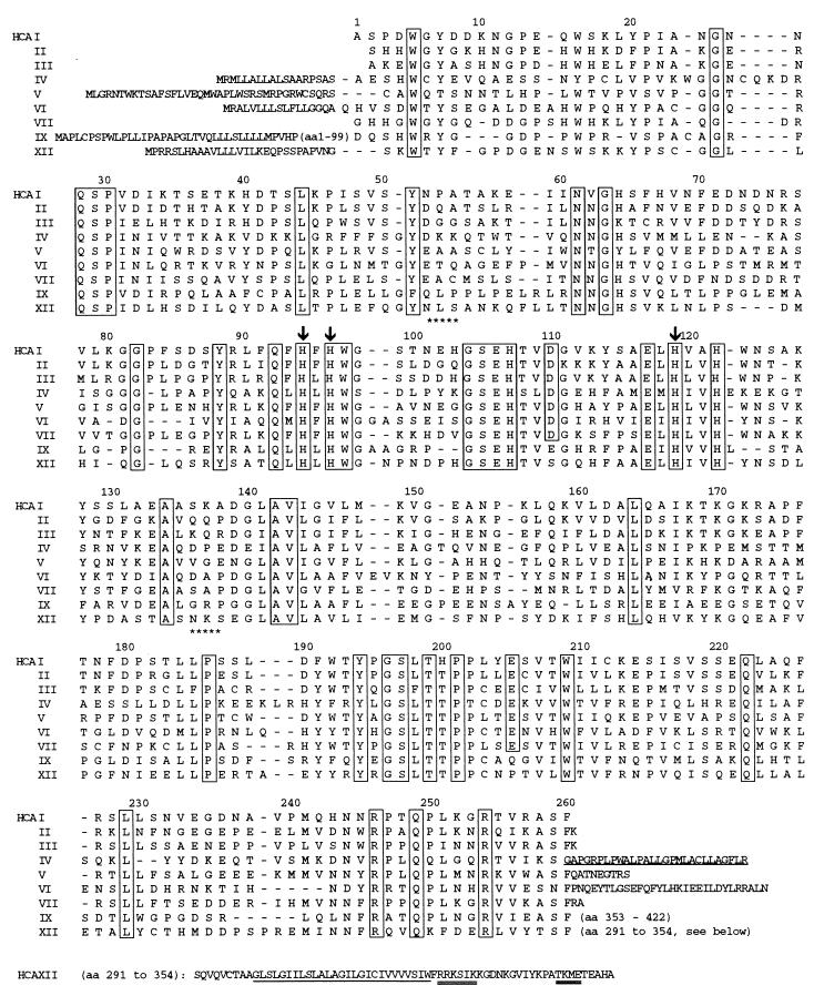
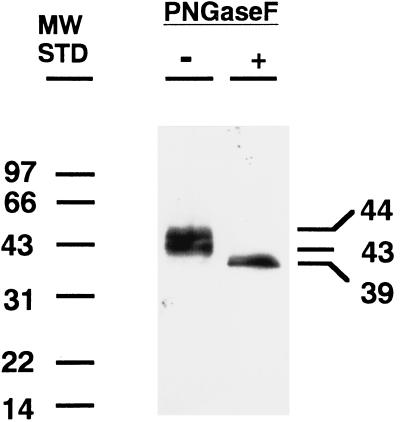
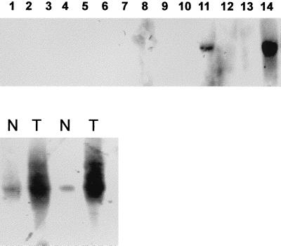
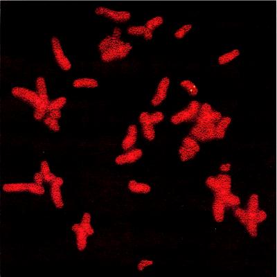
We report the cloning and characterization of a tumor-associated carbonic anhydrase (CA) that was identified in a human renal cell carcinoma (RCC) by serological expression screening with autologous antibodies. The cDNA sequence predicts a 354-amino acid polypeptide with a molecular mass of 39,448 Da that has features of a type I membrane protein. The predicted sequence includes a 29-amino acid signal sequence, a 261-amino acid CA domain, an additional short extracellular segment, a 26-amino acid hydrophobic transmembrane domain, and a hydrophilic C-terminal cytoplasmic tail of 29 amino acids that contains two potential phosphorylation sites. The extracellular CA domain shows 30-42% homology with known human CAs, contains all three Zn-binding histidine residues found in active CAs, and contains two potential sites for asparagine glycosylation. When expressed in COS cells, the cDNA produced a 43- to 44-kDa protein in membranes that had around one-sixth the CA activity of membranes from COS cells transfected with the same vector expressing bovine CA IV. We have designated this human protein CA XII. Northern blot analysis of normal tissues demonstrated a 4.5-kb transcript only in kidney and intestine. However, in 10% of patients with RCC, the CA XII transcript was expressed at much higher levels in the RCC than in surrounding normal kidney tissue. The CA XII gene was mapped by using fluorescence in situ hybridization to 15q22. CA XII is the second catalytically active membrane CA reported to be overexpressed in certain cancers. Its relationship to oncogenesis and its potential as a clinically useful tumor marker clearly merit further investigation.
Figures
References
Publication types
MeSH terms
Substances
Associated data
- Actions
Grants and funding
LinkOut - more resources
Full Text Sources
Other Literature Sources
Medical
Molecular Biology Databases
