

doi: 10.1073/pnas.95.13.7721.
Functional organization of spatial and nonspatial working memory processing within the human lateral frontal cortex
Affiliations
- PMID: 9636217
- PMCID: PMC22736
- DOI: 10.1073/pnas.95.13.7721
Item in Clipboard
Functional organization of spatial and nonspatial working memory processing within the human lateral frontal cortex
Proc Natl Acad Sci U S A.
.
Abstract
The present study used functional magnetic resonance imaging to demonstrate that performance of visual spatial and visual nonspatial working memory tasks involve the same regions of the lateral prefrontal cortex when all factors unrelated to the type of stimulus material are appropriately controlled. These results provide evidence that spatial and nonspatial working memory may not be mediated, respectively, by mid-dorsolateral and mid-ventrolateral regions of the frontal lobe, as widely assumed, and support the alternative notion that specific regions of the lateral prefrontal cortex make identical executive functional contributions to both spatial and nonspatial working memory.
Figures
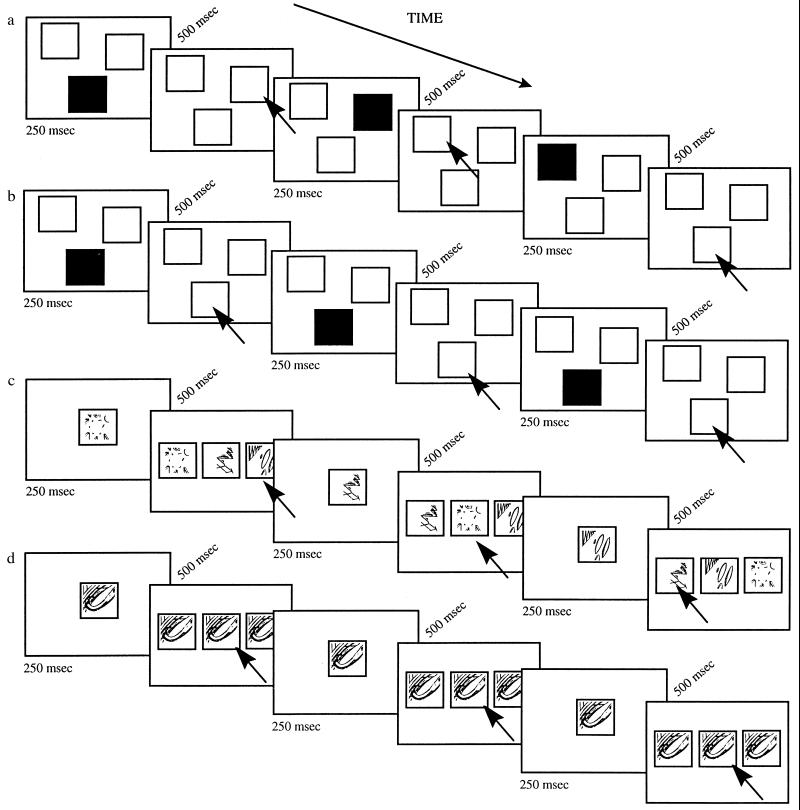
Illustrated are: (a) the spatial working memory task, (b) the spatial control task, (c) the nonspatial working memory task, and (d) the nonspatial control task. A number of trials are shown in each case. Trials were presented sequentially with a constant 500-ms interval in between. Black arrows, subjects’ responses.
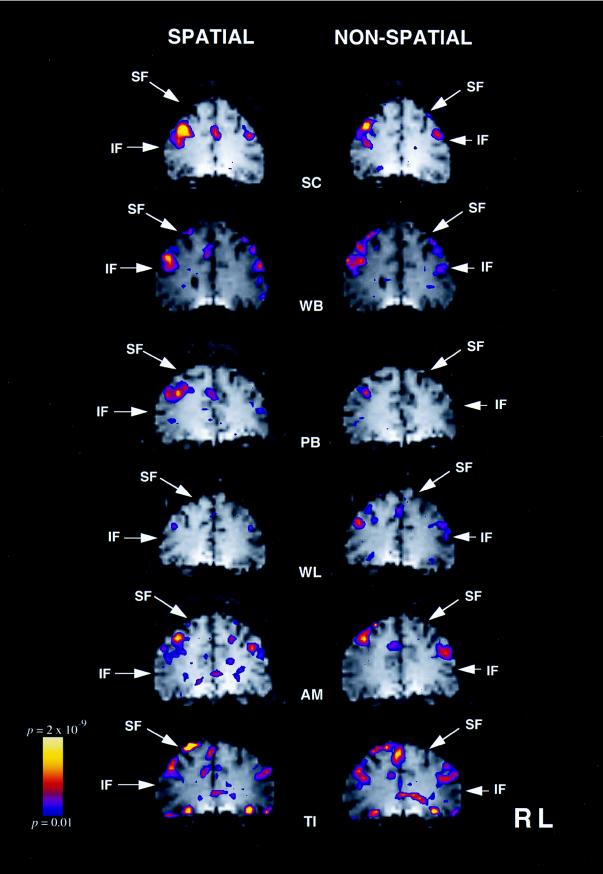
fMRI signal increases in the mid-dorsolateral frontal cortex for each of the six subjects during the spatial working memory task (left column) and during the nonspatial working memory task (right column). The color bar on the left gives the range of significance values, with blue representing P < 0.01 and yellow a level of P < 2 × 10⁻⁹. The left hemisphere appears on the right of each image. SF, superior frontal sulcus; IF, inferior frontal sulcus.
References
-
- Wilson F A W,, Scalaidhe S P O, Goldman-Rakic P S. Science. 1993;260:1955–1958. - PubMed
-
- Goldman-Rakic P S. Ann N Y Acad Sci. 1995;769:71–83. - PubMed
-
- Cohen J D, Forman S D, Braver T S, Casey B J, Servan-Schreiber D, Noll D C. Hum Brain Mapp. 1994;1:293–304. - PubMed
-
- Smith E E, Jonides J J, Koeppe R A, Awh E, Schumacher E H, Minoshima S. J Cognit Neurosci. 1995;7:337–356. - PubMed
-
- Courtney S M, Ungerlieder L G, Keil K, Haxby J V. Cereb Cortex. 1996;6:39–49. - PubMed
Publication types
MeSH terms
LinkOut - more resources
Full Text Sources
Medical
