

doi: 10.1101/gad.12.12.1769.
Myc activates telomerase
Affiliations
- PMID: 9637678
- PMCID: PMC316904
- DOI: 10.1101/gad.12.12.1769
Item in Clipboard
Myc activates telomerase
Genes Dev.
.
Abstract
Telomere maintenance has been proposed as an essential prerequisite to human tumor development. The telomerase enzyme is itself a marker for tumor cells, but the genetic alterations that activate the enzyme during neoplastic transformation have remained a mystery. Here, we show that Myc induces telomerase in both normal human mammary epithelial cells (HMECs) and normal human diploid fibroblasts. Myc increases expression of hEST2 (hTRT/TP2), the limiting subunit of telomerase, and both Myc and hEST2 can extend the life span of HMECs. The ability of Myc to activate telomerase may contribute to its ability to promote tumor formation.
Figures
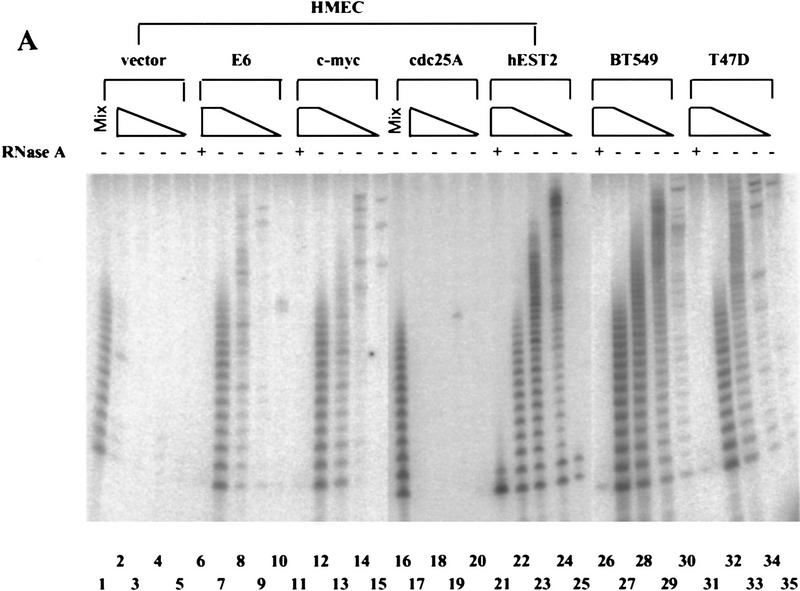
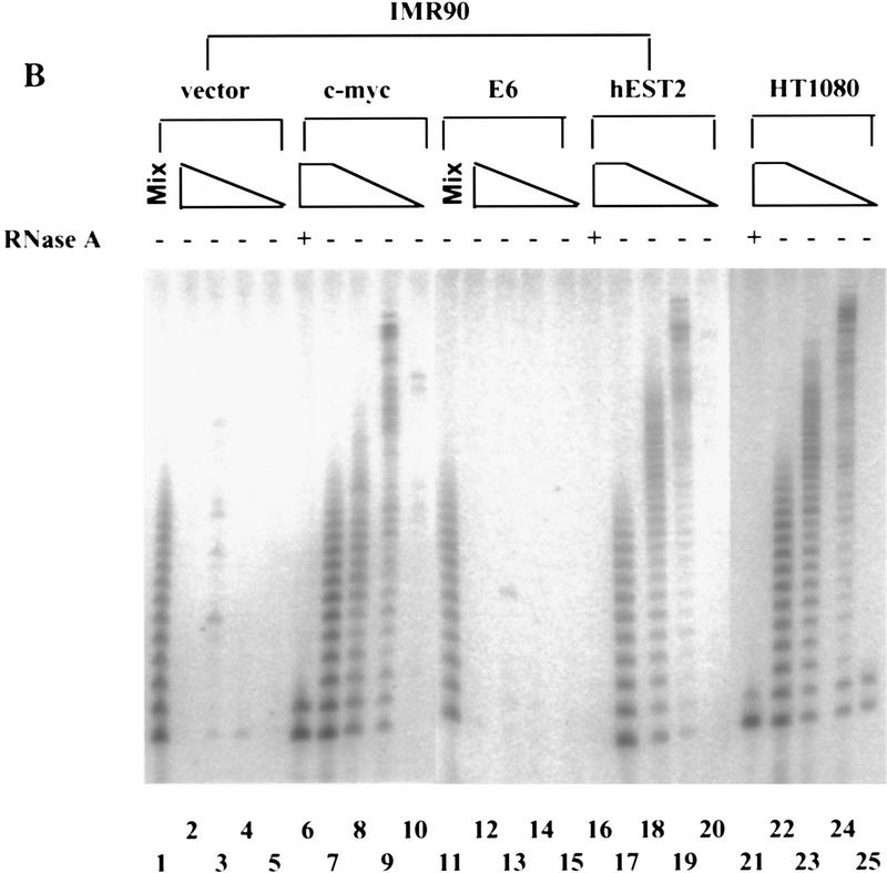
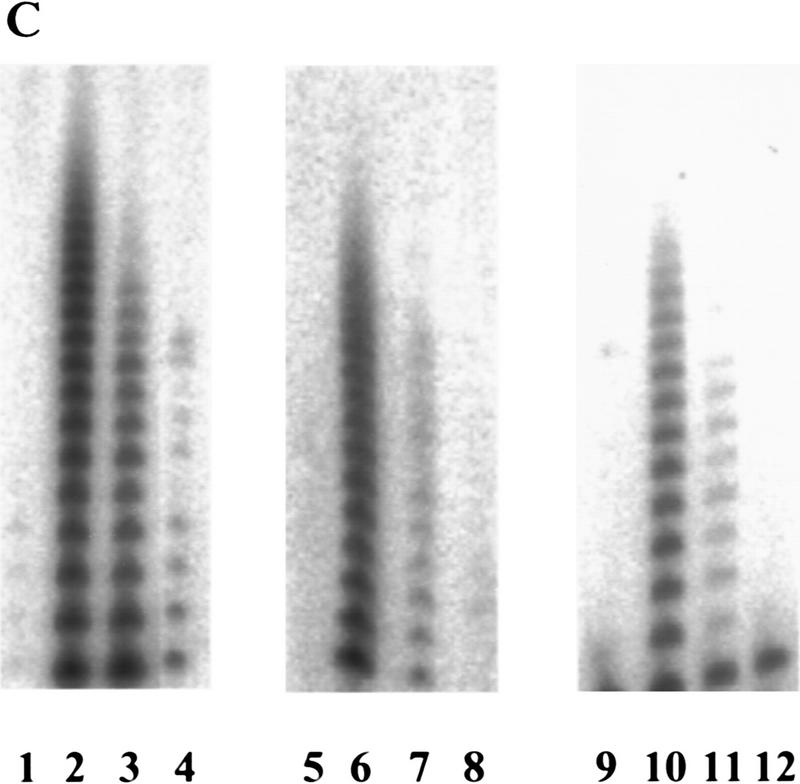
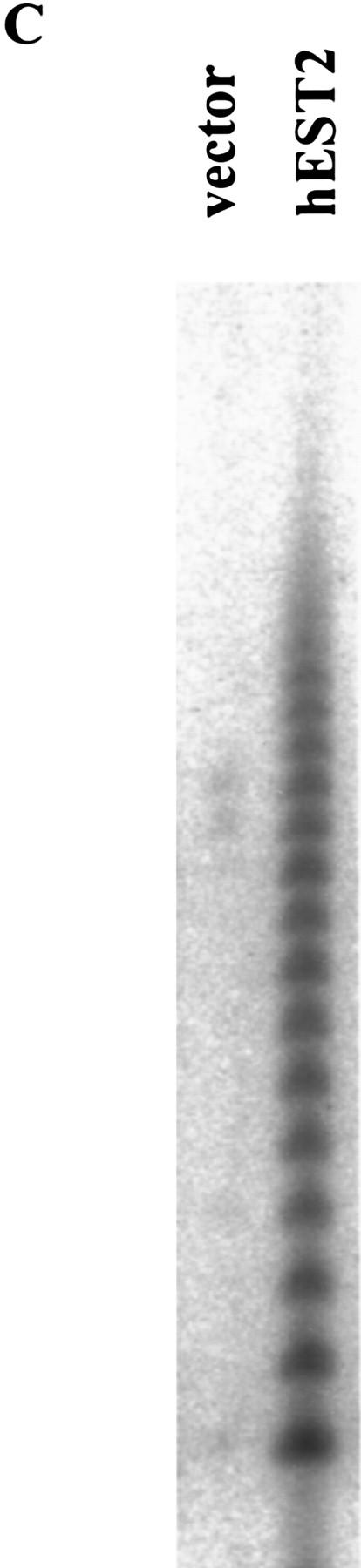
Myc activates telomerase. (A) Primary HMECs at passage 12 were infected with empty vector (lanes 1–5), E6 (lanes 6–10), c-Myc (lanes 11–15), cdc25A (lanes 16–20), or hEST2 (lanes 21–25) viruses. Breast cancer cell lines BT549 (lanes 26–30) and T47D (lanes 31–35) were included for comparison. TRAP assays contained lysates from 10,000 (lanes 2,6,7,11,12,17,21,22,26,27,31,32), 1000 (lanes 3,8,13,18,23,28,33), 100 (lanes 4,9,14,19,24,29,34), or 10 (lanes 5,10,15,20,25,30,35) cells. (− and +) Absence or presence of RNase A, respectively. (Mix; lanes 1,16) To exclude the presence of inhibitors in apparently negative lysates, lysate from 10,000 of the indicated cells was mixed with lysate from 10,000 c-Myc-expressing cells. (B) IMR90 cells at passage 14 were infected with empty vector (lanes 1–5), c-Myc (lanes 6–10), E6 (lanes 11–15), or hEST2 (lanes 16–20) viruses. HT1080 cells (lanes 21–25) were included for comparison. TRAP assays were performed with decreasing cell equivalents as in A. (C) HMEC (lanes 1–4), IMR90 (lanes 5–8), or WI38 (lanes 9–12) cells were infected with empty vector (lanes 1,5,9), hEST2 (lanes 2,6,10), c-Myc (lanes 3,7,11), or E6 viruses (lanes 4,8,12). Cells were selected for ∼5 days with puromycin or hygromycin and then lysed for telomerase assay. Each lane corresponds to 10,000 cells.
Myc activates telomerase. (A) Primary HMECs at passage 12 were infected with empty vector (lanes 1–5), E6 (lanes 6–10), c-Myc (lanes 11–15), cdc25A (lanes 16–20), or hEST2 (lanes 21–25) viruses. Breast cancer cell lines BT549 (lanes 26–30) and T47D (lanes 31–35) were included for comparison. TRAP assays contained lysates from 10,000 (lanes 2,6,7,11,12,17,21,22,26,27,31,32), 1000 (lanes 3,8,13,18,23,28,33), 100 (lanes 4,9,14,19,24,29,34), or 10 (lanes 5,10,15,20,25,30,35) cells. (− and +) Absence or presence of RNase A, respectively. (Mix; lanes 1,16) To exclude the presence of inhibitors in apparently negative lysates, lysate from 10,000 of the indicated cells was mixed with lysate from 10,000 c-Myc-expressing cells. (B) IMR90 cells at passage 14 were infected with empty vector (lanes 1–5), c-Myc (lanes 6–10), E6 (lanes 11–15), or hEST2 (lanes 16–20) viruses. HT1080 cells (lanes 21–25) were included for comparison. TRAP assays were performed with decreasing cell equivalents as in A. (C) HMEC (lanes 1–4), IMR90 (lanes 5–8), or WI38 (lanes 9–12) cells were infected with empty vector (lanes 1,5,9), hEST2 (lanes 2,6,10), c-Myc (lanes 3,7,11), or E6 viruses (lanes 4,8,12). Cells were selected for ∼5 days with puromycin or hygromycin and then lysed for telomerase assay. Each lane corresponds to 10,000 cells.
Myc activates telomerase. (A) Primary HMECs at passage 12 were infected with empty vector (lanes 1–5), E6 (lanes 6–10), c-Myc (lanes 11–15), cdc25A (lanes 16–20), or hEST2 (lanes 21–25) viruses. Breast cancer cell lines BT549 (lanes 26–30) and T47D (lanes 31–35) were included for comparison. TRAP assays contained lysates from 10,000 (lanes 2,6,7,11,12,17,21,22,26,27,31,32), 1000 (lanes 3,8,13,18,23,28,33), 100 (lanes 4,9,14,19,24,29,34), or 10 (lanes 5,10,15,20,25,30,35) cells. (− and +) Absence or presence of RNase A, respectively. (Mix; lanes 1,16) To exclude the presence of inhibitors in apparently negative lysates, lysate from 10,000 of the indicated cells was mixed with lysate from 10,000 c-Myc-expressing cells. (B) IMR90 cells at passage 14 were infected with empty vector (lanes 1–5), c-Myc (lanes 6–10), E6 (lanes 11–15), or hEST2 (lanes 16–20) viruses. HT1080 cells (lanes 21–25) were included for comparison. TRAP assays were performed with decreasing cell equivalents as in A. (C) HMEC (lanes 1–4), IMR90 (lanes 5–8), or WI38 (lanes 9–12) cells were infected with empty vector (lanes 1,5,9), hEST2 (lanes 2,6,10), c-Myc (lanes 3,7,11), or E6 viruses (lanes 4,8,12). Cells were selected for ∼5 days with puromycin or hygromycin and then lysed for telomerase assay. Each lane corresponds to 10,000 cells.
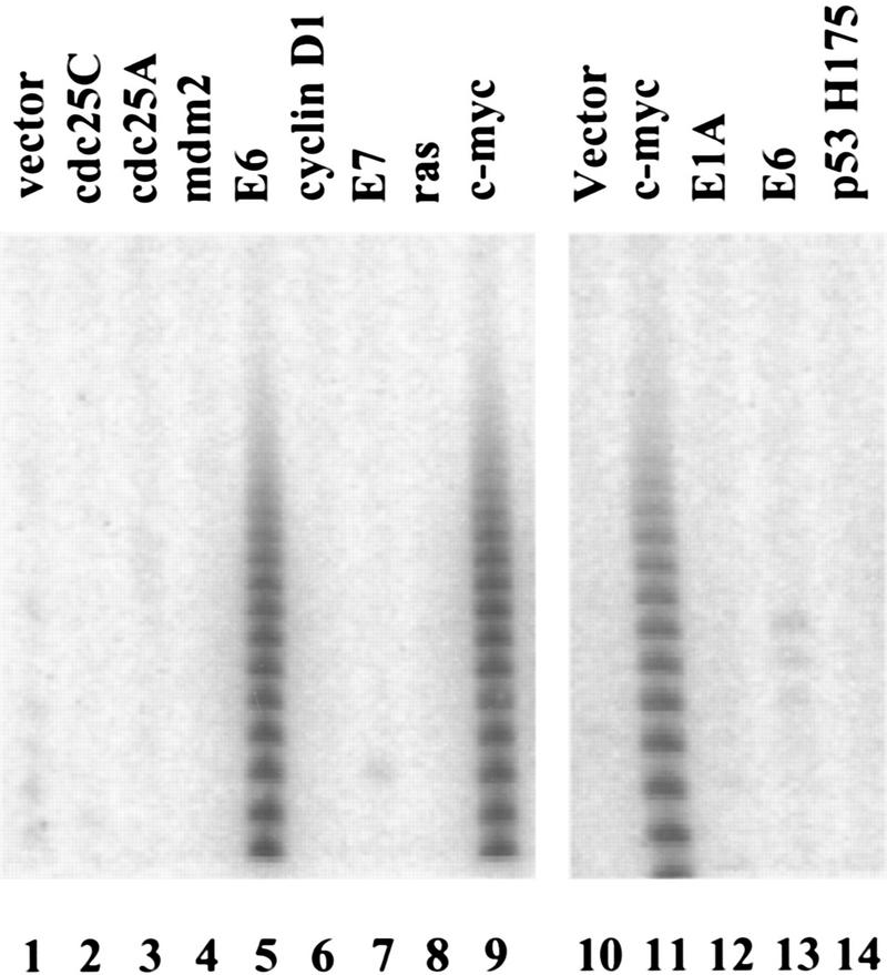
Oncogene activation of telomerase. HMECs (lanes 1–9) or IMR90 cells (lanes 10–14) were infected with viruses that direct the expression of the indicated oncogenes (lanes 2–9,11–14) or empty vector (lanes 1,10). Cell extracts were analyzed by TRAP assay.
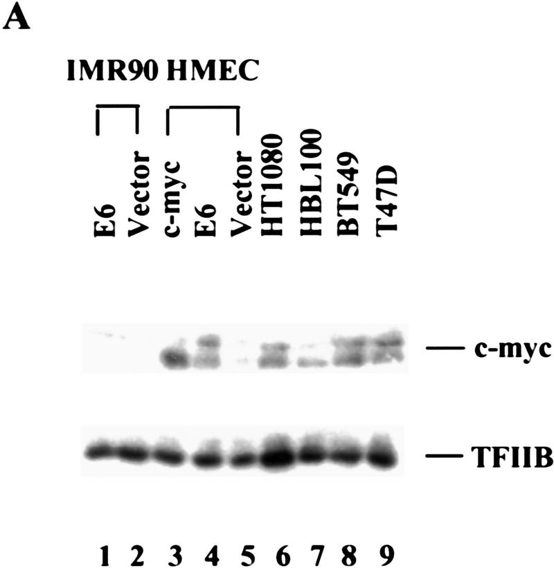
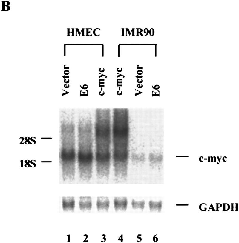
E6 increases c-Myc protein in HMECs. (A) Cell lysates from E6 (lane 1)- and vector (lane 2)-infected IMR90 cells and lysates from c-Myc (lane 3)-, E6 (lane 4)-, and vector (lane 5)-infected HMECs were analyzed by Western blot with a polyclonal Myc antibody. Tumor cell lines, HT1080 (lane 6), HBL100 (lane 7), BT549 (lane 8), and T47D (lane 9), were included for comparison. The expression of TFIIB was used to normalize loading. (B) Northern analysis of Myc RNA levels in total RNA. GAPDH was probed as a loading control.
E6 increases c-Myc protein in HMECs. (A) Cell lysates from E6 (lane 1)- and vector (lane 2)-infected IMR90 cells and lysates from c-Myc (lane 3)-, E6 (lane 4)-, and vector (lane 5)-infected HMECs were analyzed by Western blot with a polyclonal Myc antibody. Tumor cell lines, HT1080 (lane 6), HBL100 (lane 7), BT549 (lane 8), and T47D (lane 9), were included for comparison. The expression of TFIIB was used to normalize loading. (B) Northern analysis of Myc RNA levels in total RNA. GAPDH was probed as a loading control.
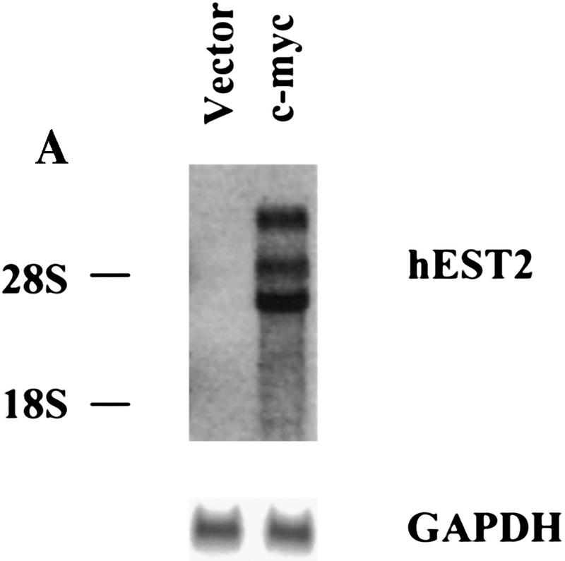
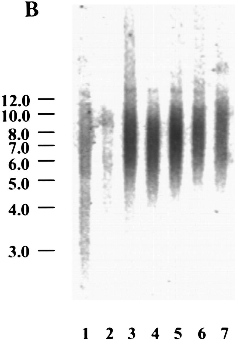
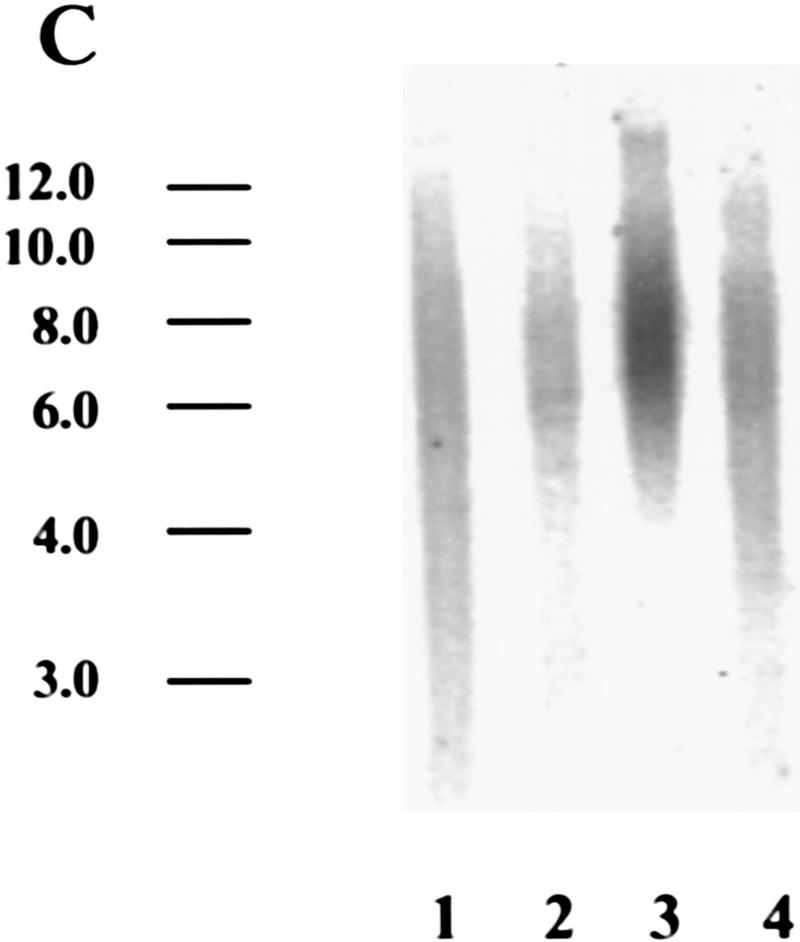
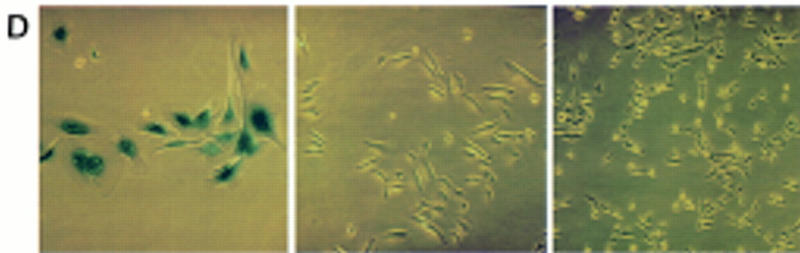
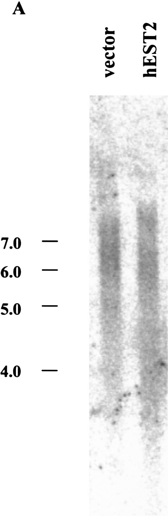
Myc regulates hEST2 and extends cellular life span in HMECs. (A) hEST2 Northern analysis of poly(A)+ RNA from normal HMECs and from HMECs that had been infected with a Myc retrovirus. A Northern blot with GAPDH was performed as a loading control. (B) Genomic DNA (3 μg) from early-passage HMECs (passage 12, lane 1), late-passage HMECs (passage 26, lane 2), and hEST2-expressing HMECs [infected at passage 12 and cultured for 3 (lane 3), 6 (lane 4), 8 (lane 5), 10 (lane 6), or 14 (lane 7) additional passages] was digested with RsaI and HinfI. Fragments were separated on a 0.8% agarose gel, and telomeric restriction fragments were visualized with a 32P-labeled human telomeric sequence (TTAGGG)3 probe. (C) Genomic DNA (3 μg) from early-passage HMECs (passage 12, lane 1), vector-infected HMEC (infected at passage 12 and cultured for six additional passages or ∼12–14 PD, lane 2), hEST2-expressing HMECs (infected at passage 12 and cultured for six additional passages or ∼12–14 PD, lane 3), and Myc-expressing cells (infected at passage 12 and cultured for six additional passages or ∼18 PD, lane 4) were digested with RsaI and HinfI. Fragments were probed with a telomeric probe as described in B. TRF intensity was quantitated on a Fuji BAS2000 PhosphorImager. Normalizing vector-containing HMECs (lane 2) to 100 units of intensity, both early passage HMECs (lane 1) and Myc-expressing HMECs (lane 4) gave ∼150 units of intensity and hEST2-expressing HMECs (lane 3) gave ∼200 units of intensity. (D) HMECs transduced with empty vector (left), hEST2 (middle), or c-Myc viruses (right) were grown to a PDL of ∼56–60. At this PDL, vector cells adopted a senescent morphology and ceased growth. Cells expressing c-Myc and hEST2 continued to proliferate. To assess the percentage of senescent cells in the population, each culture was stained for senescence-associated β-galactosidase. Greater than 95% of the vector-containing cells were β-galactosidase positive whereas <10% of cells expressing hEST2 or Myc were stained.
Myc regulates hEST2 and extends cellular life span in HMECs. (A) hEST2 Northern analysis of poly(A)+ RNA from normal HMECs and from HMECs that had been infected with a Myc retrovirus. A Northern blot with GAPDH was performed as a loading control. (B) Genomic DNA (3 μg) from early-passage HMECs (passage 12, lane 1), late-passage HMECs (passage 26, lane 2), and hEST2-expressing HMECs [infected at passage 12 and cultured for 3 (lane 3), 6 (lane 4), 8 (lane 5), 10 (lane 6), or 14 (lane 7) additional passages] was digested with RsaI and HinfI. Fragments were separated on a 0.8% agarose gel, and telomeric restriction fragments were visualized with a 32P-labeled human telomeric sequence (TTAGGG)3 probe. (C) Genomic DNA (3 μg) from early-passage HMECs (passage 12, lane 1), vector-infected HMEC (infected at passage 12 and cultured for six additional passages or ∼12–14 PD, lane 2), hEST2-expressing HMECs (infected at passage 12 and cultured for six additional passages or ∼12–14 PD, lane 3), and Myc-expressing cells (infected at passage 12 and cultured for six additional passages or ∼18 PD, lane 4) were digested with RsaI and HinfI. Fragments were probed with a telomeric probe as described in B. TRF intensity was quantitated on a Fuji BAS2000 PhosphorImager. Normalizing vector-containing HMECs (lane 2) to 100 units of intensity, both early passage HMECs (lane 1) and Myc-expressing HMECs (lane 4) gave ∼150 units of intensity and hEST2-expressing HMECs (lane 3) gave ∼200 units of intensity. (D) HMECs transduced with empty vector (left), hEST2 (middle), or c-Myc viruses (right) were grown to a PDL of ∼56–60. At this PDL, vector cells adopted a senescent morphology and ceased growth. Cells expressing c-Myc and hEST2 continued to proliferate. To assess the percentage of senescent cells in the population, each culture was stained for senescence-associated β-galactosidase. Greater than 95% of the vector-containing cells were β-galactosidase positive whereas <10% of cells expressing hEST2 or Myc were stained.
Myc regulates hEST2 and extends cellular life span in HMECs. (A) hEST2 Northern analysis of poly(A)+ RNA from normal HMECs and from HMECs that had been infected with a Myc retrovirus. A Northern blot with GAPDH was performed as a loading control. (B) Genomic DNA (3 μg) from early-passage HMECs (passage 12, lane 1), late-passage HMECs (passage 26, lane 2), and hEST2-expressing HMECs [infected at passage 12 and cultured for 3 (lane 3), 6 (lane 4), 8 (lane 5), 10 (lane 6), or 14 (lane 7) additional passages] was digested with RsaI and HinfI. Fragments were separated on a 0.8% agarose gel, and telomeric restriction fragments were visualized with a 32P-labeled human telomeric sequence (TTAGGG)3 probe. (C) Genomic DNA (3 μg) from early-passage HMECs (passage 12, lane 1), vector-infected HMEC (infected at passage 12 and cultured for six additional passages or ∼12–14 PD, lane 2), hEST2-expressing HMECs (infected at passage 12 and cultured for six additional passages or ∼12–14 PD, lane 3), and Myc-expressing cells (infected at passage 12 and cultured for six additional passages or ∼18 PD, lane 4) were digested with RsaI and HinfI. Fragments were probed with a telomeric probe as described in B. TRF intensity was quantitated on a Fuji BAS2000 PhosphorImager. Normalizing vector-containing HMECs (lane 2) to 100 units of intensity, both early passage HMECs (lane 1) and Myc-expressing HMECs (lane 4) gave ∼150 units of intensity and hEST2-expressing HMECs (lane 3) gave ∼200 units of intensity. (D) HMECs transduced with empty vector (left), hEST2 (middle), or c-Myc viruses (right) were grown to a PDL of ∼56–60. At this PDL, vector cells adopted a senescent morphology and ceased growth. Cells expressing c-Myc and hEST2 continued to proliferate. To assess the percentage of senescent cells in the population, each culture was stained for senescence-associated β-galactosidase. Greater than 95% of the vector-containing cells were β-galactosidase positive whereas <10% of cells expressing hEST2 or Myc were stained.
Myc regulates hEST2 and extends cellular life span in HMECs. (A) hEST2 Northern analysis of poly(A)+ RNA from normal HMECs and from HMECs that had been infected with a Myc retrovirus. A Northern blot with GAPDH was performed as a loading control. (B) Genomic DNA (3 μg) from early-passage HMECs (passage 12, lane 1), late-passage HMECs (passage 26, lane 2), and hEST2-expressing HMECs [infected at passage 12 and cultured for 3 (lane 3), 6 (lane 4), 8 (lane 5), 10 (lane 6), or 14 (lane 7) additional passages] was digested with RsaI and HinfI. Fragments were separated on a 0.8% agarose gel, and telomeric restriction fragments were visualized with a 32P-labeled human telomeric sequence (TTAGGG)3 probe. (C) Genomic DNA (3 μg) from early-passage HMECs (passage 12, lane 1), vector-infected HMEC (infected at passage 12 and cultured for six additional passages or ∼12–14 PD, lane 2), hEST2-expressing HMECs (infected at passage 12 and cultured for six additional passages or ∼12–14 PD, lane 3), and Myc-expressing cells (infected at passage 12 and cultured for six additional passages or ∼18 PD, lane 4) were digested with RsaI and HinfI. Fragments were probed with a telomeric probe as described in B. TRF intensity was quantitated on a Fuji BAS2000 PhosphorImager. Normalizing vector-containing HMECs (lane 2) to 100 units of intensity, both early passage HMECs (lane 1) and Myc-expressing HMECs (lane 4) gave ∼150 units of intensity and hEST2-expressing HMECs (lane 3) gave ∼200 units of intensity. (D) HMECs transduced with empty vector (left), hEST2 (middle), or c-Myc viruses (right) were grown to a PDL of ∼56–60. At this PDL, vector cells adopted a senescent morphology and ceased growth. Cells expressing c-Myc and hEST2 continued to proliferate. To assess the percentage of senescent cells in the population, each culture was stained for senescence-associated β-galactosidase. Greater than 95% of the vector-containing cells were β-galactosidase positive whereas <10% of cells expressing hEST2 or Myc were stained.
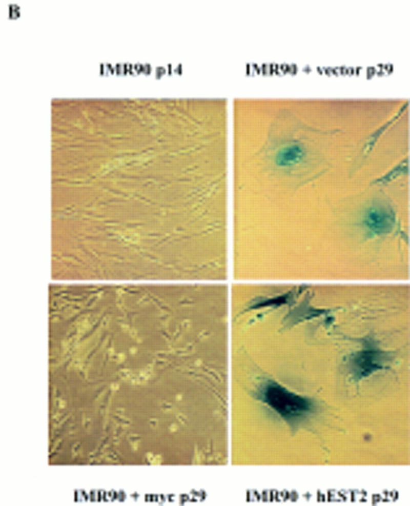
Telomerase activation does not affect life span in IMR-90 cells. (A) TRF length of senescent vector-containing IMR-90 and hEST2-expressing IMR-90 cells was analyzed as in Fig. 4. (B) Early-passage IMR-90 cells (passage 14) were infected with empty vector, a hEST2 retrovirus, or a Myc retrovirus as indicated. Cells were passaged until the vector-infected cells reached senescence (∼15 additional passages). At this time, hEST2 cells also senesced, but Myc-expressing IMR-90 cells continued to proliferate. Shown are senescence-associated β-galactosidase stains of early-passage IMR-90 cells, senescent vector-containing IMR-90 cells, senescent hEST2-expressing IMR-90 cells and Myc-expressing IMR-90 cells that have bypassed the senescence point and entered extended life span. (C) Telomerase assays of lysates derived from senescent vector-containing IMR-90 and hEST2-expressing IMR-90 populations. Each lane corresponds to 10,000 cells.
Telomerase activation does not affect life span in IMR-90 cells. (A) TRF length of senescent vector-containing IMR-90 and hEST2-expressing IMR-90 cells was analyzed as in Fig. 4. (B) Early-passage IMR-90 cells (passage 14) were infected with empty vector, a hEST2 retrovirus, or a Myc retrovirus as indicated. Cells were passaged until the vector-infected cells reached senescence (∼15 additional passages). At this time, hEST2 cells also senesced, but Myc-expressing IMR-90 cells continued to proliferate. Shown are senescence-associated β-galactosidase stains of early-passage IMR-90 cells, senescent vector-containing IMR-90 cells, senescent hEST2-expressing IMR-90 cells and Myc-expressing IMR-90 cells that have bypassed the senescence point and entered extended life span. (C) Telomerase assays of lysates derived from senescent vector-containing IMR-90 and hEST2-expressing IMR-90 populations. Each lane corresponds to 10,000 cells.
Telomerase activation does not affect life span in IMR-90 cells. (A) TRF length of senescent vector-containing IMR-90 and hEST2-expressing IMR-90 cells was analyzed as in Fig. 4. (B) Early-passage IMR-90 cells (passage 14) were infected with empty vector, a hEST2 retrovirus, or a Myc retrovirus as indicated. Cells were passaged until the vector-infected cells reached senescence (∼15 additional passages). At this time, hEST2 cells also senesced, but Myc-expressing IMR-90 cells continued to proliferate. Shown are senescence-associated β-galactosidase stains of early-passage IMR-90 cells, senescent vector-containing IMR-90 cells, senescent hEST2-expressing IMR-90 cells and Myc-expressing IMR-90 cells that have bypassed the senescence point and entered extended life span. (C) Telomerase assays of lysates derived from senescent vector-containing IMR-90 and hEST2-expressing IMR-90 populations. Each lane corresponds to 10,000 cells.
References
-
- Alitalo K, Koskinen P, Makela TP, Saksela K, Sistonen L, Winqvist R. myc oncogenes: Activation and amplification. Biochim Biophys Acta. 1987;907:1–32. - PubMed
-
- Bodnar AG, Ouellette M, Frolkis M, Holt SE, Chiu CP, Morin GB, Harley CB, Shay JW, Lichtsteiner S, Wright WE. Extension of life-span by introduction of telomerase into normal human cells. Science. 1998;279:349–352. - PubMed
-
- Bryan TM, Reddel RR. Telomere dynamics and telomerase activity in in vitro immortalised human cells. Eur J Cancer. 1997;33:767–773. - PubMed
Publication types
MeSH terms
Substances
Grants and funding
LinkOut - more resources
Full Text Sources
Other Literature Sources
Molecular Biology Databases