

doi: 10.1101/gad.12.12.1781.
Seven-up, the Drosophila homolog of the COUP-TF orphan receptors, controls cell proliferation in the insect kidney
Affiliations
- PMID: 9637680
- PMCID: PMC316909
- DOI: 10.1101/gad.12.12.1781
Item in Clipboard
Seven-up, the Drosophila homolog of the COUP-TF orphan receptors, controls cell proliferation in the insect kidney
Genes Dev.
.
Abstract
Morphogenesis of the insect kidney, the Malpighian tubules, is controlled in Drosophila by a single large cell, the tip cell. It has been postulated that this cell sends out a mitogenic signal that induces the division of neighboring cells. The signal and the molecules that receive it have remained elusive. We show that the COUP-TF-related nuclear orphan receptor Seven-up is a key component that becomes induced in response to mitogenic EGF receptor signaling activity emanating from the tip cell. Seven-up in turn is capable of regulating the transcription of cell cycle regulators.
Figures
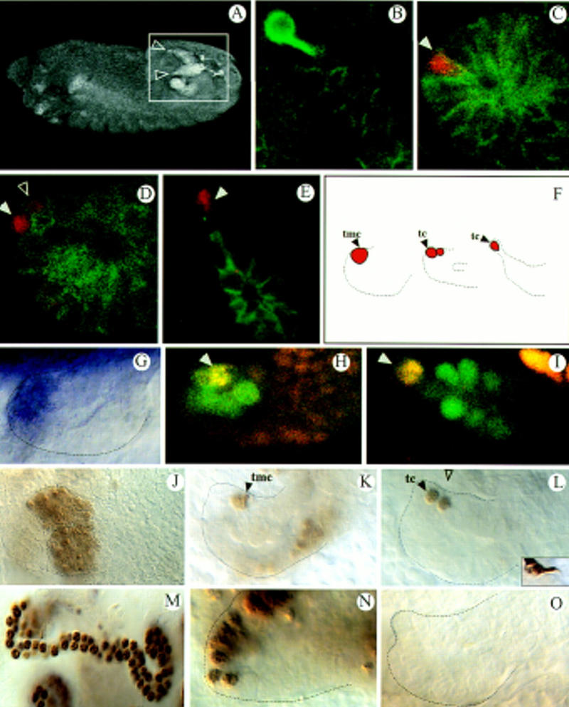
svp is required for MT growth. (A) Dorsolateral view of a stage 13 embryo visualizing two of the four everting tubules (inset, arrowheads) by mAb FascII labels tubule membranes. (B) mAb 22C10, mAb FascII double staining (both green) of a stage 16 tubule; 22C10 expression marks the neural tip cell. (C–E) Anti-Kr (red), mAb FascII (green) double stainings highlighting the tip mother cell in stage 10 (C, arrowhead), the two daughter cells shortly thereafter (D, arrowheads) and the tip cell in stage 14 (E, arrowhead). (F) Scheme of tip cell allocation. (G–I) svp expression monitored by in situ hybridization (G; stage 11 tubule) or via a svp lacZ line (H,I; late stage 11) showing the same expression pattern. Anti-β-gal (green), anti-Kr (red) double stainings. Yellow shows coexpression of svp and Kr in the tip mother cell (H; arrowhead) and the tip cell (I; arrowhead). (J–M,O) svp mutant embryos, (N) wild type. (J–L) Anti-Kr staining. The primordium (J), tip cell allocation (K,L), and tip cell differentiation (inset in L, mAb 22C10 stains) are normal. (M) Anti-Cut staining reveals a reduced tubule cell number in svp mutants (Table 1). (N,O) BrDU incorporation studies of stage 11 embryos. (tmc) Tip mother cell; (tc) tip cell.
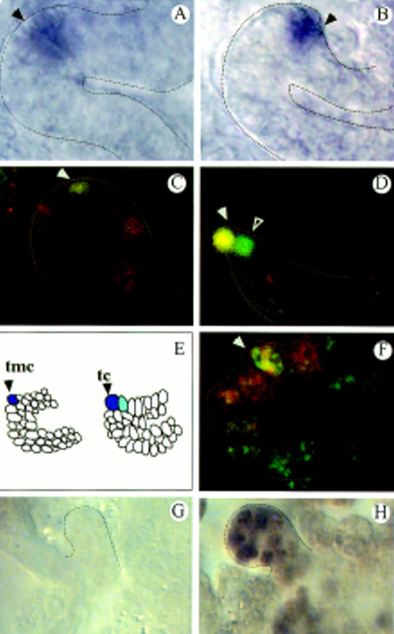
Localized rho and S activity in the tip cells of the tubules. (A,B) rho expression in the tip mother cell (A, arrowhead) and the tip cell (B, arrowhead) as revealed by in situ hybridization. (C,D) S lacZ expression; double staining of anti-β-gal (green) and anti-Kr (red) revealing S expression in the tip mother cell (C, arrowhead), the tip cell (D, arrowhead), and its sibling cell (D, open arrowhead). (E) Summary of the rho (blue) and S (light blue) expression patterns in the everting tubules. (F) pnt–lacZ expression; double staining of anti-β-gal (red) and anti-Kr (green) revealing pnt expression in the tip mother cell (arrowhead) and its neighboring cells. (G,H) BrdU incorporation studies in EGFR mutants (flbIK35; stage 12; G) and (stage 15; H).
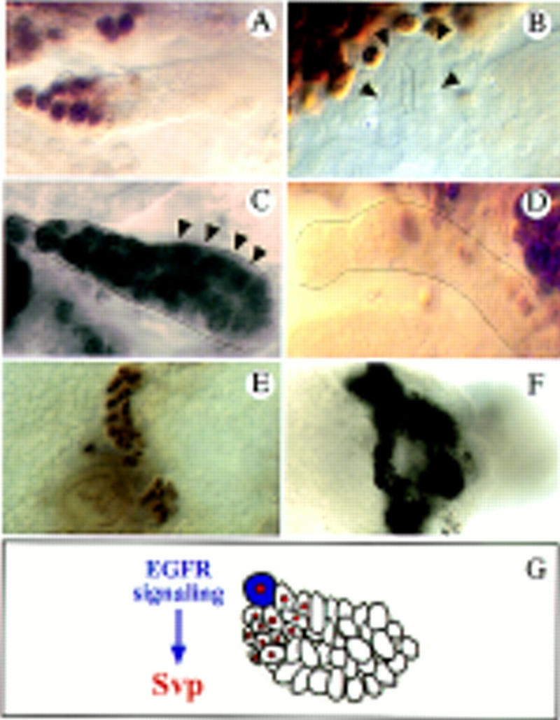
svp expression is dependent on EGFR signaling. (A–D) svp lacZ expression shown by anti-β-gal antibody stainings (the same results were obtained monitoring svp expression by in situ hybridization). (A) svp expression in the tip region of a wild-type tubule (stage 15). (B) svp lacZ expression is abolished in mutants of the EGFR (flbIK35). (C) Upon ectopic sSpi expression the svp domain is largely expanded (arrowheads point to cells that ectopically express svp; cf. A) and the cell number increases. (D) Ectopic expression of the dominant-negative Dras1N17 allele using the XB2-3-Gal4, reduces svp expression (cf. A). (E,F) Anti-Cut stainings marking all of the tubule cells. (E) Reduced tubule cell number in flbIK35 mutants. (F) Ectopic svp expression of rescues the tubules of flbIK35 mutants. Approximately three times more cells were obtained compared to the mutant condition. (G) Summary of the results. Svp (red nuclei) is activated by EGFR signaling, which emanates from the tip cell (blue) and controls cell division.
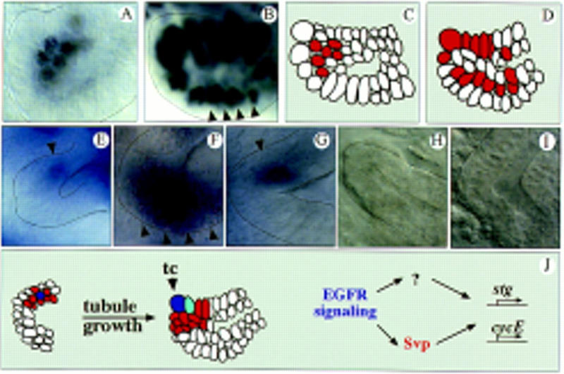
EGFR signaling and Svp control cell cycle gene expression. BrDU incorporation in stage 10 wild-type (A) and G455.2–Gal4/UAS–Svp II tubules (mediating SvpII expression in all tubule cells) (B). Cell proliferation does not occur on one side of the outgrowing tubules as in wild type (A) but throughout the everting tubules (B, arrowheads). (C,D) Schematic representation of the BrdU incorporation studies in A and B. Red marks cell division. (E–I) RNA in situ hybridization experiments of wild-type (E–G) and (flbIK35) mutant embryos (H–I). (E) stg expression in proliferating tubule cells of wild-type embryos occurs on one side of the outgrowing tubule (arrowhead) in early stage 10. (F) Upon ectopic expression of Svp, stg becomes ectopically expressed in cells that undergo extra divisions (arrowheads, cf. E and B). In wild type, cycE is also expressed asymmetrically in the outgrowing tubules in stage 10 (G; arrowhead) and in the distal proliferation zone (not shown). Localized transcription of stg (H; stage 11) and cycE (I; stage 10) is absent in flbIK35 mutants. (J) Model of how tip cell (blue) signaling might control tubule growth. Dividing cells are red; See text for details.
Similar articles
-
A genetic hierarchy establishes mitogenic signalling and mitotic competence in the renal tubules of Drosophila.Development. 2002 Feb;129(4):935-44. doi: 10.1242/dev.129.4.935. Development. 2002. PMID: 11861476
-
Modulation of retinoic acid sensitivity in lung cancer cells through dynamic balance of orphan receptors nur77 and COUP-TF and their heterodimerization.EMBO J. 1997 Apr 1;16(7):1656-69. doi: 10.1093/emboj/16.7.1656. EMBO J. 1997. PMID: 9130711 Free PMC article.
-
Stat 5b and the orphan nuclear receptors regulate expression of the alpha2-macroglobulin (alpha2M) gene in rat ovarian granulosa cells.Mol Endocrinol. 1998 Sep;12(9):1393-409. doi: 10.1210/mend.12.9.0161. Mol Endocrinol. 1998. PMID: 9731707
-
Physiological function of the orphans GCNF and COUP-TF.Trends Endocrinol Metab. 2001 Aug;12(6):247-51. doi: 10.1016/s1043-2760(01)00424-6. Trends Endocrinol Metab. 2001. PMID: 11445441 Review.
-
[Gene cascade by hormonal control during development of insects].Tanpakushitsu Kakusan Koso. 2003 Dec;48(16):2254-60. Tanpakushitsu Kakusan Koso. 2003. PMID: 14661583 Review. Japanese. No abstract available.
Cited by
-
Female-specific wing degeneration caused by ecdysteroid in the Tussock Moth, Orgyia recens: hormonal and developmental regulation of sexual dimorphism.J Insect Sci. 2003;3:11. doi: 10.1093/jis/3.1.11. Epub 2003 Apr 29. J Insect Sci. 2003. PMID: 15841227 Free PMC article.
-
Sites of Fgf signalling and perception during embryogenesis of the beetle Tribolium castaneum.Dev Genes Evol. 2008 Apr;218(3-4):153-67. doi: 10.1007/s00427-007-0192-x. Epub 2008 Apr 8. Dev Genes Evol. 2008. PMID: 18392877
-
Identification of genes controlling malpighian tubule and other epithelial morphogenesis in Drosophila melanogaster.Genetics. 1999 Feb;151(2):685-95. doi: 10.1093/genetics/151.2.685. Genetics. 1999. PMID: 9927461 Free PMC article.
-
Release and spread of Wingless is required to pattern the proximo-distal axis of Drosophila renal tubules.Elife. 2018 Aug 10;7:e35373. doi: 10.7554/eLife.35373. Elife. 2018. PMID: 30095068 Free PMC article.
-
The function of nuclear receptors in regulation of female reproduction and embryogenesis in the red flour beetle, Tribolium castaneum.J Insect Physiol. 2010 Oct;56(10):1471-80. doi: 10.1016/j.jinsphys.2010.04.004. Epub 2010 Apr 27. J Insect Physiol. 2010. PMID: 20416316 Free PMC article.
References
-
- Baumann, P. and H. Skaer. 1993. The Drosophila EGFR homolog (DER) is required for Malpighian tubule development. Development (Suppl.) 65–75. - PubMed
-
- Begemann G, Michon A-M, v.d. Voorn L, Wepf R, Mlodzik M. The Drosophila orphan nuclear receptor Seven-up requires the Ras pathway for its function in photoreceptor determination. Development. 1995;121:225–237. - PubMed
-
- Bier E, Jan LY, Jan YN. rhomboid, a gene required for dorsoventral Drosophila melanogaster. Genes & Dev. 1990;4:190–203. - PubMed
-
- Brand AH, Perrimon N. Targeted gene expression as a means of altering cell fates and generating dominant phenotypes. Development. 1993;118:401–415. - PubMed
-
- Campos-Ortega JA, Hartenstein V. The embryonic development of Drosophila melanogaster. New York, NY: Springer Verlag; 1997.
Publication types
MeSH terms
Substances
LinkOut - more resources
Full Text Sources
Molecular Biology Databases
Miscellaneous