. 1998 Jun 15;12(12):1837-46.
doi: 10.1101/gad.12.12.1837.

Miranda as a multidomain adapter linking apically localized Inscuteable and basally localized Staufen and Prospero during asymmetric cell division in Drosophila

Affiliations

Miranda as a multidomain adapter linking apically localized Inscuteable and basally localized Staufen and Prospero during asymmetric cell division in Drosophila

C P Shen et al. Genes Dev. .

Abstract

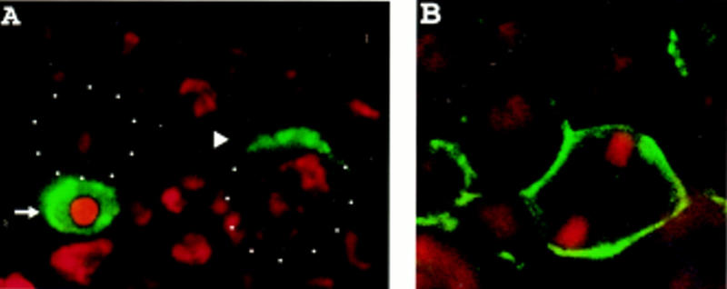
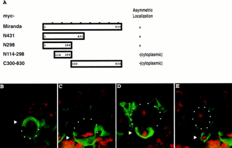
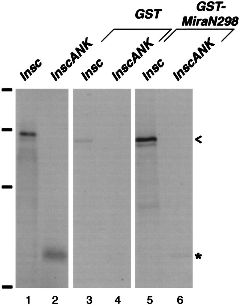
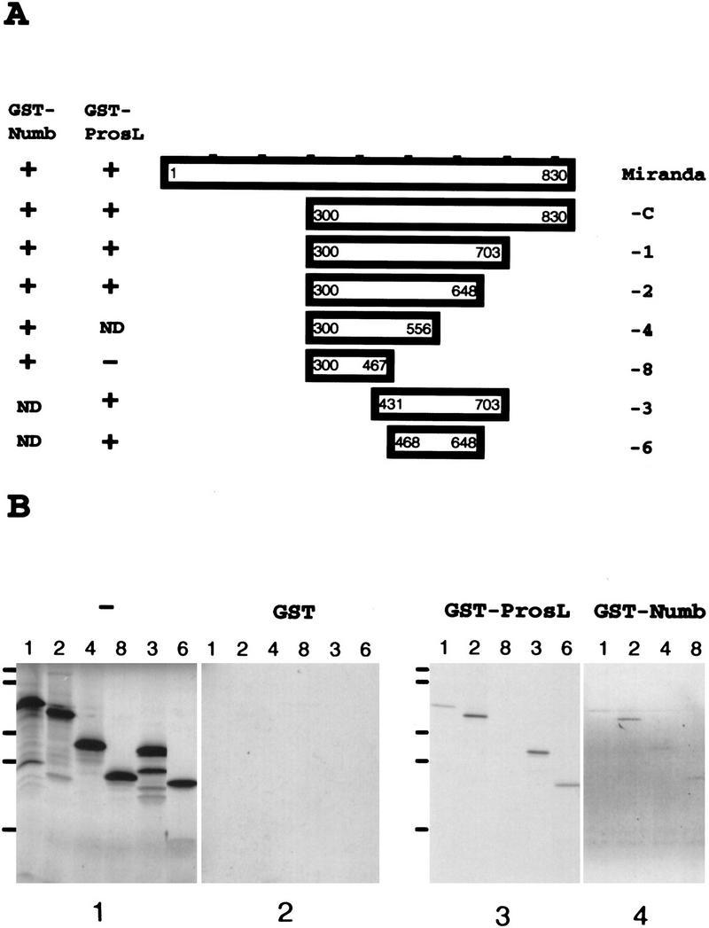
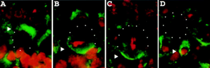
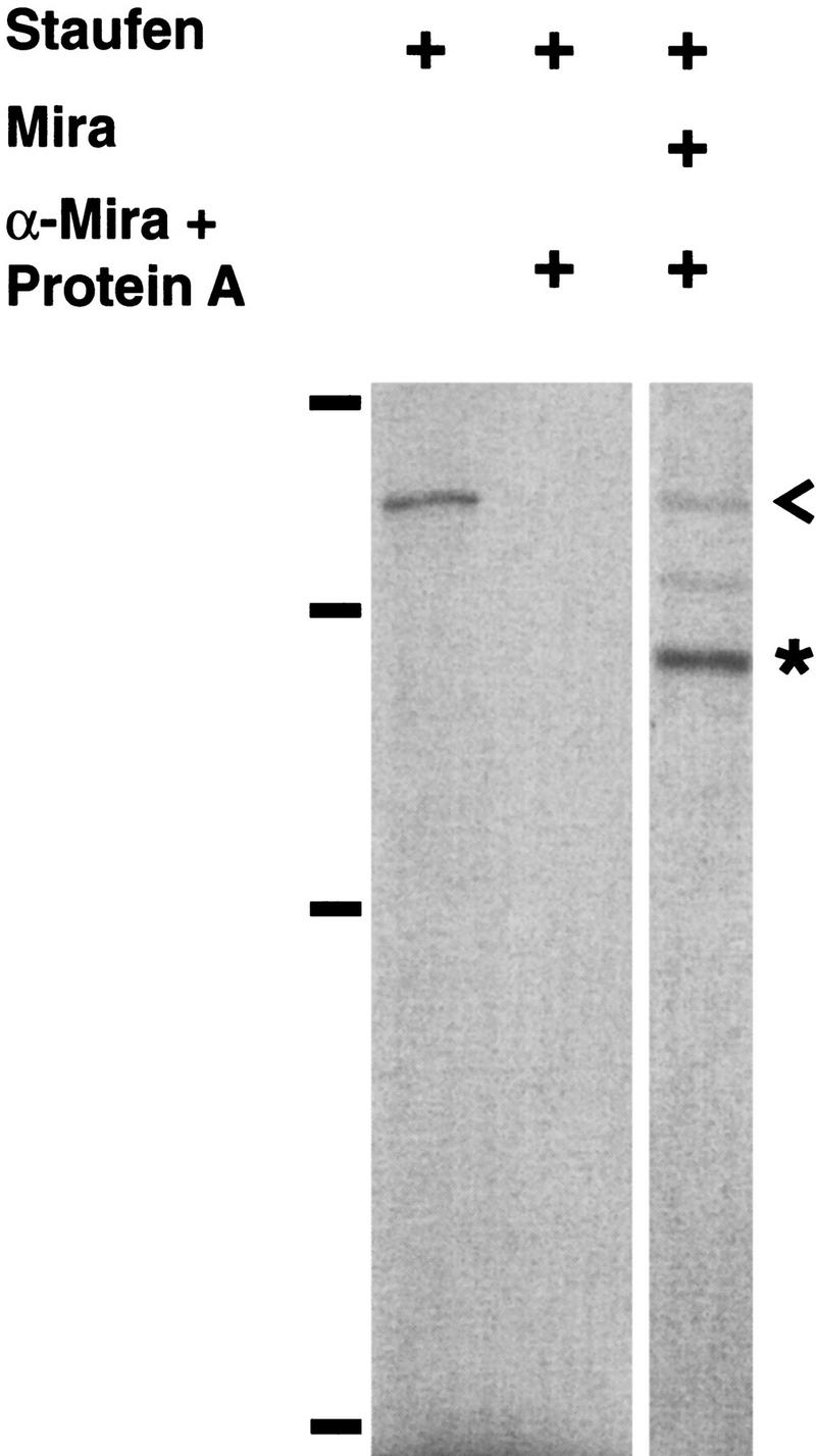
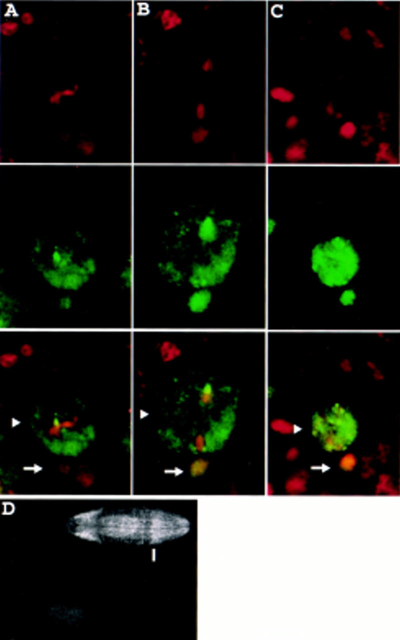
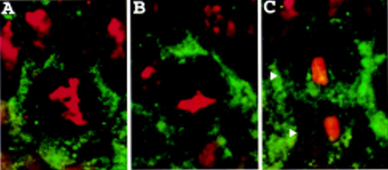
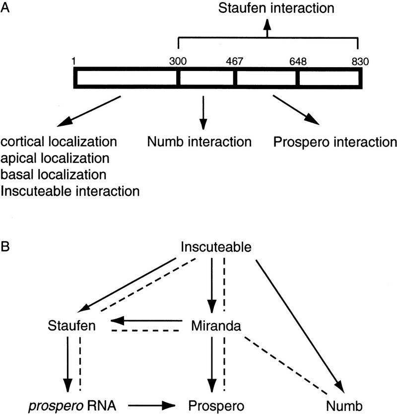
Neuroblasts in the developing Drosophila CNS asymmetrically localize the cell fate determinants Numb and Prospero as well as prospero RNA to the basal cortex during mitosis. The localization of Prospero requires the function of inscuteable and miranda, whereas prospero RNA localization requires inscuteable and staufen function. We demonstrate that Miranda contains multiple functional domains: an amino-terminal asymmetric localization domain, which interacts with Inscuteable, a central Numb interaction domain, and a more carboxy-terminal Prospero interaction domain. We also show that Miranda and Staufen have similar subcellular localization patterns and interact in vitro. Furthermore, miranda function is required for the asymmetric localization of Staufen. Miranda localization is disrupted by the microfilament disrupting agent latrunculin A. Our results suggest that Miranda directs the basal cortical localization of multiple molecules, including Staufen and prospero RNA, in mitotic neuroblasts in an actin-dependent manner.


Figures

Figure 1
Figure 1
Microfilaments are required for the localization of Miranda to the apical then basal cortex. (A) Wild-type embryos were stained with anti-Miranda antibody (green) and propidium iodide to visualize DNA (red). Apical is toward the top; white dots indicate neuroblast cell borders in this and all subsequent figures. In the neuroblast at right, which is in early prophase, Miranda is localized to the apical cell cortex (arrowhead). The neuroblast at left has nearly completed mitosis. Miranda is present exclusively in the basal daughter cell (arrow). (B) Wild-type embryos were treated with 200 μm latrunculin A for 20 min and stained for Miranda (green) and DNA (red). Under these conditions, phalloidin reactivity is virtually abolished (Knoblich et al. 1997), and Miranda is localized along the entire cortex of mitotic neuroblasts.
Figure 2
Figure 2
The amino-terminal 298 amino acids of Miranda are sufficient for asymmetric localization even in the absence of endogenous Miranda. (A) Full-length Miranda or Miranda fragments were myc-tagged and expressed in transgenic animals. Staining with an anti-myc antibody was performed to visualize their subcellular localization. The results are summarized at right. Full-length Miranda, the amino-terminal 431 amino acids, and the amino-terminal 298 amino acids of Miranda all localize to the apical cortex in early mitotic neuroblasts and then to the basal cortex later in mitosis (not shown). Amino acids 114–298 and the carboxy-terminal 530 amino acids, however, remain in the cytoplasm of dividing neuroblasts, fail to localize asymmetrically and enter both daughter cells (not shown). (B–E) Embryos expressing myc–MiraN298 but deficient for endogenous miranda were stained for myc (green) and DNA (red). In early prophase, myc–MiraN298 forms a crescent at the apical cortex (arrowhead, B). myc–MiraN298 then localizes to the basal cortex in metaphase (arrowhead, C) and is exclusively localized to the basal daughter in telophase (arrowheads, D,E).
Figure 3
Figure 3
The amino-terminal 298 amino acids of Miranda interact with the ankyrin-like repeat region of Inscuteable. Transcription and translation of full-length inscuteable in vitro produces a major band that migrates at 120 kD (lane 1; <); InscANK, a fragment from amino acids 252 to 615, migrates at 45 kD (lane 2, *). These labeled polypeptides coprecipitate with GST fused to the amino-terminal 298 amino acids of Mira in the presence of glutathione beads (lanes 5,6) but do not coprecipitate with GST alone (lanes 3,4). inscuteable constructs were in vitro translated and transcribed in the presence of [35S]methionine and directly subjected to SDS-PAGE (lanes 1,2) or incubated with bacterially expressed GST or GST–MiraN298, precipitated with glutathione beads, and subjected to SDS-PAGE. Bars at left indicate positions of molecular mass markers (from bottom to top: 42, 79, 130, and 200 kD).
Figure 4
Figure 4
Miranda interacts with Numb and Prospero through distinct domains. (A) Various Miranda constructs were tested for their ability to interact with GST fused to either the Numb amino-terminal 223 amino acids or Prospero amino acids 820–1026 (ProsL), which include the asymmetric localization domain. The results are summarized at left. (B) The labeled Miranda fragments are shown in panel 1. Fragments 1, 2, 3, and 6, which all contain amino acids 468–648 of Miranda, interact with GST–Prospero (panel 3); fragments 1, 2, 4, and 8, which all contain amino acids 300–467, interact with GST–Numb (panel 4). None of the fragments interact with GST alone (panel 2). miranda constructs were in vitro translated and transcribed in the presence of [35S]-methionine and directly subjected to SDS-PAGE (panel 1) or incubated with bacterially expressed GST (panel 2), GST–Prospero (panel 3), or GST–Numb (panel 4) fixed to glutathione beads, which were precipitated, washed, and subjected to SDS-PAGE. Bars at left indicate positions of molecular mass markers (from bottom to top: 17, 32, 41, 71, and 126 kD).
Figure 5
Figure 5
Staufen localizes to the apical and then the basal cortex in dividing neuroblasts. Wild-type embryos were stained for Staufen (green) and DNA (red). Staufen localizes to the apical cortex of neuroblasts in late interphase (arrowhead, A), to the basal cortex in metaphase (arrowhead, B) and anaphase (arrowhead, C), and enters the basal daughter in telophase (arrowhead, D).
Figure 6
Figure 6
Miranda interacts with Staufen physically. Staufen coimmunoprecipitates with anti-Miranda antiserum and protein A beads in the presence (right lane) but not the absence (middle lane) of Miranda. The left lane shows the position of labeled Staufen (<). A construct encoding full-length Staufen was transcribed and translated in vitro in the presence of [35S]methionine and directly subjected to SDS-PAGE (left lane). Staufen was either translated in vitro alone (middle lane) or cotranslated with Miranda (right lane) and incubated with anti-Miranda antiserum and protein A beads, which were then precipitated, washed, and subjected to SDS-PAGE. Bars at left indicate molecular mass markers (from bottom to top: 42, 79, 130, and 200 kD). (*) The position of Miranda.
Figure 7
Figure 7
Prospero is mislocalized in embryos homozygous for miraDEB, a protein null allele of miranda. (A–C) Homozygous miraDEB embryos were stained for DNA (red, top row) and Prospero (green, middle row). Prospero is present throughout the neuroblast cytoplasm in metaphase (arrowhead, column A) and anaphase (arrowhead, column B). After cell division Prospero enters the nuclei of both the larger apical daughter (arrowhead) and the smaller basal daughter (arrow, column C). Arrows indicate two small, basally located, Prospero-positive nuclei in column A and one nucleus in column B that belong to ganglion mother cells produced by previous neuroblast divisions. (D) Progeny from males and females of the genotype miraDEB/TM3, Ubx–lacZ were stained with anti-Miranda and anti-β-galactosidase antibodies. Ventral views of two representative embryos are shown; the top and bottom embryos are oriented with anterior to the left and right, respectively. The top embryo expresses β-galactosidase in the Ubx pattern, characterized by a sharp anterior boundary (indicated by the bar) and therefore carries the TM3, Ubx–lacZ “blue balancer” and is not homozygous for miraDEB. This embryo exhibits a wild-type staining pattern of Miranda, with characteristic strong staining in the procephalic neurogenic region and in the midgut. In contrast, the lower embryo, which is identified as homozygous for miraDEB by the lack of β-galactosidase expression, fails to stain with anti-Miranda antibody. Propidium iodide staining shows that both embryos are accessible to staining (not shown).
Figure 8
Figure 8
Staufen is mislocalized in homozygous miraDEB embryos. Homozygous miraDEB embryos were stained for Staufen (green) and DNA (red). Staufen is diffusely localized to the cell cortex in neuroblasts in prophase (A) and metaphase (B) and in telophase is present in both daughter cells (arrowheads, C).
Figure 9
Figure 9
(A) Miranda contains several independent domains. The amino-terminal 298 amino acids of Miranda is sufficient for physical interaction with Inscuteable, localization to the neuroblast cortex, apical localization in late interphase and early prophase, and basal localization later in mitosis. Amino acids 300–467 are sufficient for Numb interaction, and amino acids 468–648 for Prospero interaction. The carboxy-terminal 530 amino acids are sufficient for interaction with Staufen. (B) Summary of known interactions. Arrows indicate functional interactions. Broken lines indicate physical interactions.

Similar articles

Cited by

References

    1. Ayscough KR, Stryker J, Pokala N, Sanders M, Crews P, Drubin DG. High rates of actin filament turnover in budding yeast and roles for actin in establishment and maintenance of cell polarity revealed using the actin inhibitor latrunculin-A. J Cell Biol. 1997;137:399–416. - PMC - PubMed
    1. Brand AH, Perrimon N. Targeted gene expression as a means of altering cell fates and generating dominant phenotypes. Development. 1993;118:401–415. - PubMed
    1. Broadus J, Doe CQ. Extrinsic cues, intrinsic cues and microfilaments regulate asymmetric protein localization in Drosophila neuroblasts. Curr Biol. 1997;7:827–835. - PubMed
    1. Broadus J, Fuerstenberg S, Doe CQ. Staufen-dependent localization of prospero mRNA contributes to neuroblast daughter-cell fate. Nature. 1998;39:792–795. - PubMed
    1. Burchard S, Paululat A, Hinz U, Renkawitz-Pohl R. The mutant not enough muscles (nem) reveals reduction of the Drosophila embryonic muscle pattern. J Cell Sci. 1995;108:1443–1454. - PubMed

Publication types

MeSH terms

LinkOut - more resources