. 1998 Jun 15;12(12):1858-70.
doi: 10.1101/gad.12.12.1858.

Binary sibling neuronal cell fate decisions in the Drosophila embryonic central nervous system are nonstochastic and require inscuteable-mediated asymmetry of ganglion mother cells

Affiliations

Binary sibling neuronal cell fate decisions in the Drosophila embryonic central nervous system are nonstochastic and require inscuteable-mediated asymmetry of ganglion mother cells

M Buescher et al. Genes Dev. .

Abstract

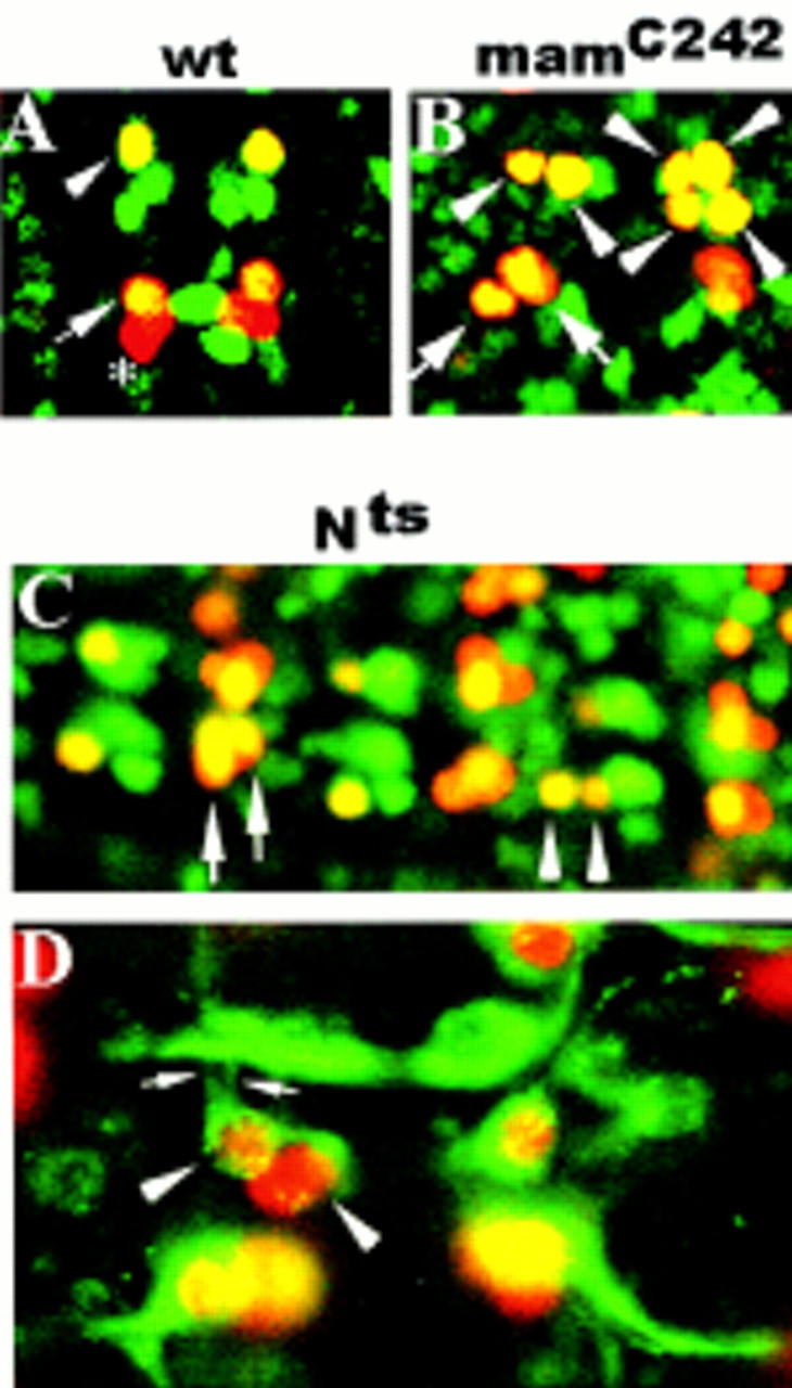
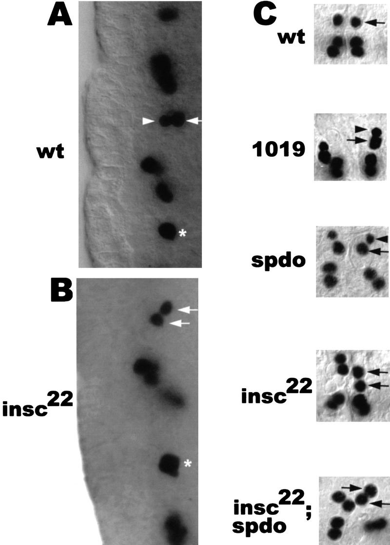
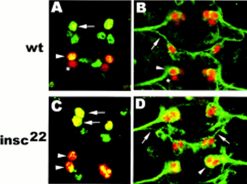
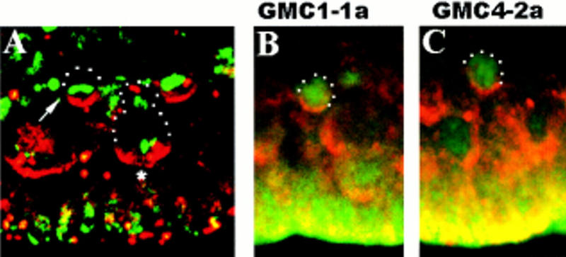
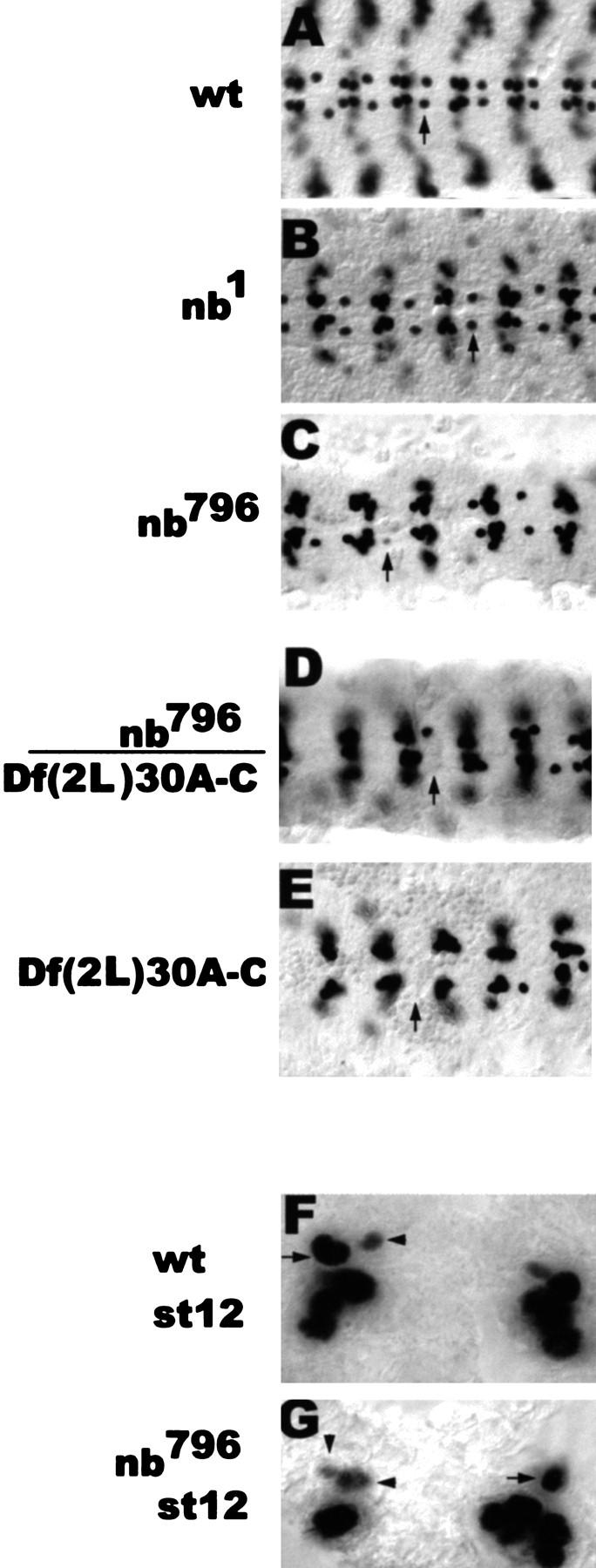
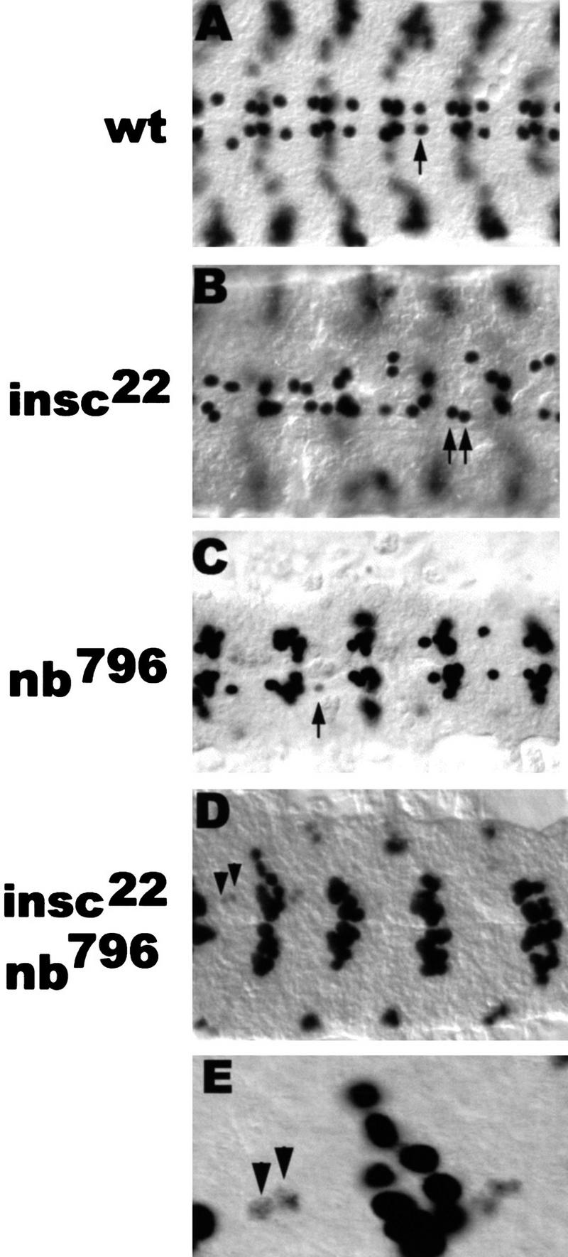
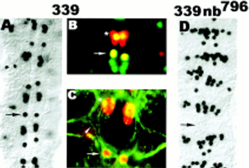
Asymmetric cell division is a widespread mechanism in developing tissues that leads to the generation of cell diversity. In the embryonic central nervous system of Drosophila melanogaster, secondary precursor cells-ganglion mother cells (GMCs)-divide and produce postmitotic neurons that take on different cell fates. In this study, we show that binary fate decision of two pairs of sibling neurons is accomplished through the interplay of Notch (N) signaling and the intrinsic fate determinant Numb. We show that GMCs have apical-basal polarity and Numb localization and the orientation of division are coordinated to segregate Numb to only one sibling cell. The correct positioning of Numb and the proper orientation of division require Inscuteable (Insc). Loss of insc results in the generation of equivalent sibling cells. Our results provide evidence that sibling neuron fate decision is nonstochastic and normally depends on the presence of Numb in one of the two siblings. Moreover, our data suggest that the fate of some sibling neurons may be regulated by signals that do not require lateral interaction between the sibling cells.


Figures

Figure 1
Figure 1
Mutations in mam and N are associated with RP2sib to RP2 and pCC to aCC fate transformations. (A,B,C) Ventral views of stage 15 embryos double stained with rabbit anti-Eve (red) and mouse anti-Zfh-1 (green). (A,B) Anterior is up; (C) anterior is left. (A) wt RP2 (arrowhead) and aCC (large arrow), but not pCC (asterisk), coexpress Eve and Zfh-1 (yellow). (B) mamC242. (C) Nts1. Note the different sizes of the RP2 nuclei in B and C (arrowheads). The right hemisegment in B depicts four RP2 neurons that presumably derive from two parental NB4-2. (D) Ventral view of a stage 15 whole-mount Nts1 embryo stained with rabbit anti-Eve (red) and mouse-mAb 22C10 (green). Anterior is up. Note the ipsilateral axon projections of both RP2s (small arrows).
Figure 2
Figure 2
Mutations in spdo result in RP2sib to RP2 fate transformation with respect to marker gene expression but not with respect to cell/nuclear size. (A,B) Lateral views of stage 11 embryos stained with anti-Eve. Ventral (apical) is to the left. (A) In wt, GMC4-2a divides into a smaller and a larger cell. The newborn siblings are oriented perpendicular to the apical surface with the larger cell (future RP2) in the more dorsal position. (B) In insc22, GMC4-2a divides into sibling cells of equal size that are rarely oriented perpendicular to the apical surface. (Arrows) RP2 neuron; (arrowheads) RP2sib; (asterisks) undivided GMC4-2a. (C) Dorsal views of dissected wild-type and various mutant stage 15 embryos stained with anti-Eve. Anterior is up. In GA1019 (1019) embryos, GMC4-2a does not undergo cytokinesis and binucleated cells with two Eve-expressing nuclei are formed. Note the difference in the sizes of the two nuclei (arrowhead vs. arrow) derived from GMC4-2a in spdo and GA1019 embryos.
Figure 3
Figure 3
Loss of insc causes RP2sib to RP2 and pCC to aCC fate transformations. Ventral view of whole-mount stage 15 embryos. Anterior is up. (A,C) Double staining with rabbit anti-Eve (red) and mouse anti-Zfh-1 (green). (B,D) Double staining with rabbit anti-Eve (red) and mouse mAb 22C10 (green). (A,B) wt: RP2 (arrow) and aCC (arrowheads) coexpress Eve and Zfh-1 (yellow in A) and form a 22C10-positive, ipsilateral axon projection (green in B); pCC (asterisk) neither expresses Zfh-1 (A) nor 22C10 (B). (C,D) insc22. Note that both GMC1-1a and GMC4-2a progeny coexpress Eve and Zfh-1 (yellow in C) and form an ipsilateral axon projection (D).
Figure 4
Figure 4
Insc protein forms apical crescents in dividing GMCs. Lateral views of wt stage 11 embryos; basal (dorsal) is up. (A) Double staining with rabbit anti-Insc (red) and DNA stain (green). (Arrow) GMC in metaphase. Note that the metaphase plate is oriented horizontally with respect to the apical surface. (Asterisk) Dividing neuroblast. (B,C) GMC1-1a and GMC4-2a double stained with anti-Insc (red) and mouse anti-Eve (green).
Figure 5
Figure 5
insc is required for the coordination of Numb localization and the orientation of the metaphase plate in GMCs. Lateral views of dividing GMCs in stage 11 embryos triple stained with anti-Numb (with rhodamine conjugated secondary antibody, red), mouse anti-Eve (with FITC conjugated secondary antibody, blue) and DNA stain (green). Basal (dorsal) is up. (A,B,E,I, left) Wild type; (C,D,G,H,I, right) insc22. Top panels (A–D) depict the anti-Numb and anti-Eve staining; middle panels (E–H) show the anti-Numb and DNA staining of the same samples; bottom panels (I,J) show the anti-Numb and DNA staining in randomly chosen GMCs. (A,B,E,F,I) wt. Note the basal Numb crescents in all samples and the horizontal orientation of the metaphase plates in E and I. (C,D,G,H,J) insc22. Note the cortical Numb staining with no distinct crescents in C,G,J and the apical crescent in D and H. The metaphase plates are not oriented horizontally with respect to the apical surface (G,J). Relevant cells are circled with dots.
Figure 6
Figure 6
Mutations in numb cause RP2 to RP2sib fate transformation. (A–E) Dorsal views of dissected stage 15 embryos stained with rabbit anti-Eve. Anterior is to the left. (Arrows) RP2 neuron (A) or the RP2 position (C–E). (F,G) Ventral views of stage 12 whole-mount embryos stained with rabbit anti-Eve. Anterior is up. (Arrowheads) RP2sib. Compare the fading Eve staining in the smaller and the larger cell in nb796 (G) with the fading Eve staining in the smaller cell only in wt (F).
Figure 7
Figure 7
In equalized sibling neurons (insc background), RP2/RP2sib fate specification is only dependent on Numb. Dorsal view of dissected stage 15 embryos stained with rabbit anti-Eve. Anterior is to the left. (Arrows) RP2 neuron; (arrowheads) RP2sib. (E) Enlarged detail of D. Note the fading Eve staining in two RPsibs of equal size in the insc22 nb796 double-mutant embryo (E). Also note the increase in the number of Eve-expressing cells at the position of the CQ neurons, see Discussion.
Figure 8
Figure 8
In the absence of cell division, GMC4-2a differentiation is dependent on Numb. Dorsal view of dissected stage 15 embryos; anterior is up. (A) GA339 stained with rabbit anti-Eve, note that each hemisegment has an Eve-positive cell at the RP2 position; note the decrease in the number of Eve-expressing cells in the position of the CQ neurons, see Discussion. (B) GA339 double stained with rabbit anti-Eve (red) and mouse anti-Zfh-1 (green). Note that the undivided GMC4-2a expresses Zfh-1, a marker gene, which in wild-type embryos is not expressed in GMC4-2a but only the mature RP2 neuron. (C) GA339 double stained with rabbit anti-Eve (red) and mouse mAb 22C10 (green). Note that the undivided GMC4-2a forms an ipsilateral axon projection characteristic for the RP2 neuron. (D) nb796 GA339 double mutant, note the loss of Eve-positive cells at the RP2 position and the increase in the number of Eve-expressing cells in the position of the CQ neurons. (Arrows) Position of the RP2 neuron; (arrowhead in C) the RP2 axon; [asterisk (*) in C] aCC neuron.

References

    1. Artavanis-Tsakonas A, Matsuno K, Fortini ME. Notch signaling. Science. 1995;268:225–232. - PubMed
    1. Bossing T, Udolph G, Doe CQ, Technau G. The embryonic CNS lineages of Drosophila melanogaster: I. Neuroblast lineages derived from the ventral half of the neuroectoderm. Dev Biol. 1996;179:41–64. - PubMed
    1. Burchard S, Paululat A, Hinz U, Renkawitz-Pohl R. The mutant not enough muscles (nem) reveals reduction of the Drosophila embryonic muscle pattern. J Cell Sci. 1995;108:1443–1454. - PubMed
    1. Campos-Ortega JA. Numb diverts Notch pathway off the Tramtrack. Neuron. 1996;17:1–4. - PubMed
    1. Campos-Ortega JA, Hartenstein V. The embryonic development of Drosophila melanogaster. Berlin, Germany: Springer-Verlag; 1985.

Publication types

MeSH terms

LinkOut - more resources