. 1998 Jun 15;12(12):1871-83.
doi: 10.1101/gad.12.12.1871.

The checkpoint protein MAD2 and the mitotic regulator CDC20 form a ternary complex with the anaphase-promoting complex to control anaphase initiation

Affiliations

The checkpoint protein MAD2 and the mitotic regulator CDC20 form a ternary complex with the anaphase-promoting complex to control anaphase initiation

G Fang et al. Genes Dev. .

Abstract

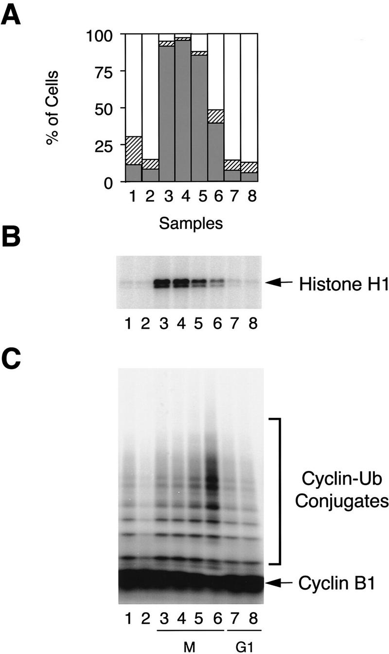
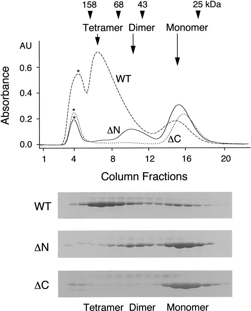
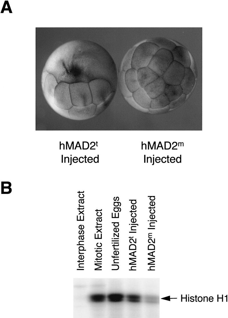
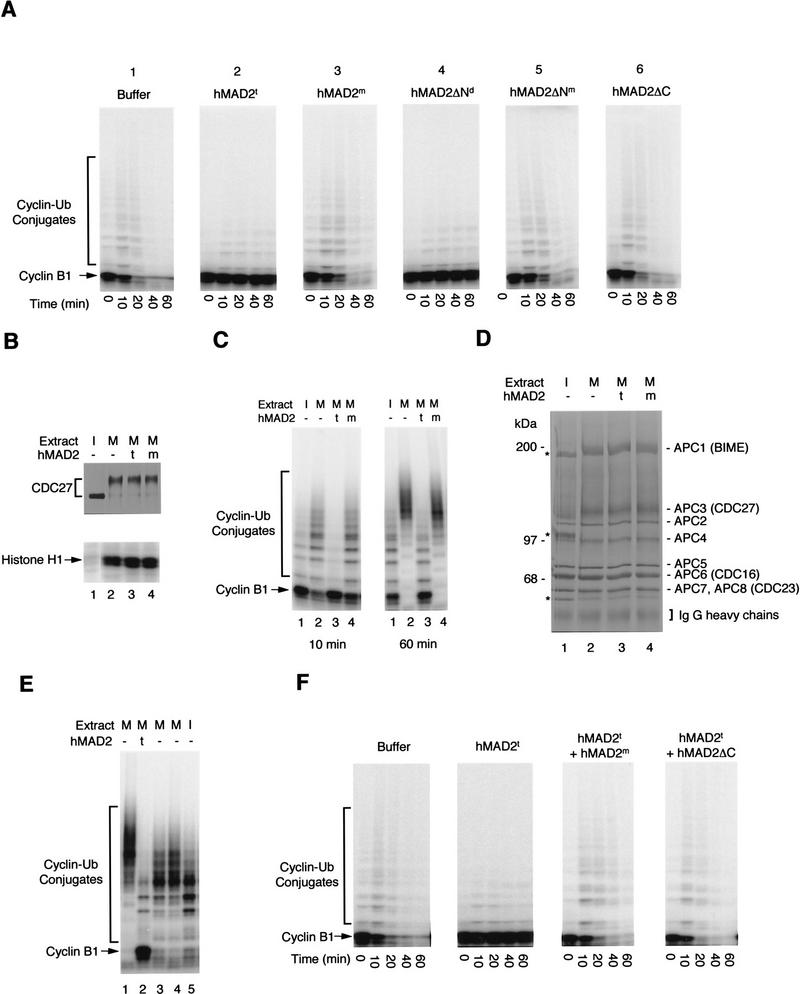
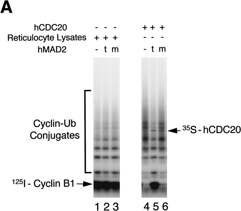
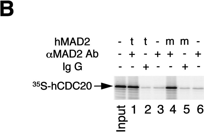
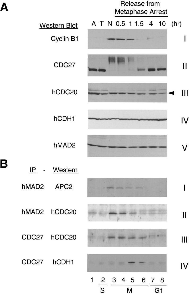
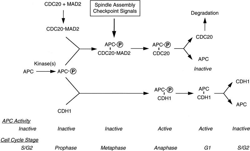
The spindle assembly checkpoint mechanism delays anaphase initiation until all chromosomes are aligned at the metaphase plate. Activation of the anaphase-promoting complex (APC) by binding of CDC20 and CDH1 is required for exit from mitosis, and APC has been implicated as a target for the checkpoint intervention. We show that the human checkpoint protein hMAD2 prevents activation of APC by forming a hMAD2-CDC20-APC complex. When injected into Xenopus embryos, hMAD2 arrests cells at mitosis with an inactive APC. The recombinant hMAD2 protein exists in two-folded states: a tetramer and a monomer. Both the tetramer and the monomer bind to CDC20, but only the tetramer inhibits activation of APC and blocks cell cycle progression. Thus, hMAD2 binding is not sufficient for inhibition, and a change in hMAD2 structure may play a role in transducing the checkpoint signal. There are at least three different forms of mitotic APC that can be detected in vivo: an inactive hMAD2-CDC20-APC ternary complex present at metaphase, a CDC20-APC binary complex active in degrading specific substrates at anaphase, and a CDH1-APC complex active later in mitosis and in G1. We conclude that the checkpoint-mediated cell cycle arrest involves hMAD2 receiving an upstream signal to inhibit activation of APC.


Figures

Figure 1
Figure 1
APC activity at different stages of cell cycle. HeLa cells were synchronized at prometaphase by a thymidine–nocodazole block. Cells were collected immediately after nocodazole treatment (lane 3), or released into fresh medium for 0.5, 1, 1.5, 4, and 10 hr (lanes 4–8). Asynchronous cells (lane 1) and cells arrested at G1/S boundary by a double thymidine block (lane 2) were included as controls. (A) Cell cycle stage was determined by FACS analysis. (Open bars) G1; (hatched bars) S; (solid bars) G2/M. (B) Cdc2 kinase was immunopurified from cell lysates and assayed for histone H1 kinase activity. (C) APC was purified from the cell lysates with anti-CDC27/protein A beads and analyzed for its ability to ubiquitinate a 125I-labeled amino-terminal fragment of Xenopus cyclin B1.
Figure 2
Figure 2
Identification of different oligomerization states of the recombinant hMAD2 protein. Oligomerization states of the recombinant hMAD2 proteins were determined by gel filtration chromatography with a S100 column. Column fractions were analyzed by SDS-PAGE. The elution peaks of molecular mass standards are labeled at the top of the UV trace. (*) Peak corresponding to void volume. (WT) Wild-type hMAD2; (ΔN) hMAD2ΔN; (ΔC) hMAD2ΔC.
Figure 3
Figure 3
Block of cell division in Xenopus embryos by the tetrameric hMAD2. (A) Twenty-five nanoliters of 10 mg/ml recombinant hMAD2t and hMAD2m proteins were microinjected into one of the blastomeres in two-cell stage Xenopus embryos. The embryos were collected and analyzed 2 hr after injection. Cell cycle arrest by hMAD2t is stable for at least 8 hr before cells degenerate. (B) To assay histone H1 kinase assays, both blastomeres in two-cell stage embryos were injected with hMAD2, and extracts from injected embryos were analyzed for histone H1 kinase activity. Unfertilized eggs and equivalent aliquots of interphase and mitotic extracts were also assayed as controls.
Figure 4
Figure 4
Inhibition of APC by hMAD2 oligomers in Xenopus mitotic extracts. (A) Effect of hMAD2 on cyclin degradation in Xenopus extracts. hMAD2t, hMAD2m, hMAD2ΔNd, hMAD2ΔNm, and hMAD2ΔC were incubated with Xenopus mitotic extracts for 20 min at a final concentration of 0.5 mg/ml before addition of the radioactive amino-terminal fragment of the Xenopus cyclin B1. Aliquots of the extracts were sampled at different times and the stability of the radioactive cyclin B1 was analyzed by SDS-PAGE. (B–D) The state of extracts and of APC in extracts treated with hMAD2 tetramer and monomer. hMAD2t and hMAD2m were incubated with Xenopus mitotic extracts for 20 min at a final concentration of 0.5 mg/ml. Aliquots of extracts were then assayed for histone H1 kinase activity and for the phosphorylation state of CDC27 by Western blot analysis (B). APC was immunopurified from the hMAD2-treated extracts with anti-CDC27 antibody beads and analyzed for the cyclin ubiquitination activity in the presence of recombinant E1 and E2 (C) and for subunit composition by silver staining (D). The mitotic APC subunits are labeled as APC1–APC8 in (D). Because of phosphorylation, the electrophoretic mobility of APC1(BIME), APC3(CDC27), APC8(CDC23) is retarded in mitotic APC; the corresponding interphase subunits are labeled by stars. (I) Interphase extracts; (M) mitotic extracts; (t) hMAD2 tetramer; (m) hMAD2 monomer. (E) Reactivation of hMAD2t-inhibited APC. hMAD2t was incubated with Xenopus mitotic extracts for 20 min at a final concentration of 0.5 mg/ml. APC was immunopurified from extracts with anti-CDC27 antibody beads and incubated with fresh mitotic extracts (lane 3) or interphase extracts (lane 5) in the absence of hMAD2. As a negative control, the APC beads was also incubated with mitotic extracts that have been preincubated with hMAD tetramer (lane 2). As a positive control, APC was immunopurified directly from mitotic extracts in the absence of hMAD2, incubated with mitotic extracts again, and then assayed for cyclin ubiquitination activity (lane 1). The APC beads were then washed stringently and assayed for cyclin ubiquitination activity. Activity detected in lane 3 is not attributable to an exchange of hMAD2t-treated APC with mitotic APC in extracts, as hMAD2t-treated APC is also reactivated in APC-depleted mitotic extracts (lane 4). (F) The dominant effect of monomeric hMAD2 on the inhibition by hMAD2 tetramer. hMAD2t was mixed with either hMAD2m or hMAD2ΔC and then added to Xenopus mitotic extracts. After addition of the radioactive amino-terminal fragment of cyclin B, aliquots of extracts were sampled at different times, and the stability of radioactive cyclin B was analyzed by SDS-PAGE.
Figure 5
Figure 5
Inhibition of hCDC20-mediated activation of APC by hMAD2t. (A) 35S-labeled hCDC20 (lanes 4–6) was incubated for 30 min with hMAD2t (lane 5), hMAD2ΔC (lane 6), or with buffer (lane 4). As controls, rabbit reticulocyte lysates alone were incubated with hMAD2t (lane 2), hMAD2ΔC (lane 3), or with buffer (lane 1). APC was immunopurified from Xenopus interphase extracts with anti-CDC27 antibody beads, and then incubated with the hMAD2/hCDC20 mixture for 60 min. The APC beads were washed with buffer and assayed for cyclin ubiquitination activity. hCDC20 bound to APC beads was marked on the right. (t) hMAD2 tetramer; (m) hMAD2ΔC monomer. (B) 35S-labeled hCDC20 were incubated with either hMAD2t (lanes 1,2) or hMAD2ΔC (lanes 4,5) for 30 min. Affinity purified anti-hMAD2 antibody (lanes 1,3,4,6) or rabbit total IgG (lanes 2,5) were added to the hMAD2/hCDC20 mixture. After 90-min incubation, Affi-Prep protein A beads (Bio-Rad) were added and incubated for another 90 min. The beads were then washed stringently, and amount of hCDC20 bound to the beads was analyzed by SDS-PAGE. Input lane contains one-tenth amount of hCDC20 added to the binding reactions. For experiments described in A and B, similar results were achieved with purified hCDC20 protein. Briefly, in vitro translated HA-tagged hCDC20 was immunoprecipitated from reticulocyte lysates with anti-HA antibody/protein A beads. hCDC20 protein bound to antibody beads was eluted with the HA peptide and purified to apparent homogeneity as analyzed by silver staining (data not shown).
Figure 5
Figure 5
Inhibition of hCDC20-mediated activation of APC by hMAD2t. (A) 35S-labeled hCDC20 (lanes 4–6) was incubated for 30 min with hMAD2t (lane 5), hMAD2ΔC (lane 6), or with buffer (lane 4). As controls, rabbit reticulocyte lysates alone were incubated with hMAD2t (lane 2), hMAD2ΔC (lane 3), or with buffer (lane 1). APC was immunopurified from Xenopus interphase extracts with anti-CDC27 antibody beads, and then incubated with the hMAD2/hCDC20 mixture for 60 min. The APC beads were washed with buffer and assayed for cyclin ubiquitination activity. hCDC20 bound to APC beads was marked on the right. (t) hMAD2 tetramer; (m) hMAD2ΔC monomer. (B) 35S-labeled hCDC20 were incubated with either hMAD2t (lanes 1,2) or hMAD2ΔC (lanes 4,5) for 30 min. Affinity purified anti-hMAD2 antibody (lanes 1,3,4,6) or rabbit total IgG (lanes 2,5) were added to the hMAD2/hCDC20 mixture. After 90-min incubation, Affi-Prep protein A beads (Bio-Rad) were added and incubated for another 90 min. The beads were then washed stringently, and amount of hCDC20 bound to the beads was analyzed by SDS-PAGE. Input lane contains one-tenth amount of hCDC20 added to the binding reactions. For experiments described in A and B, similar results were achieved with purified hCDC20 protein. Briefly, in vitro translated HA-tagged hCDC20 was immunoprecipitated from reticulocyte lysates with anti-HA antibody/protein A beads. hCDC20 protein bound to antibody beads was eluted with the HA peptide and purified to apparent homogeneity as analyzed by silver staining (data not shown).
Figure 6
Figure 6
Cell cycle-regulated association of APC with hCDC20, hCDH1, and hMAD2. HeLa cells were synchronized at the prometaphase by a thymidine–nocodazole block. Cells were collected immediately after nocodazole treatment (lane 3, N), or released into fresh medium for 0.5, 1, 1.5, 4, and 10 hr (lanes 4–8). Asynchronous cells (lane 1, A) and cells arrested at G1/S boundary by a double thymidine block (lane 2, T) were included as controls. (A) HeLa cells were lysed with the SDS sample buffer and the levels of cyclin B1, CDC27, hCDC20, hCDH1, and hMAD2 proteins were determined by Western blot analysis. The arrowhead in panel III points to hCDC20. The band above is a cross-reacting protein. (B) Extracts from HeLa cells were immunoprecipitated with either anti-hMAD2 antibody beads or with anti-CDC27 antibody beads, and the immunoprecipitates were analyzed by Western blotting with anti-APC2, anti-hCDC20, or anti-hCDH1 antibodies.
Figure 7
Figure 7
hMAD2 and hCDC20 colocalize to kinetochores at prometaphase. (A) HeLa cells were stained with anti-hMAD2, CREST, and DAPI. (B) Hela cells were stained with anti-hCDC20, CREST, and DAPI. (C) Hela cells were stained with anti-hCDC20, anti-hMAD2, and DAPI.
Figure 8
Figure 8
A model for a regulatory network of APC in the cell cycle.

Similar articles

Cited by

References

    1. Amon A. Regulation of B-type cyclin proteolysis by Cdc28-associated kinases in budding yeast. EMBO J. 1997;16:2693–2702. - PMC - PubMed
    1. Amon A, Irniger S, Nasmyth K. Closing the cell cycle circle in yeast: G2 cyclin proteolysis initiated at mitosis persists until the activation of G1 cyclins in the next cycle. Cell. 1994;77:1037–1050. - PubMed
    1. Brandeis M, Hunt T. The proteolysis of mitotic cyclins in mammalian cells persists from the end of mitosis until the onset of S phase. EMBO J. 1996;15:5280–5289. - PMC - PubMed
    1. Chen RH, Waters JC, Salmon ED, Murray AW. Association of spindle assembly checkpoint component XMAD2 with unattached kinetochores. Science. 1996;274:242–246. - PubMed
    1. Clute P, Masui Y. Regulation of the appearance of division asynchrony and microtubule-dependent chromosome cycles in Xenopus laevis embryos. Dev Biol. 1995;171:273–285. - PubMed

Publication types

MeSH terms