Comparative Study
. 1998 Jul;180(13):3432-40.
doi: 10.1128/JB.180.13.3432-3440.1998.

Clustered genes encoding the methyltransferases of methanogenesis from monomethylamine

Affiliations
Comparative Study

Clustered genes encoding the methyltransferases of methanogenesis from monomethylamine

S A Burke et al. J Bacteriol. 1998 Jul.

Abstract

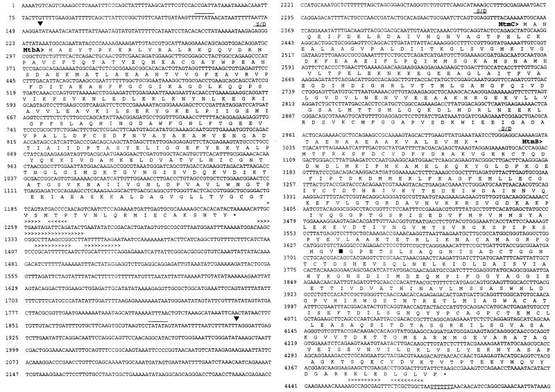
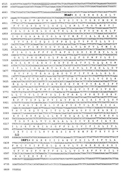
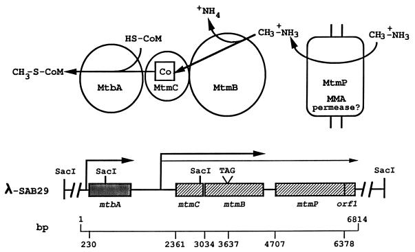
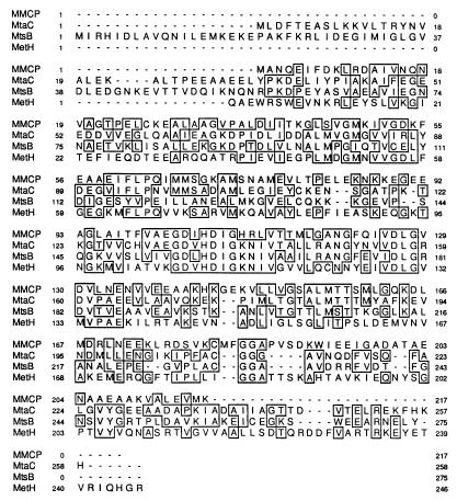
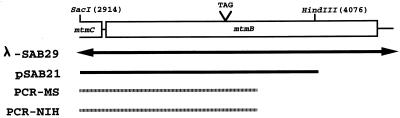
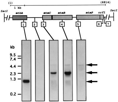
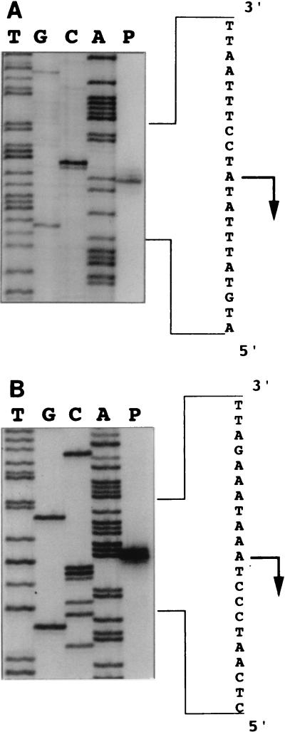
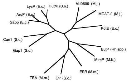
Coenzyme M (CoM) is methylated during methanogenesis from monomethyamine in a reaction catalyzed by three proteins. Using monomethylamine, a 52-kDa polypeptide termed monomethylamine methyltransferase (MMAMT) methylates the corrinoid cofactor bound to a second polypeptide, monomethylamine corrinoid protein (MMCP). Methylated MMCP then serves as a substrate for MT2-A, which methylates CoM. The genes for these proteins are clustered on 6.8 kb of DNA in Methanosarcina barkeri MS. The gene encoding MMCP (mtmC) is located directly upstream of the gene encoding MMAMT (mtmB). The gene encoding MT2-A (mtbA) was found 1.1 kb upstream of mtmC, but no obvious open reading frame was found in the intergenic region between mtbA and mtmC. A single monocistronic transcript was found for mtbA that initiated 76 bp from the translational start. Separate transcripts of 2.4 and 4.7 kb were detected, both of which carried mtmCB. The larger transcript also encoded mtmP, which is homologous to the APC family of cationic amine permeases and may therefore encode a methylamine permease. A single transcriptional start site was found 447 bp upstream of the translational start of mtmC. MtmC possesses the corrinoid binding motif found in corrinoid proteins involved in dimethylsulfide- and methanol-dependent methanogenesis, as well as in methionine synthase. The open reading frame of mtmB was interrupted by a single in-frame, midframe, UAG codon which was also found in mtmB from M. barkeri NIH. A mechanism that circumvents UAG-directed termination of translation must operate during expression of mtmB in this methanogen.


Figures

FIG. 1
FIG. 1
Nucleic acid sequences and predicted amino acid sequences of mtbA, mtmC, mtmB, mtmP, and orf1 of the mtm operon from M. barkeri. The predicted amino acid sequences are shown in single-letter code directly below the second base of each codon. Position numbers are given to the left of each row of the sequence. Overlined bases represent the predicted promoter sequences. Symbols and abbreviations: ▪, transcript start site determined from primer extension; ∗, predicted stop codon; ▾, position of UAG in reading frame of mtmB; > and < (shown above bases included in repeat sequences), direction of the repeat (indicated by the direction of the arrow head); S/D, predicted Shine-Dalgarno sequence.
FIG. 1
FIG. 1
Nucleic acid sequences and predicted amino acid sequences of mtbA, mtmC, mtmB, mtmP, and orf1 of the mtm operon from M. barkeri. The predicted amino acid sequences are shown in single-letter code directly below the second base of each codon. Position numbers are given to the left of each row of the sequence. Overlined bases represent the predicted promoter sequences. Symbols and abbreviations: ▪, transcript start site determined from primer extension; ∗, predicted stop codon; ▾, position of UAG in reading frame of mtmB; > and < (shown above bases included in repeat sequences), direction of the repeat (indicated by the direction of the arrow head); S/D, predicted Shine-Dalgarno sequence.
FIG. 2
FIG. 2
Organization of genes that mediate the initial steps of methanogenesis from MMA. The physical map of the 15-kb insert of M. barkeri genomic DNA from the λ-SAB29 clone shows the organization of the genes encoding the catalyzing polypeptides involved in the methylation of CoM with MMA. The transcripts containing these genes, designated by the arrows, are drawn to scale to indicate the identified 5′ termini and estimated 3′ termini. Base positions of initiation codons and the in-frame amber codon of mtmB are indicated below and correspond to the nucleic acid sequence shown in Fig. 1. The biochemical reactions and catalyzing polypeptides involved in CoM methylation with MMA are shown in the diagram at the top, along with the predicted function of the mtmP gene product.
FIG. 3
FIG. 3
Sequence alignment of the monomethylamine corrinoid protein (MtmC) with the corrinoid-binding subunits of methylthiol:CoM methyltransferase (MtsB) (38) and methyltransferase I (MtaC) (43) as well as the corrinoid-binding tryptic fragment of methionine synthase (MetH) (12). Residues shared between sequences are boxed. Position numbers are given to the left and right of each row of each sequence.
FIG. 4
FIG. 4
Confirmation of the mtmB sequence containing the in-frame UAG codon. The figure illustrates the regions that were cloned or amplified by PCR from M. barkeri genomic DNA and sequenced. λ-SAB29 is the original phage clone in which the amber codon was first observed. Nucleotide numbers are the same as those in Fig. 1. pSAB21 is a pUC19 derivative which contains the indicated 1.1-kb SacI-HindIII fragment cloned directly from M. barkeri MS genomic DNA. PCR-MS and PCR-NIH are fragments amplified from M. barkeri MS or NIH genomic DNA and directly sequenced.
FIG. 5
FIG. 5
Identification of the mtbA, mtmCB, and mtmCBPorf1 transcripts. Northern analyses of total RNA isolated from M. barkeri grown on MMA were performed with the same blot and with probes complementary to the regions designated in the diagram by the boxed numbers. The probes were complementary to the following positions of the contiguous nucleotide sequence: probe 1, 933 to 1010; probe 2, 1775 to 1792; probe 3, 2384 to 2403; probe 4, 3723 to 3741; probe 5, 4777 to 4794; probe 6, 6549 to 6566; and probe 7, 6750 to 6768. Molecular size standards are indicated to the left, while arrows designating the migration positions of the three transcripts are located to the right. Probes 6 and 7 gave essentially the same autoradiograms as those indicated for probe 5 and 2, respectively.
FIG. 6
FIG. 6
Identification of initiation sites of mtbA, mtmCB, and mtmCBPorf1 transcripts. Primers were annealed with total RNA isolated from M. barkeri grown on MMA. The products of primer extension for the mtbA (A) and mtm (B) transcripts are shown adjacent to the DNA sequencing ladders generated with the same primers. The antisense nucleotide sequence is shown to the right with the 5′ terminus and direction of transcription indicated by an arrow. The positions of the transcriptional starts are indicated in Fig. 1.
FIG. 7
FIG. 7
MtmP belongs to the APC family of cationic amine permeases. A phylogenetic tree was constructed with the TreeGen program (21) from the CLUSTALW (49)-generated multiple sequence alignment of MtmP of M. barkeri with members of the APC family of transporters (40). The abbreviations for the gene products are as follows: EutP (Rh. spp.), putative ethanolamine permease from Rhodococcus sp. strain NI86/21 (GenBank accession no. L24492); Ctr (S.c.), choline transporter from Saccharomyces cerevisiae (J05603); ERR (M.m.), ecotropic retroviral receptor from Musculus musculus (M26687); TEA (M.m.), T-cell early activator from M. musculus (D83596); PotE (E.c.), putative putrescine/ornithine permease from E. coli (M64495); MJ0609 (M.j.) and MJ1196 [MCAT-2 (M.j.)], both putative cation transporters from M. jannaschii (3); Gap1 (S.c.), general amino acid permease from S. cerevisiae (X52633); Can1 (S.c.), arginine permease from S. cerevisiae (X03784); Gabp (E.c.), γ-aminobutyrate from E. coli (X65104); AroP (E.c.), general aromatic amino acid permease from E. coli (X17333); LysP (E.c.), lysine-specific permease from E. coli (M89774); HutM (B.s.), putative histidine permease from Bacillus subtilis (D31856).

References

    1. Altschul S F, Gish W, Miller W, Myers E W, Lipman D J. Basic local alignment search tool. J Mol Biol. 1990;215:403–410. - PubMed
    1. Boone D R, Whitman W B, Rouvière P. Methanogenesis. Ecology, physiology, biochemistry, and genetics. New York, N.Y: Chapman & Hall; 1993. Diversity and taxonomy of methanogens; pp. 35–80.
    1. Bult C J, White O, Olsen G J, Zhou L, Fleischmann R D, Sutton G G, Blake J A, Fitzgerald L M, Clayton R A, Gocayne J D, Kerlavage A R, Dougherty B A, Tomb J-F, Adams M D, Reich C I, Overbeek R, Kirkness E F, Weinstock K G, Merrick J M, Glodeck A, Scott J L, Georghagen N S M, Weidman J F, Fuhrman J L, Nguyen D, Utterback T R, Kelly J M, Peterson J D, Sadow P W, Hanna M C, Cotton M D, Roberts K M, Hurst M A, Kaine B P, Borodovsky M, Klenk H-P, Fraser C F, Smith H O, Woese C R, Venter J C. Complete genome sequence of the methanogenic archaeon, Methanococcus jannaschii. Science. 1996;273:1058–1073. - PubMed
    1. Burgett S G, Rosteck P R. Use of dimethylsulfoxide in fluorescent tag sequencing. In: Adams M D, Fields C, Venter J C, editors. Automated DNA sequencing and analysis. San Diego, Calif: Academic Press; 1994. pp. 211–215.
    1. Burke S A, Krzycki J A. Involvement of the “A” isozyme of methyltransferase II and the 29-kilodalton corrinoid protein in methanogenesis from monomethylamine. J Bacteriol. 1995;177:4410–4416. - PMC - PubMed

Publication types

MeSH terms

Associated data

LinkOut - more resources