. 1998 Jun 29;141(7):1489-502.
doi: 10.1083/jcb.141.7.1489.

Localization, dynamics, and protein interactions reveal distinct roles for ER and Golgi SNAREs

Affiliations

Localization, dynamics, and protein interactions reveal distinct roles for ER and Golgi SNAREs

J C Hay et al. J Cell Biol. .

Erratum in

  • J Cell Biol 1998 Aug 10;142(3):following 881

Abstract

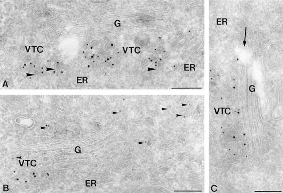
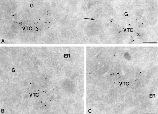
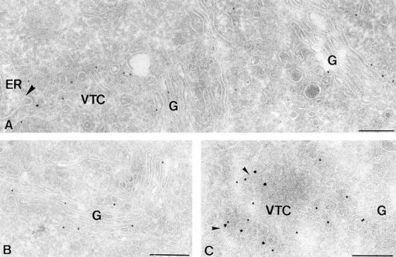
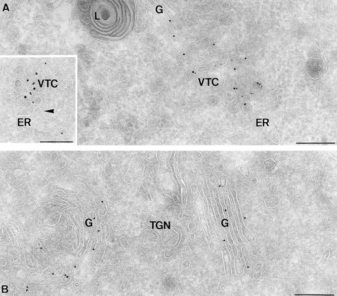
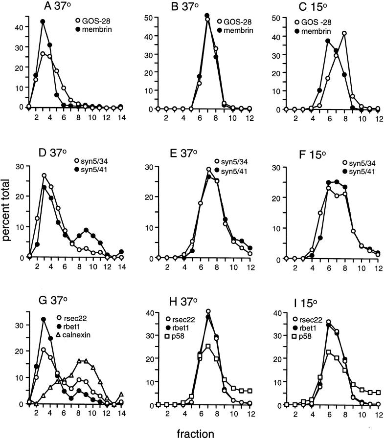
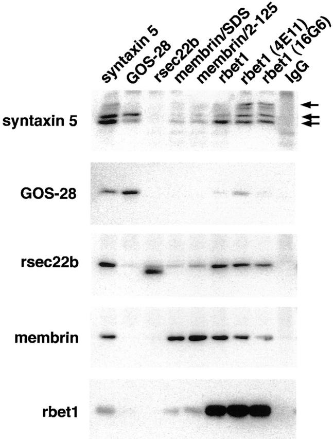
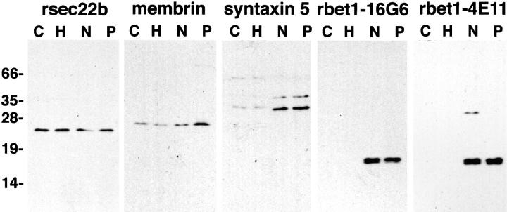
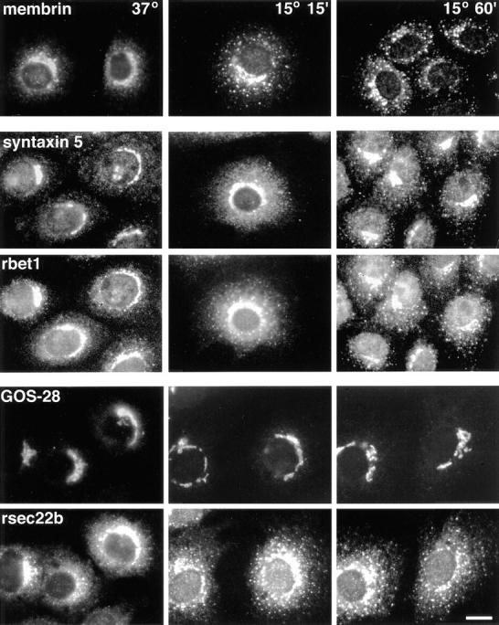
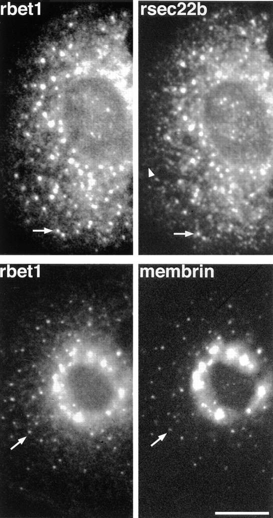
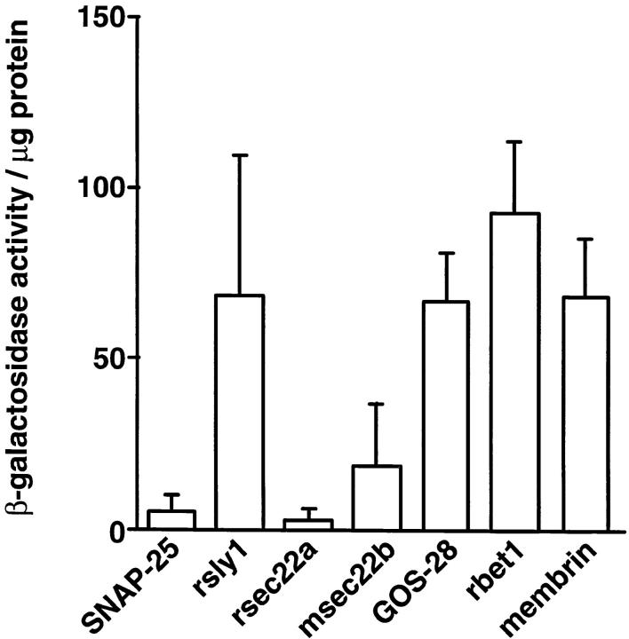
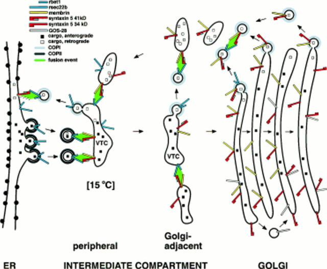
ER-to-Golgi transport, and perhaps intraGolgi transport involves a set of interacting soluble N-ethylmaleimide-sensitive factor attachment protein receptor (SNARE) proteins including syntaxin 5, GOS-28, membrin, rsec22b, and rbet1. By immunoelectron microscopy we find that rsec22b and rbet1 are enriched in COPII-coated vesicles that bud from the ER and presumably fuse with nearby vesicular tubular clusters (VTCs). However, all of the SNAREs were found on both COPII- and COPI-coated membranes, indicating that similar SNARE machinery directs both vesicle pathways. rsec22b and rbet1 do not appear beyond the first Golgi cisterna, whereas syntaxin 5 and membrin penetrate deeply into the Golgi stacks. Temperature shifts reveal that membrin, rsec22b, rbet1, and syntaxin 5 are present together on membranes that rapidly recycle between peripheral and Golgi-centric locations. GOS-28, on the other hand, maintains a fixed localization in the Golgi. By immunoprecipitation analysis, syntaxin 5 exists in at least two major subcomplexes: one containing syntaxin 5 (34-kD isoform) and GOS-28, and another containing syntaxin 5 (41- and 34-kD isoforms), membrin, rsec22b, and rbet1. Both subcomplexes appear to involve direct interactions of each SNARE with syntaxin 5. Our results indicate a central role for complexes among rbet1, rsec22b, membrin, and syntaxin 5 (34 and 41 kD) at two membrane fusion interfaces: the fusion of ER-derived vesicles with VTCs, and the assembly of VTCs to form cis-Golgi elements. The 34-kD syntaxin 5 isoform, membrin, and GOS-28 may function in intraGolgi transport.


Figures

Figure 2
Figure 2
Ultrathin cryosections of HepG2 cells showing the subcellular distribution of human sec22b (hsec22b). (A and C) Double immunogold labeling for hsec22b (15 nm gold) and COPII (10 nm gold). hsec22b colocalizes to a high extent with COPII to VTC membranes. Label in the Golgi (G), if detectable, is restricted to the cis-most cisterna (arrow in C). Note that the membrane buds evolving from the ER (large arrowhead in A) are coated with COPII. (B) Double immunogold labeling for hsec22b (15 nm gold) and COPI (10 nm gold). COPI-coated membranes (small arrowheads) show a much more widespread distribution than hsec22b-positive membranes. Bars, 200 nm.
Figure 3
Figure 3
Ultrathin cryosections of PC12 cells showing the localization of rbet1. (A) Double immunogold labeling for rsec22b (10 nm gold) and rbet1 (15 nm gold). Both SNARE proteins localize to the same VTC membranes. Label in the Golgi (G) is restricted to the cis-most cisterna (arrow). (B) Double immunogold labeling for rbet1 (10 nm gold) and COPII (15 nm gold) showing colocalization on VTC membranes. (C) Double immunogold labeling of rbet1 (10 nm gold) and COPI (15 nm gold). The arrowhead points to a tubular VTC membrane that contains rbet1, and is coated with COPI. Bars, 200 nm.
Figure 4
Figure 4
Ultrathin cryosections of PC12 cells showing the subcellular localization of membrin. (A and B) Immunogold labeling of membrin (10 nm gold) is dispersed over VTC membranes and the Golgi stack (G). The large arrowhead in A points to a membrane bud on the ER. (C) Double immunogold labeling of membrin (15 nm gold) and COPI (10 nm gold) showing partial colocalization (small arrowheads) on VTC membranes. Bars, 200 nm.
Figure 5
Figure 5
Ultrathin cryosections of PC12 cells showing immunogold labeling of syntaxin 5. (A) Syntaxin 5 (10 nm gold) partially colocalizes with COPII (5 nm gold) on VTC membranes. The inset shows rsec22b (10 nm gold) and syntaxin 5 (15 nm gold) colocalization in membrane profiles of a VTC that is in close proximity to the ER (arrowhead points to an ER membrane bud). (B) Syntaxin 5 (10 nm gold) is present throughout the entire Golgi stack (G). L, lysosome, TGN, trans Golgi network. Bars, 200 nm.
Figure 8
Figure 8
Localization and dynamics of ER/Golgi SNAREs by subcellular fractionation. NRK cells were incubated at 37°C or 15°C for 2 h, and were then homogenized and fractionated on sucrose equilibrium density gradients. (A, D, G) 0.75–1.75 M sucrose gradients that resolve Golgi/IC membranes (fractions 2, 3, and 4) from ER membranes (fractions 8, 9, and 10). Material that pelleted during centrifugation is included with fraction 14. (B, C, E, F, H, I) 0.75–1.125 M sucrose gradients that partially resolve Golgi and IC membranes at 15°C. On these shallow gradients, ER membranes pellet and are not included above. Gradient fractions were immunoblotted to detect the indicated proteins, and the amounts of each protein were quantitated by densitometry and plotted on the ordinates above. The 41-kD and 34-kD isoforms of syntaxin 5 are presented separately in D, E, and F. Fractions are displayed from least dense to most dense.
Figure 9
Figure 9
Syntaxin 5 isoforms immunoprecipitate in two major distinct complexes. Detergent extracts of salt-stripped rat liver membranes were subjected to immunoprecipitation using the antibodies shown along the top. The immunoprecipitates were then electrophoresed, transferred to nitrocellulose, and immunoblotted using the antibodies shown along the left. The three arrows mark the positions of the 41-kD, 34-kD, and 31-kD (degradation product of 41 kD) isoforms of syntaxin 5. Membrin/SDS rabbit antisera was prepared identically to membrin/2-125 (see Materials and Methods), except that SDS-denatured membrin was used as the immunogen.
Figure 1
Figure 1
Specific antibodies. COS, HepG2, NRK, and PC12 cells were solubilized in SDS sample buffer, electrophoresed, and transferred to nitrocellulose for immunoblotting with affinity-purified rabbit polyclonal antisera for rsec22b, membrin, and syntaxin 5, or with culture medium from the anti-rbet1 hybridomas 16G6 and 4E11. Molecular masses in kD are indicated on the left.
Figure 6
Figure 6
The ER/Golgi SNAREs exhibit differing dynamics when incubated at low temperatures. NRK cells incubated at 37°C or 15°C for 15 or 60 min were fixed, permeabilized, and stained for immunofluorescence microscopy with affinity-purified polyclonal (syntaxin 5, rsec22b, membrin) or monoclonal (GOS-28 and rbet1/16G6) antibodies. Panels in the second and third rows from the top represent the same fields of cells, as do panels in the fourth and fifth rows. Bar, 20 μm.
Figure 7
Figure 7
Dynamic SNAREs colocalize to the same VTCs. NRK cells were incubated at 15°C for 1 h, fixed, and double-stained for immunofluorescence as in Fig. 6. The arrows indicate individual VTCs clearly visible in both panels. The arrowhead marks tubular/reticular staining characteristic of the ER, a pattern not observed for membrin staining. Bar, 20 μm.
Figure 10
Figure 10
Multiple ER/Golgi SNAREs bind directly to syntaxin 5. HF7c reporter yeast cells were transformed with syntaxin 5 cloned into the Gal4-activation domain vector (pGAD424) in combination with different SNAREs and control proteins cloned into the Gal4-binding domain vector (pGBT9) as indicated. Transformants were allowed to grow in restrictive media, lysed, and analyzed for β-galactosidase activity using o-nitrophenyl- β-d-galactoside as a substrate. Enzyme activity was expressed in arbitrary units per μg protein as mean value ± SEM obtained with three independent transformants. None of the pGBT9 constructs activated β-galactosidase expression when tested in combination with the parental pGAD424 vector lacking a test protein insert (not shown).
Figure 11
Figure 11
Model of ER/Golgi SNARE function and trafficking. ER/Golgi transport. rsec22b and rbet1 bud from specialized ER exit sites on COPII-coated vesicles. These COPII-coated vesicles fuse with peripheral VTCs by virtue of SNARE complexes involving vesicle-bound rsec22b and rbet1, and VTC-bound membrin and syntaxin 5 (primarily the 41-kD isoform). Most rsec22b and rbet1 are recycled from peripheral VTCs to the ER on COPI-coated vesicles that fuse with the ER. The SNARE complex for the COPI vesicle/ER fusion is unknown, but may involve ER-localized syntaxin 5 (41-kD isoform) and possibly membrin or uncharacterized mammalian SNAREs (dashed outline, an unknown SNARE on the ER). Peripheral VTCs are the punctate intermediate observed at 15°C. At 15°C, peripheral VTCs may become enlarged and change density (not shown). Under normal conditions at 37°C, peripheral VTCs are transported along microtubules to an accumulation of VTCs adjacent to the cis Golgi. Here VTCs fuse with one another and elongate to form new cis Golgi cisternae. This VTC/VTC fusion is mediated by SNARE complexes involving syntaxin 5 (either isoform), membrin, and remaining rsec22b and rbet1. By the time newly formed cisternae have been incorporated into the Golgi, COPI-mediated retrieval of rsec22b and rbet1, begun in the periphery, has gone to completion. COPI-dependent retrieval of membrin and the 34-kD syntaxin 5 is weaker, allowing these proteins to penetrate deeply into the Golgi stacks. Around the Golgi, retrograde COPI vesicles aggregate and fuse by virtue of vesicle-bound rsec22b, rbet1, syntaxin 5 (both isoforms), and membrin. These fusions result in larger vesicular structures (pictured) or tubules (not pictured) containing primarily retrograde cargo that is transported along microtubules to peripheral sites. Fusion of these retrograde structures with peripheral VTCs using the rsec22b/rbet1/membrin/syntaxin 5 (primarily 41 kD) SNARE complex completes the ER/Golgi transport cycle. IntraGolgi transport. We illustrate cisternal maturation as well as interstack vesicular transport models for completeness. Intra-Golgi transport may result from one or the other or both of these mechanisms. Golgi cisternae, initially formed by coalescence of Golgi-adjacent VTCs (see above), gradually mature as they progress from cis to trans. cis and medial Golgi components, e.g,. resident Golgi enzymes, are maintained in their position despite maturation to later cisternae by vesicle-mediated retrograde transport (pictured above Golgi). This retrograde transport may be mediated by COPI-coated vesicles bearing recycling membrin and syntaxin 5 (both isoforms). Although these retrograde vesicles are depicted fusing only with other retrograde vesicles, they could also fuse with earlier stacks or nascent stacks of the Golgi. Rapid transport of anterograde cargo, e.g., secretory products, may be accomplished by interstack vesicle transfers (pictured below Golgi). One possibility is that interstack vesicles containing GOS-28 fuse with Golgi cisternae by virtue of a SNARE complex involving GOS-28 and the 34-kD syntaxin 5.

References

    1. Banfield DK, Lewis MJ, Rabouille C, Warren G, Pelham HR. Localization of Sed5, a putative vesicle targeting molecule, to the cis-Golgi network involves both its transmembrane and cytoplasmic domains. J Cell Biol. 1994;127:357–371. - PMC - PubMed
    1. Bannykh SI, Rowe T, Balch WE. The organization of endoplasmic reticulum export complexes. J Cell Biol. 1996;135:19–35. - PMC - PubMed
    1. Bennett MK, Garcia-Arraras JE, Elferink LA, Peterson K, Fleming AM, Hazuka CD, Scheller RH. The syntaxin family of vesicular transport receptors. Cell. 1993;74:863–873. - PubMed
    1. Bennett MK, Scheller RH. The molecular machinery for secretion is conserved from yeast to neurons. Proc Natl Acad Sci USA. 1993;90:2559–2563. - PMC - PubMed
    1. Burkhardt JK, Echeverri CJ, Nilsson T, Vallee RB. Overexpression of the dynamitin (p50) subunit of the dynactin complex disrupts dynein-dependent maintenance of organelle distribution. J Cell Biol. 1997;139:469–484. - PMC - PubMed

MeSH terms