. 1998 Jun 29;141(7):1515-27.
doi: 10.1083/jcb.141.7.1515.

The mammalian calcium-binding protein, nucleobindin (CALNUC), is a Golgi resident protein

Affiliations

The mammalian calcium-binding protein, nucleobindin (CALNUC), is a Golgi resident protein

P Lin et al. J Cell Biol. .

Abstract

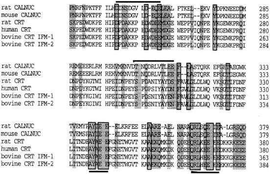
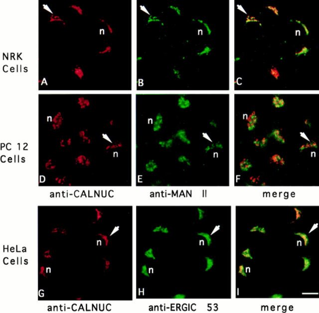
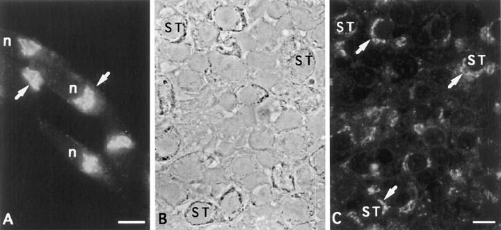
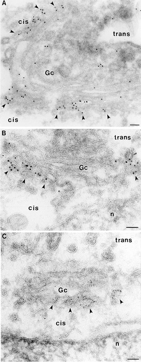
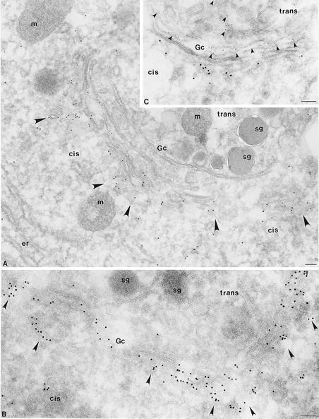
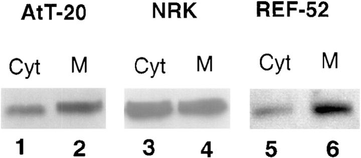
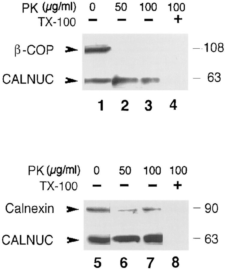
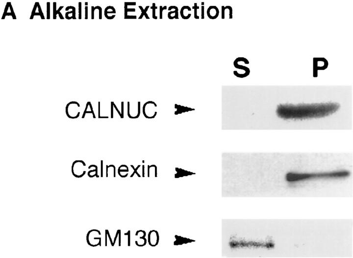
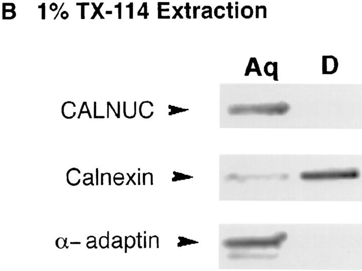
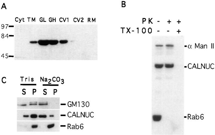
We have identified CALNUC, an EF-hand, Ca2+-binding protein, as a Golgi resident protein. CALNUC corresponds to a previously identified EF-hand/calcium-binding protein known as nucleobindin. CALNUC interacts with Galphai3 subunits in the yeast two-hybrid system and in GST-CALNUC pull-down assays. Analysis of deletion mutants demonstrated that the EF-hand and intervening acidic regions are the site of CALNUC's interaction with Galphai3. CALNUC is found in both cytosolic and membrane fractions. The membrane pool is tightly associated with the luminal surface of Golgi membranes. CALNUC is widely expressed, as it is detected by immunofluorescence in the Golgi region of all tissues and cell lines examined. By immunoelectron microscopy, CALNUC is localized to cis-Golgi cisternae and the cis-Golgi network (CGN). CALNUC is the major Ca2+-binding protein detected by 45Ca2+-binding assay on Golgi fractions. The properties of CALNUC and its high homology to calreticulin suggest that it may play a key role in calcium homeostasis in the CGN and cis-Golgi cisternae.


Figures

Figure 1
Figure 1
Characterization of GST-CALNUC and affinity-purified anti-CALNUC IgG. (A) 5 ng purified GST-CALNUC was separated by 10% SDS-PAGE, transferred to a PVDF membrane, and immunoblotted with 0.67 μg/ml affinity-purified anti-CALNUC IgG (lane 2) or preimmune serum (lane 1). The antibody was capable of detecting 5 ng GST-CALNUC (90 kD). (B) PNS prepared from NRK cells (50 μg/lane) was immunoblotted with 0.38 (lane 2) and 0.17 (lane 3) μg/ml affinity-purified anti-CALNUC IgG or preimmune serum (lane 1). Endogenous CALNUC (63 kD) could be detected with as little as 0.17 μg/ml of the purified antibody. (C) 45Ca2+-overlay. 50 μg GST (lane 1) or GST-CALNUC (lane 2) was separated by SDS-PAGE, transferred to a PVDF membrane and overlaid with 4 μCi/ml 45CaCl2. After exposure of the washed and dried membrane to Kodak X-Omat AR film, a ∼90-kD signal that corresponds to the fusion protein was detected.
Figure 2
Figure 2
(A) Structure of CALNUC. Rat CALNUC is a 434-aa protein with an NH2-terminal signal sequence, a putative DNA-binding domain (BASIC REGION) with a nuclear localization signal (NLS), an acidic region flanked by two EF-hand motifs (EF-1, EF-2), and a leucine zipper motif. (B) aa 227–287 are required for interaction with Gαi3. Those deletion mutants spanning aa 227–287 containing two EF-hands and a central acidic region (CALNUC227–372, CALNUC227–320) bind to Gαi3, whereas, mutants that lack this region (CALNUC287–434, CALNUC320–372) do not. pGBT9Gαi3 “bait” vector was cotransformed with various pACT2-CALNUC mutants into yeast strain HF7c. The transformed colonies were scored for β-gal activity by a colony lift assay. (C) Verification of the site of CALNUC's interaction with Gαi3 by an in vitro binding assay. [35S]Gαi3 (arrows) binds to GST-CALNUC116–434 (lane 6), GST-CALNUC227–372 (lane 4), and GST-CALNUC227–320 (lane 3), but not to GST-CALNUC287-434 (lane 5), GST-CALNUC320–372 (lane 2), or to control beads with GST alone (lane 1). GST-CALNUC fusion proteins bound to glutathione-agarose beads were incubated with 35S-labeled, in vitro translated Gαi3 for 2 h, the bound proteins were separated by 10% SDS-PAGE and detected by autoradiography.
Figure 3
Figure 3
Sequence comparison of rat and mouse CALNUC, and rat, human, and bovine CRT (isoform-1 and -2). CALNUC shares significant sequence homology to the P-domain (rat CRT238–297) and C-domain (rat CRT298–416) of CRT. Regions of aa identity are boxed and regions of aa similarity are shaded. EF-hand loop structures in CALNUC are indicated above (upper line). Two conserved sequences AY(I/A)EE and QRLX(Q/E)E(I/E)E, found in both CALNUC and CRT, are underlined.
Figure 4
Figure 4
CALNUC is localized by immunofluorescence in the Golgi region in cultured cells. CALNUC is found in the Golgi region (arrows) in NRK, PC-12, and HeLa cells where it partially overlaps with the Golgi marker, α-Man II, and cis-Golgi/ERGIC marker, ERGIC-53. Cells were fixed in 2% paraformaldehyde in phosphate buffer, permeabilized, and doubly incubated with affinity-purified rabbit polyclonal CALNUC antibody (5 μg/ml) and mouse mAb Man II or ERGIC-53 followed by cross-absorbed Texas red–conjugated donkey anti–rabbit and FITC-conjugated donkey anti–mouse F(ab′)2. Bar, 10 μm.
Figure 5
Figure 5
Localization of CALNUC to the Golgi region in pituitary cells. (A) AtT-20 cells, prepared as in Fig. 4, showing CALNUC concentrated in the Golgi region near the nucleus (arrows). (B and C) Semithin section of rat pituitary showing phase contrast (B) and immunofluorescence localization (C) of CALNUC in the Golgi region (arrows) of several somatotrophs (ST) as well as other cell types. Preparation was the same as in Fig. 4 except that fixation was by perfusion with PLP. Bar, 10 μm.
Figure 6
Figure 6
Localization of CALNUC to Golgi cisternae in NRK cells. Gold particles are found largely or exclusively on 1–2 cisternae (Gc) on one side of the Golgi stack (arrowheads). Typically, >80% of the label is on the side facing the nucleus (n) assumed to be the cis side of the Golgi stack. Cells were fixed in 8% paraformaldehyde (PFA; 15 min) and 4% PFA (1 h). Ultrathin cryosections were prepared and incubated sequentially with affinity-purified anti-CALNUC IgG and 5- or 10-nm colloidal gold conjugated to goat anti–rabbit IgG. Sections were postfixed and stained as described in Materials and Methods. Bar, 0.1 μm.
Figure 7
Figure 7
CALNUC is concentrated in cis-Golgi cisternae and the CGN in pituitary cells. (A and B) By immunogold labeling CALNUC (10 nm gold) is concentrated in cis-Golgi cisternae and associated vesicles (arrowheads) in a secretory cell from the rat pituitary. Counts of gold particles reveal that ∼80% of the gold is found on the 1–2 cis-most cisternae and associated vesicles of the CGN. Little gold is found over ER (er), mitochondria (m) or secretion granules (sg). (C) Double immunogold labeling for CALNUC and TGN38 in a cell from the rat pituitary. CALNUC (10 nm gold) is localized exclusively to cis-Golgi cisternae and associated vesicles. TGN38 (5 nm gold) marks the TGN located on the opposite (trans) side of the Golgi stack. There is little or no overlap between the two. Preparation and staining was the same as in Fig. 6 except that fixation was by perfusion with PLP fixative, and “C” was doubly stained for CALNUC and TGN38 followed by gold-conjugated anti–rabbit and anti–mouse gold conjugates. Bar, 0.1 μm.
Figure 8
Figure 8
CALNUC is found in both membrane and cytosolic fractions. Membrane (M) and cytosolic (Cyt) fractions were prepared by centrifugation of PNS at 100,000 g for 1 h. Membrane pellets were resuspended in homogenization buffer to the same volume as supernatants. 50-μl membrane or cystolic fractions were separated by 10% SDS-PAGE and immunoblotted with affinity-purified rabbit anti-CALNUC. CALNUC (63 kD) is detected in both membrane (M) and cytosolic (Cyt) fractions of all cell types tested. The percent of the total CALNUC associated with membrane fractions is 80% in AtT-20 cells (lane 2), 50% in NRK cells (lane 4), and 90% in REF-52 cells (lane 6).
Figure 9
Figure 9
Membrane-associated CALNUC is a luminal protein. Membranes (100,000 g pellet) prepared from AtT-20 cells (see Fig. 8) were treated with the indicated concentrations of proteinase K (PK) at room temperature for 30 min in the presence and absence of detergent followed by immunoblotting with polyclonal antibodies to β-COP, calnexin, or CALNUC. β-COP, a peripheral coat protein, was completely digested by 50 (lane 2) or 100 (lane 3) μg/ml PK whereas CALNUC (lanes 2, 3, 6, and 7) and calnexin (lanes 6 and 7), a membrane protein that faces the ER lumen, were resistant to PK digestion. In the presence of detergent (1% Triton X-100) both CALNUC and calnexin were digested (lanes 4 and 8).
Figure 10
Figure 10
Characterization of membrane-associated CALNUC. (A) Alkaline extraction: membranes prepared from AtT-20 cells (see Fig. 8) were incubated with 0.2 M Na2CO3 (pH 11.5) for 30 min and immunoblotted with antibodies against CALNUC, calnexin or GM130. Both CALNUC and calnexin, an integral membrane protein, remained associated with the membrane pellet (P), whereas GM130, a peripheral membrane protein, was released into the supernatant. (B) Triton X-114 phase separation: AtT-20 cell membrane fractions were treated with 1% Triton X-114. After phase partitioning, proteins in the aqueous (Aq) and detergent (D) phases were separated by SDS-PAGE and immunoblotted with the indicated antibodies. CALNUC and α-adaptin, a peripheral coat protein, partitioned into the aqueous phase, whereas calnexin was found in the detergent phase.
Figure 10
Figure 10
Characterization of membrane-associated CALNUC. (A) Alkaline extraction: membranes prepared from AtT-20 cells (see Fig. 8) were incubated with 0.2 M Na2CO3 (pH 11.5) for 30 min and immunoblotted with antibodies against CALNUC, calnexin or GM130. Both CALNUC and calnexin, an integral membrane protein, remained associated with the membrane pellet (P), whereas GM130, a peripheral membrane protein, was released into the supernatant. (B) Triton X-114 phase separation: AtT-20 cell membrane fractions were treated with 1% Triton X-114. After phase partitioning, proteins in the aqueous (Aq) and detergent (D) phases were separated by SDS-PAGE and immunoblotted with the indicated antibodies. CALNUC and α-adaptin, a peripheral coat protein, partitioned into the aqueous phase, whereas calnexin was found in the detergent phase.
Figure 11
Figure 11
CALNUC is enriched in Golgi fractions from rat liver. (A) CALNUC is detected mainly in Golgi light (GL) and Golgi heavy (GH) fractions. CALNUC was not detected in cytosolic (Cyt), carrier vesicle 2 (CV2), or residual microsome (RM) fractions. A small amount was seen in CV1. Total microsomes (TM) were adjusted to 1.24 M sucrose, loaded onto the bottom of a discontinuous sucrose gradient and centrifuged at 82,000 g for 1 h. Fractions were collected, and 50 μg protein from each was separated by SDS-PAGE and immunoblotted with affinity-purified anti-CALNUC IgG. (B) CALNUC is resistant to proteinase K (PK) digestion. Rab6, a membrane anchored protein facing the cytoplasm was digested, whereas CALNUC and Man II, a membrane protein facing the Golgi lumen, were protected from PK digestion. In the presence of detergent (Triton X-100) both Man II and CALNUC were digested. (C) Alkaline extraction: when Golgi membranes (GL and GH) were treated with 200 mM Na2CO3, ∼80% of the CALNUC was released into the supernatant (S) and the remainder remained associated with the membrane pellet (P). Rab6, a membrane-anchored protein, was not released, and all of the GM130, a Golgi peripheral membrane protein, was released into the supernatant after this treatment.
Figure 12
Figure 12
Membrane-associated CALNUC is a Golgi resident protein. 100 μg each of Golgi fractions S-I (lightest fraction, lane 1), S-II (lane 2), and S-III (heaviest fraction, lane 3) obtained from livers of rats treated with CHX (lanes 1–3) for 90 min or pooled Golgi fractions from controls (lane 4) were separated by SDS-PAGE and immunoblotted for CALNUC as described in Materials and Methods. Endogenous CALNUC can be identified as a 63-kD band in both controls and Golgi fractions depleted of proteins in transit by CHX treatment.
Figure 13
Figure 13
45Ca2+-overlay on pooled Golgi fractions (S-I, S-II, and S-III), demonstrating Ca2+-binding to endogenous CALNUC. Golgi proteins (200 μg) were separated by SDS-PAGE and transferred onto a PVDF membrane. The membrane was overlaid with 45CaCl2 (200 μCi/ml), and calcium binding was quantitated on a PhosphorImager. Lane 1, Immunoblot of pooled Golgi fractions indicating the position of CALNUC. Lane 2, A single band, 63 kD, with the same mobility as CALNUC binds 45Ca2+ in this assay.

Similar articles

Cited by

References

    1. Baksh, S., and M. Michalak. 1996. Basic characteristics and ion binding to calreticulin. In Calreticulin. M. Michalak, editor. R.G. Landes Company, Georgetown. 11–26.
    1. Ballif BA, Mincek NV, Barratt JT, Wilson ML, Simmons DL. Interaction of cyclooxygenases with an apoptosis- and autoimmunity-associated protein. Proc Natl Acad Sci USA. 1996;93:5544–5549. - PMC - PubMed
    1. Booth C, Koch GLE. Perturbation of cellular calcium induces secretion of luminal ER proteins. Cell. 1989;59:729–737. - PubMed
    1. Bordier C. Phase separation of integral membrane proteins in Triton X-114 solution. J Biol Chem. 1991;256:1604–1607. - PubMed
    1. Burke B, Griffiths G, Reggio H, Louvard D, Warren G. A monoclonal antibody against a 135-K Golgi membrane protein. EMBO (Eur Mol Biol Organ) J. 1982;1:1621–1628. - PMC - PubMed

Publication types

MeSH terms