. 1998 Jun 29;141(7):1575-87.
doi: 10.1083/jcb.141.7.1575.

A role for NIMA in the nuclear localization of cyclin B in Aspergillus nidulans

Affiliations

A role for NIMA in the nuclear localization of cyclin B in Aspergillus nidulans

L Wu et al. J Cell Biol. .

Abstract

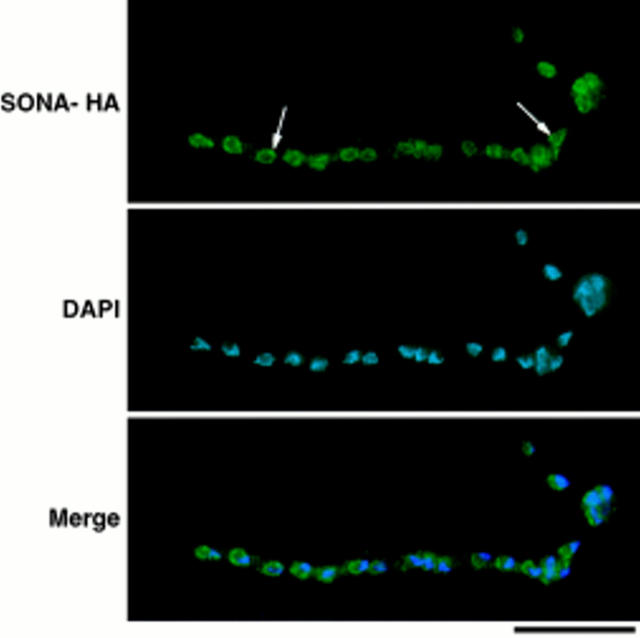
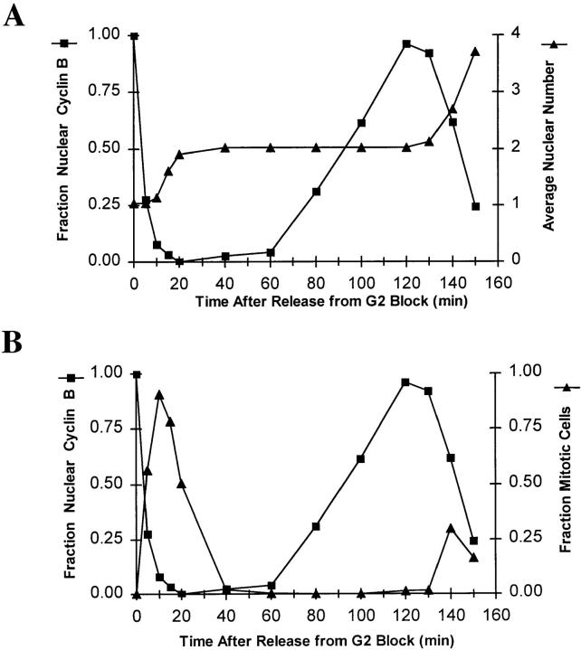
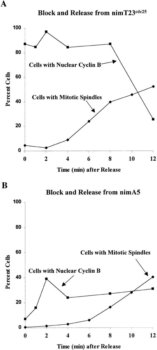
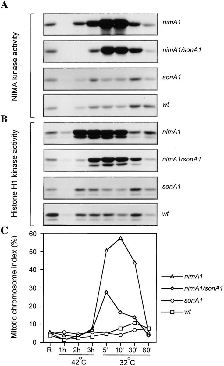
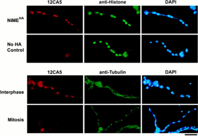
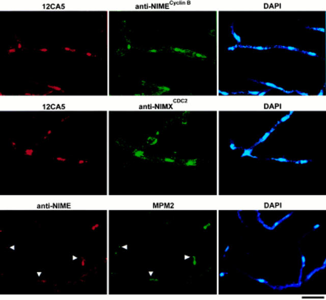
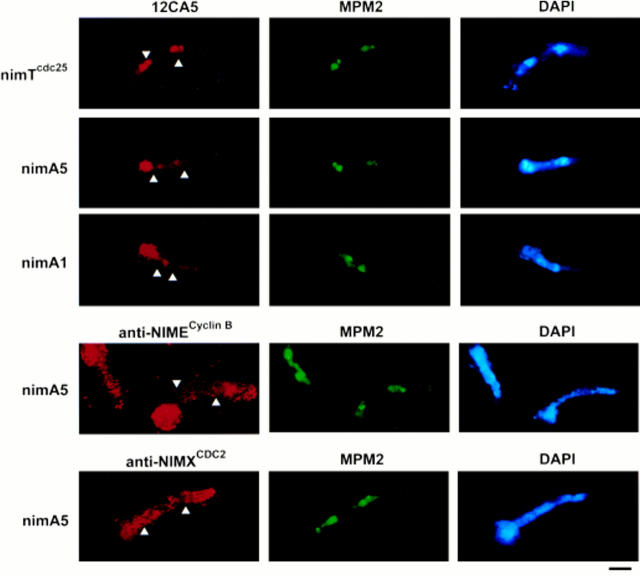
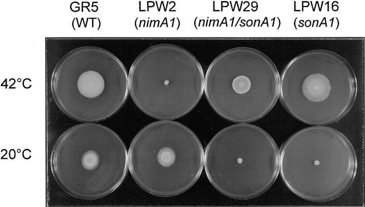
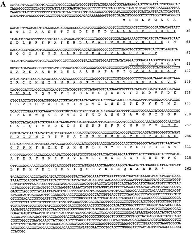
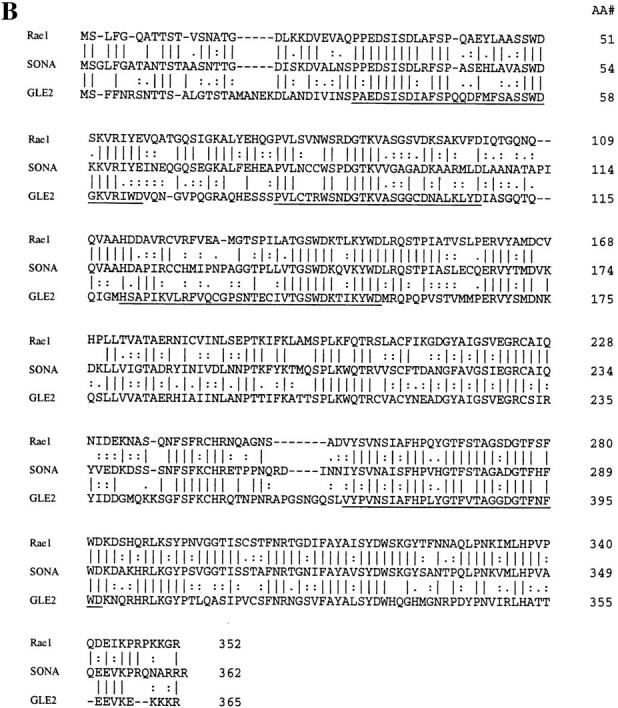
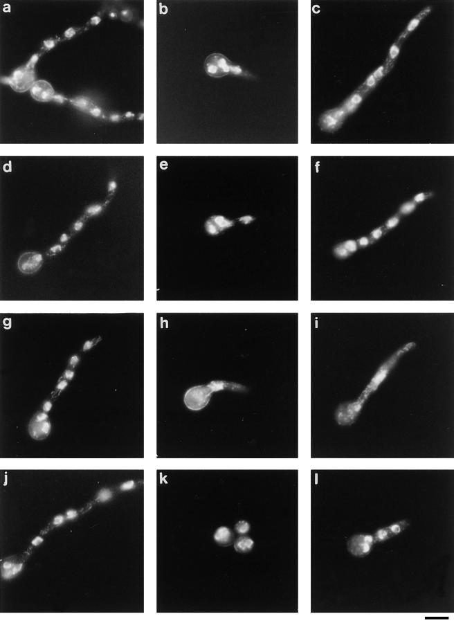
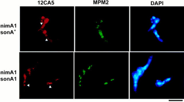
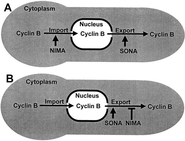
NIMA promotes entry into mitosis in late G2 by some mechanism that is after activation of the Aspergillus nidulans G2 cyclin-dependent kinase, NIMXCDC2/NIMECyclin B. Here we present two independent lines of evidence which indicate that this mechanism involves control of NIMXCDC2/NIMECyclin B localization. First, we found that NIMECyclin B localized to the nucleus and the nucleus-associated organelle, the spindle pole body, in a NIMA-dependent manner. Analysis of cells from asynchronous cultures, synchronous cultures, and cultures arrested in S or G2 showed that NIMECyclin B was predominantly nuclear during interphase, with maximal nuclear accumulation in late G2. NIMXCDC2 colocalized with NIMECyclin B in G2 cells. Although inactivation of NIMA using either the nimA1 or nimA5 temperature-sensitive mutations blocked cells in G2, NIMXCDC2/NIMECyclin B localization was predominantly cytoplasmic rather than nuclear. Second, we found that nimA interacts genetically with sonA, which is a homologue of the yeast nucleocytoplasmic transporter GLE2/RAE1. Mutations in sonA were identified as allele-specific suppressors of nimA1. The sonA1 suppressor alleviated the nuclear division and NIMECyclin B localization defects of nimA1 cells without markedly increasing NIMXCDC2 or NIMA kinase activity. These results indicate that NIMA promotes the nuclear localization of the NIMXCDC2/ NIMECyclin B complex, by a process involving SONA. This mechanism may be involved in coordinating the functions of NIMXCDC2 and NIMA in the regulation of mitosis.


Figures

Figure 8
Figure 8
SONA-HA localizes to the nuclear periphery. Cells of the SONA-HA–expressing strain, LPW42, were cultured, fixed, and then prepared for immunocytology as described in Materials and Methods. The image is identified to the left (SONA-HA shows 12CA5 staining). Arrows, example nuclei within the multinucleate cell shown. Digital images were captured using a Sensys Photometrics CCD camera and were merged using Phase 3 Imaging Systems software. Bar, 10 μm.
Figure 3
Figure 3
Kinetic analysis of NIMECyclin B localization in cells from a synchronized culture. Cells of the nimTcdc25 mutant, SFC4-21, were cultured, fixed, and then prepared for immunocytology as described in Materials and Methods. The cells were stained for HA-NIMECyclin B, α-tubulin, and DAPI. A and B present data from the same samples plotted to show the fraction of cells with nuclear NIMEHA (▪) relative to nuclear division (▴) in A or relative to spindle formation (▴) in B. Each value was determined by counting more than 300 cells.
Figure 5
Figure 5
Cells released from the nimA5 G2 arrest accumulate nuclear NIMECyclin B before entering mitosis. Cells of the nimTcdc25 mutant, SFC4-21 (A), and the nimA5 mutant, PMC654-19 (B), were arrested in late G2 by incubation at restrictive temperature and then released from the G2 arrest by shift to permissive temperature (refer to Materials and Methods). The cells were stained for HA-NIMECyclin B to determine the percent cells with nuclear cyclin B (▪) and with anti–α-tubulin to determine the percent cells with mitotic spindles (♦). Each value was determined by counting at least 100 cells.
Figure 10
Figure 10
NIMA and NIMXCDC2 kinase activities in wild-type–, nimA1-, and sonA1-containing strains. Cells of a wild-type strain (GR5), a nimA1 containing strain (LPW2), a sonA1-containing strain (LPW16), and a nimA1, sonA1 double mutant strain (LPW29) were cultured and then sampled for kinase activity (A and B) and percentage of mitotic cells (C) as described in Materials and Methods. Samples were taken from exponentially growing, asynchronous cultures (R) or from cultures shifted to 42°C for 1, 2, or 3 h, or from cultures returned to 32°C for 5, 10, 30, or 60 min. A and B show autoradiographs representing NIMA (A) and NIMXCDC2 (B) kinase activities measured in immune complexes isolated from whole cell extracts from the strains indicated at the right.
Figure 1
Figure 1
In situ localization of NIMECyclin B in wild-type cells. Cells of PMC654-4, the strain expressing HA-NIMECyclin B (first, third, and fourth rows) and R153, the no-HA control strain (second row), were cultured, fixed, and then prepared for immunocytology as described in Materials and Methods. Each row shows three images of the same cell. The images corresponding to Ab or DAPI staining are labeled above the panels. Note on cell morphology: A. nidulans is a filamentous fungus that undergoes polar growth and nuclear division without cytokinesis during much of its life cycle. Each elongated, multinucleate cell, or hypha, shown here is the result of the polarized growth and multiple, synchronous nuclear divisions of a uninucleate spore. Hyphal compartments are formed by growth and septation, with all nuclei within one compartment being synchronous. The large bulge at one end of each cell is the spore from which the cell originated. Bar, 10 μm.
Figure 2
Figure 2
Localization of NIMECyclin B and NIMXCDC2 on nuclei and SPBs in cells arrested in late G2 by a nimTcdc25 mutation. SFC4-21 cells were cultured, fixed, and then prepared for immunocytology as described in Materials and Methods. The cells contained 4–8 elongated, well separated nuclei characteristic of cells arrested in late G2 by a nimTcdc25 mutation (James et al., 1995). The images corresponding to Ab or DAPI staining are labeled above each image. Arrows point to coincident staining of SPBs by anti-NIMECyclin B and MPM2. Bar, 10 μm.
Figure 4
Figure 4
Nuclear-specific NIMECyclin B and NIMXCDC2 localization was prevented by nimA mutations. Cells of the nimTcdc25 mutant, SFC4-21, the nimA5 mutant, PMC654-19, and the nimA1 mutant, SFC403-19, were cultured, fixed, and then prepared for immunocytology as described in Materials and Methods. Left, strain identity.The images corresponding to Ab or DAPI staining are labeled at the top of the panels. Arrows, position of nuclei in the images showing NIMECyclin B and NIMXCDC2 localization. Bar, 10 μm.
Figure 6
Figure 6
Colony growth phenotype of the following strains: wild-type (GR5), nimA1 (LPW2), sonA1 (LPW16), and nimA1, sonA1 (LPW29). Strains are identified by their relevant genotype indicated at the top. Each strain was center-point inoculated on MAG medium and then incubated at 42°C for 3 d or at 20°C for 10 d, as indicated.
Figure 7
Figure 7
Sequence of sonA and alignment of SONA to GLE2 and RAE1. (A) The nucleotide sequence and predicted ORF in the 2.6-kb genomic sonA clone is shown. Right, amino acid residues of the SONA ORF; underline, predicted WD repeats; boldface at the amino terminus, a single GLFG sequence, found in multiple copies of many nuclear pore proteins; boldface at the carboxyl terminus, a potential nuclear localization sequence. The position of the intron was confirmed by sequencing a cDNA clone. (B) An alignment of SONA to GLE2 and RAE1 is shown. Right, amino acid residue numbers; solid lines, identical residues. Highly conserved and conserved residues, as determined by the Applied Biosystems Sequence Analysis Software, are indicated by the double dots, and single dots, respectively. The sonA cDNA sequence is available from EMBL/GenBank/DDBJ under accession number AF069492.
Figure 7
Figure 7
Sequence of sonA and alignment of SONA to GLE2 and RAE1. (A) The nucleotide sequence and predicted ORF in the 2.6-kb genomic sonA clone is shown. Right, amino acid residues of the SONA ORF; underline, predicted WD repeats; boldface at the amino terminus, a single GLFG sequence, found in multiple copies of many nuclear pore proteins; boldface at the carboxyl terminus, a potential nuclear localization sequence. The position of the intron was confirmed by sequencing a cDNA clone. (B) An alignment of SONA to GLE2 and RAE1 is shown. Right, amino acid residue numbers; solid lines, identical residues. Highly conserved and conserved residues, as determined by the Applied Biosystems Sequence Analysis Software, are indicated by the double dots, and single dots, respectively. The sonA cDNA sequence is available from EMBL/GenBank/DDBJ under accession number AF069492.
Figure 9
Figure 9
Nuclear division in wild-type–, nimA1-, and sonA1-containing strains. Cells of the wild-type strain, GR5 (a–c), the sonA1 mutant, LPW16 (d–f), the nimA1 mutant, LPW2 (g–i), and the nimA1, sonA1 double mutant, LPW19 (j–l) were stained with DAPI as described in Materials and Methods. Cells shown in panels a, d, g, and j were incubated at 32°C for 8 h. Other panels show cells incubated at 42°C for 8 h (b, e, h, and k) or 10 h (c, f, i, and l). Bar, 10 μm.
Figure 11
Figure 11
The sonA1 mutation suppresses the nuclear NIMECyclin B localization defect of nimA1. Cells of the nimA1 strain, SFC403-19, and the nimA1, sonA1 double mutant, SFC444-1, were cultured, fixed, and then prepared for immunocytology as described in Materials and Methods. Left, strain identity; top, images corresponding to Ab or DAPI staining; top row arrows, (nimA1 single mutant) position of nuclei in cells showing general cytoplasmic staining; bottom row arrows, position of example nuclei showing NIMECyclin B staining. Bar, 10 μm.
Figure 12
Figure 12
Model for NIMA and SONA function in the nucleocytoplasmic transport of NIMECyclin B.

References

    1. Alfa CE, Ducommun B, Beach D, Hyams JS. Distinct nuclear and spindle pole body populations of cyclincdc2in fission yeast. Nature. 1990;347:680–682. - PubMed
    1. Alfa CE, Gallagher IM, Hyams JS. Subcellular localization of the p34cdc2/p63cdc13protein kinase in fission yeast. Cold Spring Harbor Symp Quant Biol. 1991;56:489–494. - PubMed
    1. Altschul SF, Gish W, Miller W, Myers EW, Lipman DJ. Basic local alignment search tool. J Mol Biol. 1990;215:403–410. - PubMed
    1. Audit M, Barbier M, Soyer-Gobillard MO, Albert M, Geraud ML, Nicolas G, Lenaers G. Cyclin B (p56cdc13) localization in the yeast Schizosaccharomyces pombe: an ultrastructural and immunocytochemical study. Biol Cell. 1996;86:1–10. - PubMed
    1. Bergen LG, Upshall A, Morris NR. S phase, G2 and nuclear division mutants of Aspergillus nidulans. . J Bact. 1984;159:114–119. - PMC - PubMed

Publication types

MeSH terms

Associated data