. 1998 Jun 29;141(7):1601-11.
doi: 10.1083/jcb.141.7.1601.

Activity-dependent regulation of dendritic BC1 RNA in hippocampal neurons in culture

Affiliations

Activity-dependent regulation of dendritic BC1 RNA in hippocampal neurons in culture

I A Muslimov et al. J Cell Biol. .

Abstract

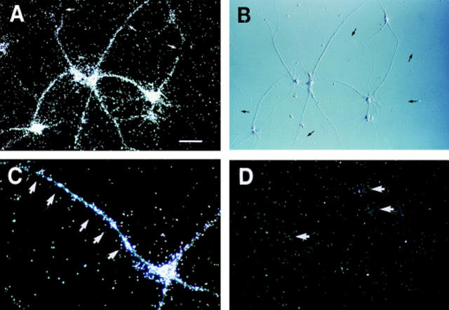
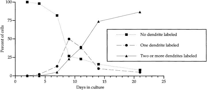
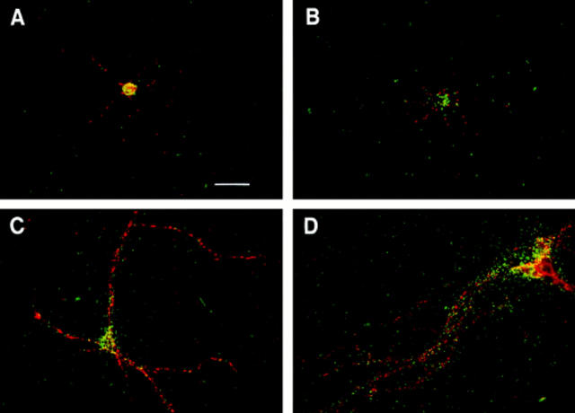
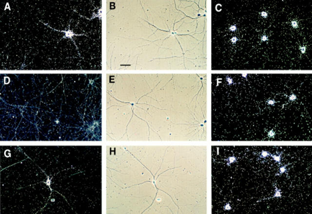
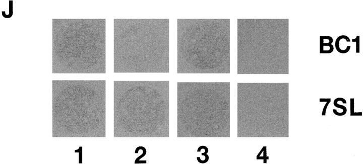
Several neuronal RNAs have been identified in dendrites, and it has been suggested that the dendritic location of these RNAs may be relevant to the spatiotemporal regulation of mosaic postsynaptic protein repertoires through transsynaptic activity. Such regulation would require that dendritic RNAs themselves, or at least some of them, be subject to physiological control. We have therefore examined the functional regulation of somatodendritic expression levels of dendritic BC1 RNA in hippocampal neurons in culture. BC1 RNA, an RNA polymerase III transcript that is a component of a ribonucleoprotein particle, became first detectable in somatodendritic domains of developing hippocampal neurons at times of initial synapse formation. BC1 RNA was identified only in such neurons that had established synapses on cell bodies and/or developing dendritic arbors. When synaptic contact formation was initiated later in low-density cultures, BC1 expression was coordinately delayed. Inhibition of neuronal activity in hippocampal neurons resulted in a substantial but reversible reduction of somatodendritic BC1 expression. We conclude that expression of BC1 RNA in somatic and dendritic domains of hippocampal neurons is regulated in development, and is dependent upon neuronal activity. These results establish (for the first time to our knowledge) that an RNA polymerase III transcript can be subject to control through physiological activity in nerve cells.


Figures

Figure 1
Figure 1
Localization of BC1 RNA to dendrites of mature hippocampal neurons in culture. Cells were probed for BC1 RNA by in situ hybridization; labeling signal is indicated by autoradiographic silver grains (white in dark-field photomicrographs A, C, and D). (A and B) BC1 labeling signal is observed over somata and dendrites, but not over axons (black arrows in DIC photomicrograph B) of hippocampal neurons in culture. Labeling was discontinuous over some dendrites: gaps are indicated by white arrows in A. A higher power photomicrograph (C) reveals clustering (white arrowheads) of the BC1 labeling signal over dendrites. (D) Sense strand control experiments did not produce significant labeling over either cell bodies (white arrowheads) or neurites. (A, B, and D) Bar, 50 μm; (C) bar, 25 μm.
Figure 2
Figure 2
Development of somatodendritic BC1 expression in hippocampal neurons in culture. Cells were grown at medium density (6,000 cells per cm2) for 2 d in vitro (DIV; A and B); 4 DIV (C and D); 7 DIV (E and F); 9 DIV (G and H); and 14 DIV (I and J). (Left) Dark-field photomicrographs; (right) DIC photomicrographs. Arrows indicate axonal (B) or dendritic (D) growth cones. Bar, 50 μm.
Figure 3
Figure 3
Quantitative analysis of dendritic BC1 delivery in hippocampal neurons developing in culture. The number of cells analyzed for each time point was as follows: 2 DIV, 214; 4 DIV, 187; 7 DIV, 171; 9 DIV, 200; 11 DIV, 298; 14 DIV, 219; 21 DIV, 238.
Figure 4
Figure 4
Expression of BC1 RNA in cultured hippocampal neurons developing at different cell densities. Hippocampal neurons were grown for 7 d in culture and were double-labeled for BC1 RNA (in situ hybridization; green) and synaptophysin (immunocytochemistry; red). A green filter was used for visualizing autoradiographic silver grains in double-exposure photomicrographs in order to differentiate them from the red immunofluorescence signal. (A and B) 1,000 cells per cm2; (C) 4,000 cells per cm2; (D) 16,000 cells per cm2. At low density, beginning BC1 expression can be observed in a few cells (B) while the majority shows no labeling (A). At medium density (C), BC1 labeling was often observed over proximal segments of dendrites that were lined with synaptophysin puncta. At high density (D), BC1 labeling was frequently observed over dendrites at considerable distances from somata. Bar, 25 μm.
Figure 5
Figure 5
Regulation of BC1 expression by neuronal activity. Hippocampal neurons were grown in culture for 14 d in the absence of TTX (A–C), in the presence of 1 μM TTX (D–F), or in the presence of 1 μM TTX for the first 9 d, and in the absence of TTX for the following 5 d (G–I). Left (A–G): BC1 RNA, dark-field photomicrographs; middle (B–H), BC1 RNA, phase contrast photomicrographs, corresponding to dark-field photomicrographs in left column; right (C–I): 7SL RNA, dark-field photomicrographs. Dark field photomicrograph D (BC1 RNA in the presence of TTX) was overexposed to reveal absence of any significant labeling over cells or neurites. All cultures were grown at medium density. Bar, 50 μm. (J) Cultured hippocampal neurons on coverslips were exposed to autoradiographic film. Autoradiographs show BC1- and 7SL-labeling signals for cells that were grown for 14 d in the absence of TTX (1), in the presence of 1 μM TTX (2), or in the presence of 1 μM TTX for the first 9 d and in the absence of TTX for the following 5 d (3). (4) Sense strand control.
Figure 5
Figure 5
Regulation of BC1 expression by neuronal activity. Hippocampal neurons were grown in culture for 14 d in the absence of TTX (A–C), in the presence of 1 μM TTX (D–F), or in the presence of 1 μM TTX for the first 9 d, and in the absence of TTX for the following 5 d (G–I). Left (A–G): BC1 RNA, dark-field photomicrographs; middle (B–H), BC1 RNA, phase contrast photomicrographs, corresponding to dark-field photomicrographs in left column; right (C–I): 7SL RNA, dark-field photomicrographs. Dark field photomicrograph D (BC1 RNA in the presence of TTX) was overexposed to reveal absence of any significant labeling over cells or neurites. All cultures were grown at medium density. Bar, 50 μm. (J) Cultured hippocampal neurons on coverslips were exposed to autoradiographic film. Autoradiographs show BC1- and 7SL-labeling signals for cells that were grown for 14 d in the absence of TTX (1), in the presence of 1 μM TTX (2), or in the presence of 1 μM TTX for the first 9 d and in the absence of TTX for the following 5 d (3). (4) Sense strand control.

Similar articles

Cited by

References

    1. Ainger K, Avossa D, Morgan F, Hill SJ, Barry C, Barbarese E, Carson JC. Transport and localization of exogenous myelin basic protein mRNA microinjected into oligodendrocytes. J Cell Biol. 1993;123:431–441. - PMC - PubMed
    1. Banker, G.A., and A.B. Waxman. 1988. Hippocampal neurons generate natural shapes in cell culture. In Intrinsic determinants of neuronal form and function. R.J. Lasek and M.M. Black, editors. Alan R. Liss, New York. 61–82.
    1. Banker, G., and K. Goslin, editors. 1998. Culturing Nerve Cells. MIT Press, Cambridge, MA. In press.
    1. Basarsky TA, Parpura V, Haydon PG. Hippocampal synaptogenesis in cell culture: developmental time course of synapse formation, calcium influx, and synaptic protein distribution. J Neurosci. 1994;14:6402–6411. - PMC - PubMed
    1. Benson DL, Cohen PA. Activity-independent segregation of excitatory and inhibitory synaptic terminals in cultured hippocampal neurons. J Neurosci. 1996;16:6424–6432. - PMC - PubMed

Publication types