. 1998 Jun 29;141(7):1625-36.
doi: 10.1083/jcb.141.7.1625.

Molecular dissection of the Rho-associated protein kinase (p160ROCK)-regulated neurite remodeling in neuroblastoma N1E-115 cells

Affiliations

Molecular dissection of the Rho-associated protein kinase (p160ROCK)-regulated neurite remodeling in neuroblastoma N1E-115 cells

M Hirose et al. J Cell Biol. .

Abstract

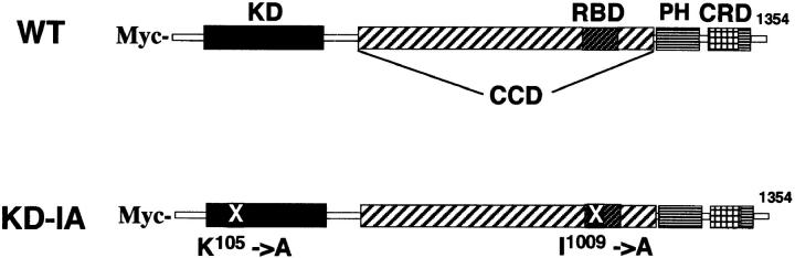
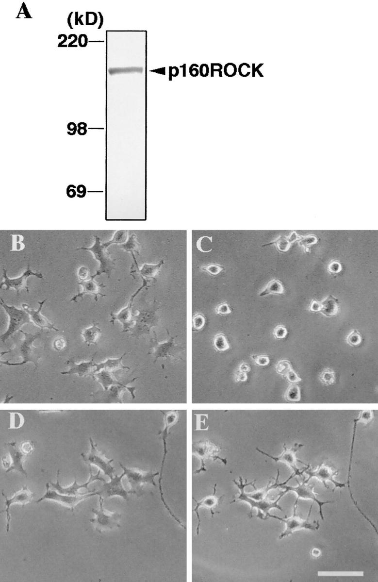
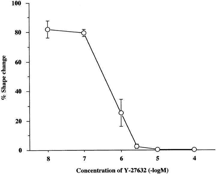
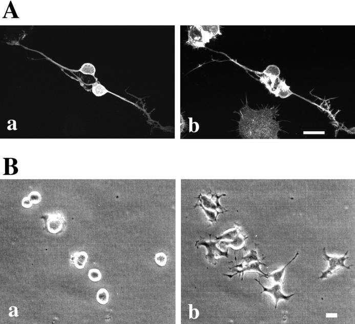
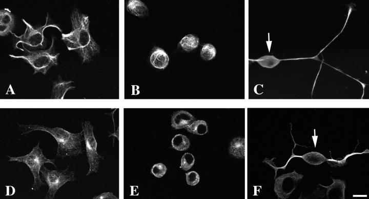
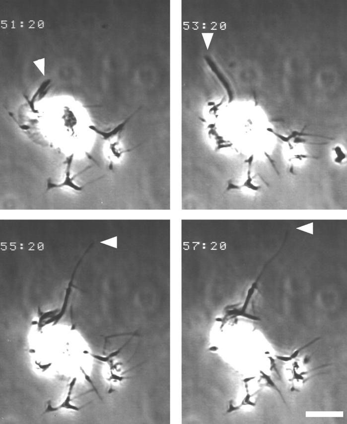
A critical role for the small GTPase Rho and one of its targets, p160ROCK (a Rho-associated coiled coil-forming protein kinase), in neurite remodeling was examined in neuroblastoma N1E-115 cells. Using wild-type and a dominant-negative form of p160ROCK and a p160ROCK-specific inhibitor, Y-27632, we show here that p160ROCK activation is necessary and sufficient for the agonist-induced neurite retraction and cell rounding. The neurite retraction was accompanied by elevated phosphorylation of myosin light chain and the disassembly of the intermediate filaments and microtubules. Y-27632 blocked both neurite retraction and the elevation of myosin light chain phosphorylation in a similar concentration-dependent manner. On the other hand, suppression of p160ROCK activity by expression of a dominant-negative form of p160ROCK induced neurites in the presence of serum by inducing the reassembly of the intermediate filaments and microtubules. The neurite outgrowth by the p160ROCK inhibition was blocked by coexpression of dominant-negative forms of Cdc42 and Rac, indicating that p160ROCK constitutively and negatively regulates neurite formation at least in part by inhibiting activation of Cdc42 and Rac. The assembly of microtubules and intermediate filaments to form extended processes by inhibitors of the Rho-ROCK pathway was also observed in Swiss 3T3 cells. These results indicate that Rho/ROCK-dependent tonic inhibition of cell process extension is exerted via activation of the actomysin-based contractility, in conjunction with a suppression of assembly of intermediate filaments and microtubules in many cell types including, but not exclusive to, neuronal cells.


Figures

Figure 1
Figure 1
Schematic representation of the wild-type and the KD-IA mutant of p160ROCK. Domains of p160ROCK and the point mutations in the KD-IA mutant are illustrated. KD, serine/threonine protein kinase domain; CCD, coiled-coil forming amphiphatic α-helical domain; RBD, Rho-binding domain; PH, pleckstrin homology domain; CRD, cysteine-rich zinc-finger domain; X, positions of mutations with amino acid numbers.
Figure 2
Figure 2
Western blotting of p160ROCK in N1E-115 cells (A) and inhibition of LPA-induced neurite retraction and cell rounding by Y-27632 (B–E). (A) N1E-115 cells were lysed in Laemmli– SDS-PAGE sample buffer and then subjected to immunoblotting using anti-p160ROCK antibody. (B–E), N1E-115 cells were maintained in serum-free DME for 24 h and allowed to extend neurites. The cells were pretreated without (B and C) or with 10 μM Y-27632 (D and E) for 30 min, and exposed to 1 μM LPA for 10 min. The same fields were photographed before the drug addition (Band D) and after LPA stimulation (C and E). Bar, 100 μm.
Figure 3
Figure 3
Concentration-dependent inhibition by Y-27632 of LPA-induced neurite retraction in N1E-115 cells. Serum-starved N1E-115 cells were pretreated with the indicated concentrations of Y-27632 for 30 min and then exposed to 1 μM LPA for 10 min. The percentages of cells with complete neurite retraction are shown. Results of three experiments are shown as mean ± SEM.
Figure 4
Figure 4
Morphology of N1E-115 cells expressing active Rho and p160ROCK. N1E-115 cells were transfected with V14Rho (A and B) or wild-type p160ROCK (C and D), and were cultured in serum-free DME for 12 h. Overexpressing cells were identified by the positive Myc-staining (A and C) and their F-actin structures were examined (B and D). Note that the transfected cells do not extend neurites even under serum-starved conditions. In addition, marked bleb formation was also noted in some of p160ROCK-expressing cells. Bar, 20 μm.
Figure 5
Figure 5
Morphology of N1E-115 cells expressing the KD-IA mutant and of N1E-115 cells treated with Y-27632. (A) N1E-115 cells were transfected with the KD-IA mutant, a dominant-negative form of p160ROCK, and were cultured for 16 h in the presence of 10% FBS. The cells were double-stained for the Myc-tag (a) and F-actin (b). Note that the transfected cells extend long neurites in the presence of serum. (B) N1E-115 cells were cultured in the presence of 10% FBS, and treated with 10 μM Y-27632, a specific ROCK inhibitor. Phase-contrast photomicrographs were taken at 0 (a) and 60 min (b) after the addition of the compound. Bar, 20 μm.
Figure 6
Figure 6
Inhibition by N17Cdc42 and N17Rac of the neurite generation induced by the KD-IA mutant of p160ROCK. N1E-115 cells were transfected with KD-IA p160ROCK alone (A and B), with N17Cdc42 and KD-IA p160ROCK (C and D) or with N17Rac and KD-IA p160ROCK (E and F). The transfected cells were cultured in DME containing 10% FBS for 16 h. Overexpressing cells were identified with p160ROCK staining (A, C, and E) and F-actin was stained with phalloidin (B, D, and F). Bar, 20 μm. (G) Quantification of cells bearing no neurites, short neurites, and long neurites. N1E cells expressing KD-IA alone, KD-IA and N17Cdc42, or KD-IA and N17Rac were identified as above, and the numbers of cells bearing no neurites, or neurites shorter or longer than 100 μm were determined. *, P < 0.001 compared with KD-IA.
Figure 6
Figure 6
Inhibition by N17Cdc42 and N17Rac of the neurite generation induced by the KD-IA mutant of p160ROCK. N1E-115 cells were transfected with KD-IA p160ROCK alone (A and B), with N17Cdc42 and KD-IA p160ROCK (C and D) or with N17Rac and KD-IA p160ROCK (E and F). The transfected cells were cultured in DME containing 10% FBS for 16 h. Overexpressing cells were identified with p160ROCK staining (A, C, and E) and F-actin was stained with phalloidin (B, D, and F). Bar, 20 μm. (G) Quantification of cells bearing no neurites, short neurites, and long neurites. N1E cells expressing KD-IA alone, KD-IA and N17Cdc42, or KD-IA and N17Rac were identified as above, and the numbers of cells bearing no neurites, or neurites shorter or longer than 100 μm were determined. *, P < 0.001 compared with KD-IA.
Figure 7
Figure 7
LPA-induced MLC phosphorylation and its inhibition by Y-27632 in N1E-115 cells. (A) Time course of LPA-induced MLC phosphorylation. Serum straved N1E-115 cells were incubated with 1 μM LPA for the indicated times. Cells were collected in Laemmli-SDS-PAGE sample buffer and then subjected to immunoblotting using anti-phosphoMLC antibody. (B) Concentration-dependent inhibition of MLC phosphorylation by Y-27632. Serum-starved N1E-115 cells were treated with the indicated concentrations of Y-27632 for 30 min, and then stimulated with LPA for 2 min. The cells were lysed and analyzed as above. The content of MLC in the cells did not change during LPA stimulation or Y-27632 treatment.
Figure 8
Figure 8
Peripherin and tubulin staining of serum-starved, serum-fed, and KD-IA–transfected N1E-115 cells. N1E-115 cells were cultured in the absence (A and D) or presence (B and E) of serum, or were transfected with the KD-IA mutant of p160ROCK and cultured in DME containing 10% serum (C and F). The cells were stained with either anti-peripherin (A, B, and C) or anti-tubulin (D, E, and F) antibodies. In C and F, the cells were also stained with either anti-myc antibody (C), or anti-ROCK antibody (F), and the cells expressing KD-IA protein were identified (indicated by arrows). Images built from optical sections by a confocal imaging system are shown. Bar, 20 μm.
Figure 10
Figure 10
Vimentin staining of Swiss 3T3 cells. Swiss 3T3 cells were cultured in the presence (A and B) or absence (C and D) of serum, or with 30 μg/ml C3 exoenzyme for 72 h (E and F) or 10 μM Y-27632 for 2 h (G and H) in the presence of serum, and were stained with OregonGreen phalloidin (A, C, E, and G) or with anti-vimentin antibody (B, D, F, and H). Images built up from optical sections by a confocal imaging system are shown. Bar, 20 μm.
Figure 11
Figure 11
Tubulin staining of Swiss 3T3 cells. Swiss 3T3 cells were cultured in the presence (A and B) or absence (C and D) of serum, or with C3 exoenzyme (E and F) or Y-27632 (G and H) in the presence of serum, and were stained with OregonGreen phalloidin (A, C, E, and G) or with anti-tubulin antibody (B, D, F, and H). Images built from optical sections by a confocal imaging system are shown. Bar, 20 μm.
Figure 9
Figure 9
Video-microscopy of Swiss 3T3 fibroblast microinjected with C3 exoenzyme. Swiss cells were maintained on a glass coverslip in Hepes-buffered DME containing 10% FBS and microinjected with C3 exoenzyme. Morphology of the injected cells was monitored by time-lapse video-microscopy. Four images taken every two min are shown. Time after the injection is indicated in the top left corner of each image. Note cell processes change in their morphology in a time-dependent manner; arrowhead, time-dependent elongation of one process. Bar, 20 μm.
Figure 12
Figure 12
Model of signal transduction and cross-talks of the Rho–ROCK pathway in the neurite remodeling. p160ROCK is activated downstream of Rho in response to agonist stimulation, and in turn induces the actomyosin-based contractility and the disassembly of the microtubules and the intermediate filaments, leading to the neurite retraction. p160ROCK also transmits a negative signal to the Cdc42/Rac pathways and suppresses the neurite outgrowth by tonically inhibiting their actions. The activation of the cAMP–A kinase pathway is likely to inhibit the Rho–ROCK pathway and to release the suppression of neurite outgrowth. Disassembly of intermediate filaments may be caused by direct phosphorylation by p160ROCK at specific amino acid residue(s) of this cytoskeletal proteins as shown for GFAP (Kosako et al., 1997) or may well be a secondary consequence of ROCK's effect on the actomyosin system.

References

    1. Amano M, Itoh M, Kimura K, Fukata Y, Chihara K, Nakano T, Mastuura Y, Kaibuchi K. Phosphorylation and activation of myosin by Rho-associated kinase (Rho-kinase) J Biol Chem. 1996;271:20246–20249. - PubMed
    1. Amano M, Chihara K, Kimura K, Fukata Y, Nakamura N, Mastuura Y, Kaibuchi K. Formation of actin stress fibers and focal adhesions enhanced by Rho-kinase. Science. 1997;275:1308–1311. - PubMed
    1. Aletta JM, Shelanski ML, Greene LA. Phosphorylation of the peripherin 58-kD, a neuronal intermediate filament protein. Regulation by nerve growth factor and other agents. J Biol Chem. 1989;264:4619–4627. - PubMed
    1. Bently D, O'Connor TP. Cytoskeletal events in growth cone steering. Curr Opin Neurobiol. 1994;4:43–48. - PubMed
    1. Daniels M. The role of microtubules in the growth and stabilization of nerve fibers. Annu NY Acad Sci. 1975;253:535–544. - PubMed

Publication types

MeSH terms