. 1998 Jun 29;141(7):1659-73.
doi: 10.1083/jcb.141.7.1659.

Fibroblast growth factor-2 (FGF-2) induces vascular endothelial growth factor (VEGF) expression in the endothelial cells of forming capillaries: an autocrine mechanism contributing to angiogenesis

Affiliations

Fibroblast growth factor-2 (FGF-2) induces vascular endothelial growth factor (VEGF) expression in the endothelial cells of forming capillaries: an autocrine mechanism contributing to angiogenesis

G Seghezzi et al. J Cell Biol. .

Abstract

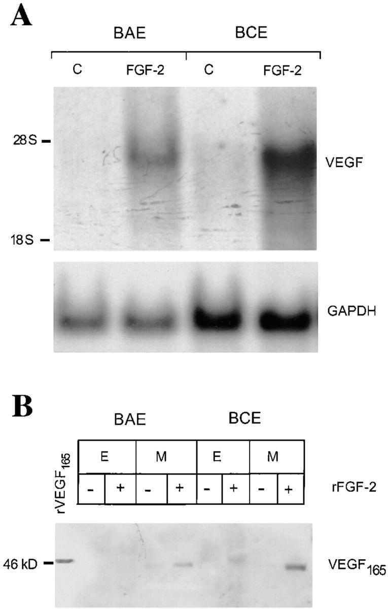
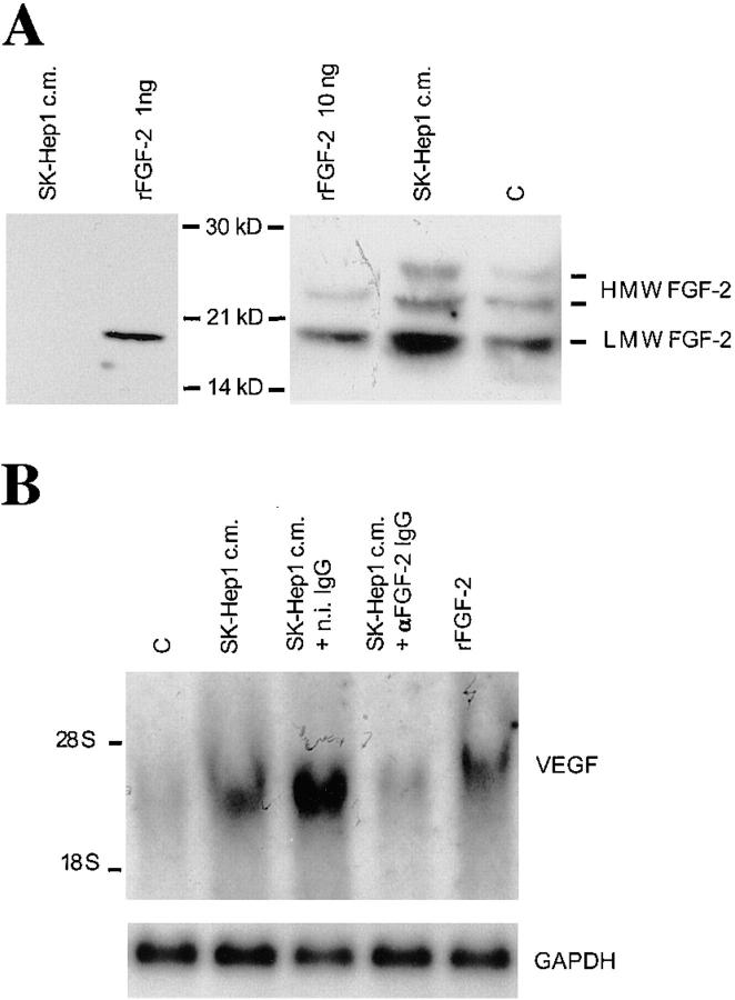
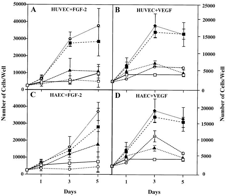
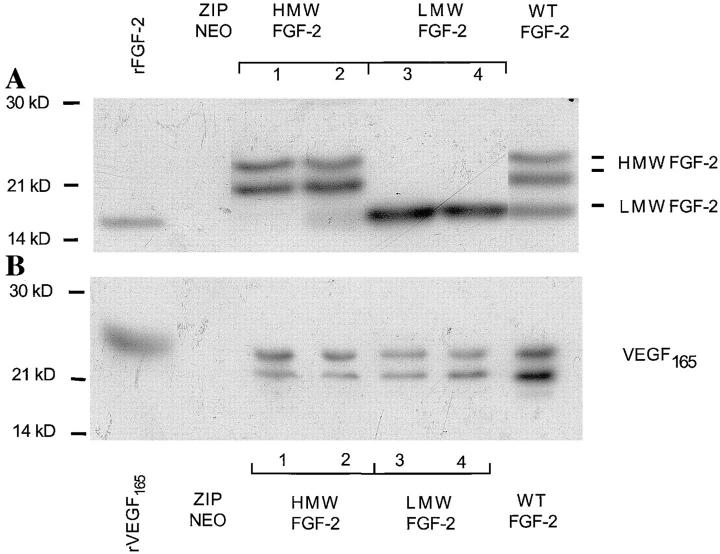
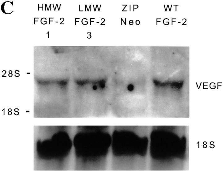
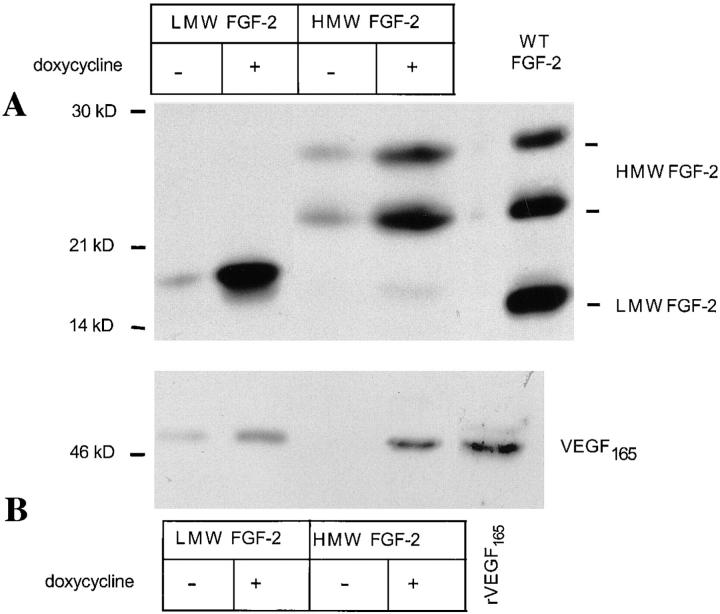
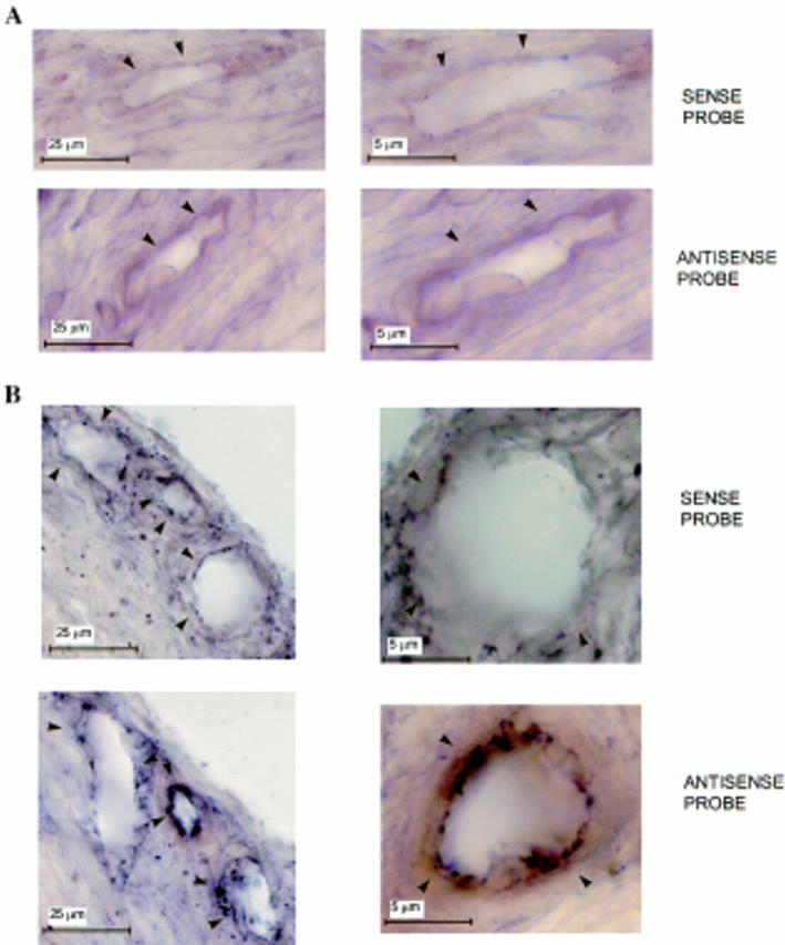
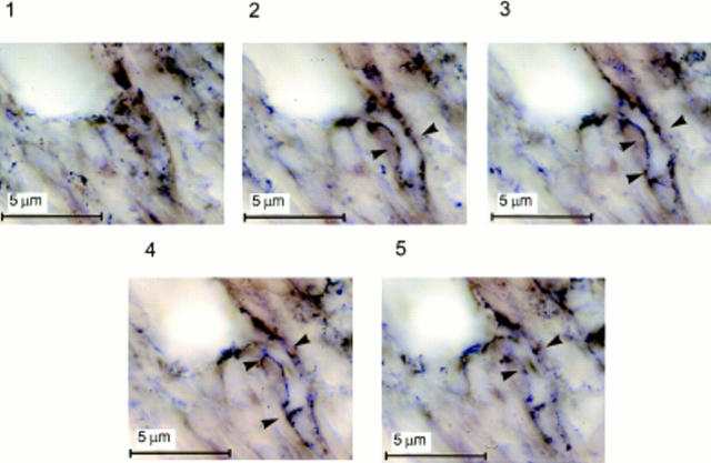
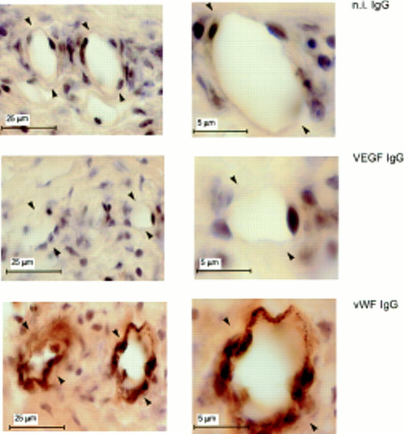
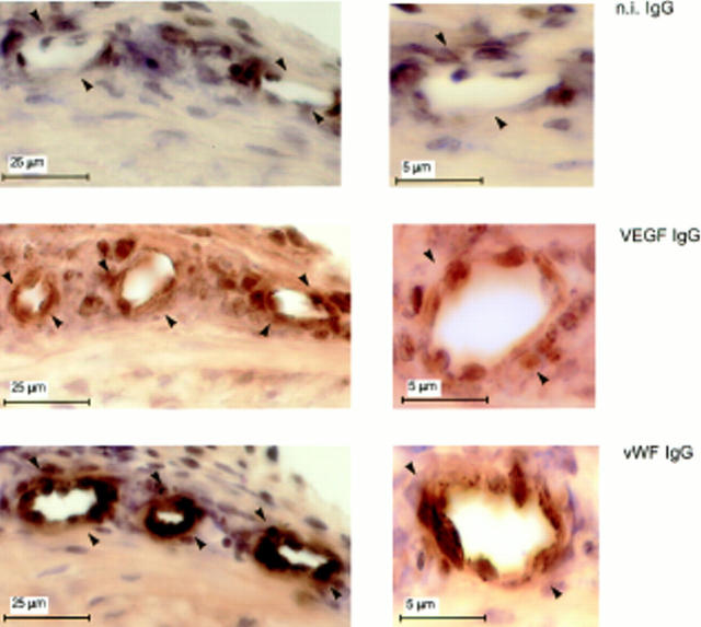
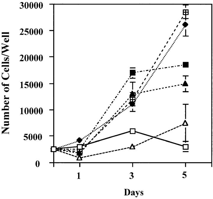
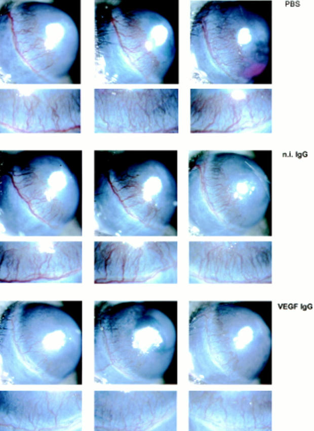
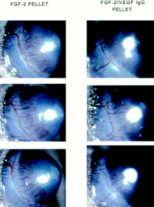
FGF-2 and VEGF are potent angiogenesis inducers in vivo and in vitro. Here we show that FGF-2 induces VEGF expression in vascular endothelial cells through autocrine and paracrine mechanisms. Addition of recombinant FGF-2 to cultured endothelial cells or upregulation of endogenous FGF-2 results in increased VEGF expression. Neutralizing monoclonal antibody to VEGF inhibits FGF-2-induced endothelial cell proliferation. Endogenous 18-kD FGF-2 production upregulates VEGF expression through extracellular interaction with cell membrane receptors; high-Mr FGF-2 (22-24-kD) acts via intracellular mechanism(s). During angiogenesis induced by FGF-2 in the mouse cornea, the endothelial cells of forming capillaries express VEGF mRNA and protein. Systemic administration of neutralizing VEGF antibody dramatically reduces FGF-2-induced angiogenesis. Because occasional fibroblasts or other cell types present in the corneal stroma show no significant expression of VEGF mRNA, these findings demonstrate that endothelial cell-derived VEGF is an important autocrine mediator of FGF-2-induced angiogenesis. Thus, angiogenesis in vivo can be modulated by a novel mechanism that involves the autocrine action of vascular endothelial cell-derived FGF-2 and VEGF.


Figures

Figure 1
Figure 1
FGF-2 induces VEGF expression in endothelial cells. (A) Northern blotting analysis of total RNA (20 μg) from BAE or BCE cells treated with rFGF-2 (10 ng/ml) or with control medium (C) for 4 h. The RNA blot was hybridized with a DIG-labeled cDNA probe for human VEGF as described in Materials and Methods. GAPDH mRNA is shown as a control. The position of 28 S and 18 S ribosomal RNA is shown on the left. This experiment was repeated four times with comparable results. (B) Western blotting analysis of Triton X-100 extracts (E; 200 μg) or conditioned medium (M) from BAE or BCE cells treated with 10 ng/ml of rFGF-2 (+) or with control medium (−) for 17 h. The samples were electrophoresed under nonreducing conditions. The protein blot was hybridized with human VEGF IgG; antigen–antibody complexes were detected as described in Materials and Methods. Recombinant VEGF165 (10 ng) was run as a control in the leftmost lane. Molecular masses are shown in kD on the left. This experiment was repeated three times with comparable results.
Figure 2
Figure 2
SK-Hep1–conditioned medium induces FGF-2 and VEGF expression in endothelial cells. (A) Right, Western blotting analysis of Triton X-100 extracts (200 μg) from BCE cells incubated for 17 h with SK-Hep1 cell-conditioned medium (SK-Hep1 c.m.) or with control medium (C). Recombinant LMW FGF-2 (rFGF-2; 10 ng) was run as a control in the leftmost lane. Left, 4 ml of SK-Hep1 cell-conditioned medium was concentrated 80-fold and analyzed by Western blotting as described in Materials and Methods. 1 ng of rFGF-2 was run in the same blot as a control. Antigen–antibody complexes were detected as described in Materials and Methods. Molecular masses are shown in kD between the two panels. The positions of LMW and HMW FGF-2 bands are shown on the right. This experiment was repeated three times with similar results. (B) Northern blotting analysis of total RNA (20 μg) from BCE cells incubated for 2 h with either control medium (C) or with SK-Hep1 cell-conditioned medium (SK-Hep1 c.m.) or 10 ng/ ml of rFGF-2 in the presence or absence of 10 μg/ml of neutralizing antibody to rFGF-2 (αFGF-2 IgG) or n.i. IgG (n.i. IgG). The RNA blots were hybridized with a DIG-labeled cDNA probe to human VEGF as described in Materials and Methods. GAPDH mRNA is shown as a control. This experiment was repeated three times with similar results.
Figure 3
Figure 3
Antibody to VEGF inhibits FGF-2–induced endothelial cell proliferation. Growth curves of HUVE (A and B) or HAE (C and D) cells in the absence (□) or in the presence of 10 ng/ml of FGF-2 (⋄) (A and C) or 30 ng/ml of VEGF (▴) (B and D) without or with addition of either 10 μg/ml of anti-VEGF (▴) or anti–FGF-2 (○) or n.i. IgG (▪). The cells were grown and counted as described in Materials and Methods. Each point represents mean and standard deviation of triplicate samples from a representative experiment.
Figure 4
Figure 4
FGF-2 and VEGF expression in NIH 3T3 cells transfected with FGF-2 cDNA. (A) FGF-2 expression. Triton X-100 extracts (50 μg) of clones of NIH 3T3 cells transfected with the cDNAs for HMW (clones 1 and 2), LMW (clones 3 and 4) or WT FGF-2, or from control cells transfected with the vector alone (ZIPNEO) were analyzed by Western blotting with antibody to human FGF-2. Recombinant LMW FGF-2 (rFGF-2; 10 ng) was run as a control in the leftmost lane. (B) VEGF expression. Medium conditioned by the clones of FGF-2 transfectants shown in A was analyzed by Western blotting with antibody to VEGF. Recombinant VEGF165 (10 ng) was run as a control in the leftmost lane. Western blotting was carried out under reducing conditions as described in Materials and Methods. Molecular masses are shown in kD on the left. This experiment was repeated three times with similar results. (C) Northern blotting analysis of total RNA (20 μg) from clones of NIH 3T3 cells transfected with the cDNAs for HMW (clone 1), LMW (clone 3) or WT FGF-2, or from control cells transfected with the vector alone (ZIPNEO). The RNA blot was hybridized with a DIG-labeled cDNA probe to human VEGF as described in Materials and Methods. The position of 28 S and 18 S ribosomal RNA is shown on the left. 18S ribosomal RNA is shown as a control. This experiment was repeated three times with comparable results.
Figure 4
Figure 4
FGF-2 and VEGF expression in NIH 3T3 cells transfected with FGF-2 cDNA. (A) FGF-2 expression. Triton X-100 extracts (50 μg) of clones of NIH 3T3 cells transfected with the cDNAs for HMW (clones 1 and 2), LMW (clones 3 and 4) or WT FGF-2, or from control cells transfected with the vector alone (ZIPNEO) were analyzed by Western blotting with antibody to human FGF-2. Recombinant LMW FGF-2 (rFGF-2; 10 ng) was run as a control in the leftmost lane. (B) VEGF expression. Medium conditioned by the clones of FGF-2 transfectants shown in A was analyzed by Western blotting with antibody to VEGF. Recombinant VEGF165 (10 ng) was run as a control in the leftmost lane. Western blotting was carried out under reducing conditions as described in Materials and Methods. Molecular masses are shown in kD on the left. This experiment was repeated three times with similar results. (C) Northern blotting analysis of total RNA (20 μg) from clones of NIH 3T3 cells transfected with the cDNAs for HMW (clone 1), LMW (clone 3) or WT FGF-2, or from control cells transfected with the vector alone (ZIPNEO). The RNA blot was hybridized with a DIG-labeled cDNA probe to human VEGF as described in Materials and Methods. The position of 28 S and 18 S ribosomal RNA is shown on the left. 18S ribosomal RNA is shown as a control. This experiment was repeated three times with comparable results.
Figure 5
Figure 5
FGF-2 and VEGF expression in NIH 3T3 cells transfected with FGF-2 cDNA under control by the tetracycline- dependent transactivator. NIH 3T3 cells transfected with the cDNAs for either LMW FGF-2 or HMW FGF-2 cDNA under control by the tetracycline-dependent transactivator were grown for 24 h in serum-free medium with (+) or without (−) addition of 1 μg/ml of doxycycline. The conditioned medium was collected and the cells were lysed with Triton X-100. Cell extracts and concentrated media were analyzed by Western blotting under nonreducing conditions as described in Materials and Methods. (A) Western blotting analysis of cell extracts (50 μg) with antibody to FGF-2. Extract of NIH 3T3 cells transfected with wt FGF-2 cDNA (WT FGF-2) was run as a control in the rightmost lane. (B) Western blotting analysis of conditioned media with antibody to VEGF. The samples were electrophoresed under nonreducing conditions. Recombinant VEGF165 (10 ng) was run as a control in the rightmost lane. Molecular masses are shown in kD on the left of each panel. This experiment was repeated twice with comparable results.
Figure 6
Figure 6
Effect of anti–FGF-2 antibody on VEGF expression by NIH 3T3 cells transfected with LMW FGF-2 or HMW FGF-2 cDNA. Western blotting analysis of medium conditioned by LMW FGF-2 or HMW FGF-2 trans-fectants (clones 1 and 3 shown in Fig. 4) in the absence or in the presence of the indicated concentrations of anti-FGF-2 antibody (αFGF-2) or n.i. (n.i.) IgG (50 μg/ml). (A) LMW FGF-2 transfectants (clone 3). (B) HMW FGF-2 transfectants (clone 1). FGF-2 expression by these clones is shown in Fig. 4 A. The cells were preincubated for 3 h in serum-free medium with or without the indicated concentrations of anti– FGF-2 antibody or n.i. IgG; the medium was replaced with fresh, serum-free medium with or without anti–FGF-2 antibody or n.i. IgG and the incubation was continued for 9 h. The conditioned medium was analyzed by Western blotting under reducing conditions with anti-VEGF antibody as described in Materials and Methods. Recombinant VEGF165 (10 ng) was run as a control in the leftmost lane of the gels shown in A and B. This experiment was repeated three times with similar results.
Figure 7
Figure 7
Expression of VEGF mRNA by vascular endothelium in vivo.In situ hybridization with DIG-labeled sense or antisense VEGF riboprobes of adjacent 30-μm sections of mouse corneas that received either (A) sham pellets or (B) pellets containing 50 ng of rFGF-2. Implantation of pellets in the cornea, preparation of the probes, and in situ hybridization were carried out as described in Materials and Methods. Contrast was enhanced by computer to increase the appearance of the reaction product. Arrowheads, vessel's wall. (A) Sections of limbic vessels show no hybridization with the probes. (B) Sections of newly forming capillaries in the stroma of the cornea show hybridization of the endothelium with the antisense but not with the sense probe. Hybridization signals (brown-black staining) are present only in the endothelium of newly formed vessels in FGF-2–treated eyes.
Figure 8
Figure 8
Expression of VEGF mRNA by the endothelial cells of branching capillaries. Photomicrographs taken in a through-focus series (1-μm steps) of a 30 μm-thick section of mouse cornea hybridized in situ with a DIG-labeled antisense riboprobe to VEGF. A hydron pellet containing 50 ng of rFGF-2 was implanted in the cornea 5 d before sectioning. Implantation of the pellet in the cornea, preparation of the probes, and in situ hybridization were carried out as described in Materials and Methods. Contrast was enhanced by computer to increase the appearance of the reaction product. Arrowheads, the wall of a capillary branching out of a larger vessel (top left corner of each panel). A homogenous hybridization signal is associated with the endothelium of the branching capillary (arrowheads, panels 2–5) but not with the endothelium of the larger vessel. In panels 1 and 5 the capillary is below and above the focus plane, respectively.
Figure 9
Figure 9
Lack of VEGF expression in quiescent endothelium. Adjacent 30-μm sections of mouse corneas that received pellets with no FGF-2 were immunostained with antibody to mouse VEGF (VEGF IgG) or to vWF (vWF IgG) or with n.i. IgG (n.i. IgG) as described in Materials and Methods. The endothelium of the limbic vessels (arrowheads) stains positively for vWF but not for VEGF.
Figure 10
Figure 10
VEGF expression in the endothelium of newly formed capillaries. Adjacent 30-μm sections of mouse corneas that received pellets containing 50 ng of rFGF-2 were immunostained with antibody to mouse VEGF (VEGF IgG) or to vWF (vWF IgG) or with n.i. IgG (n.i. IgG) as described in Materials and Methods. The endothelium of newly formed capillaries (arrowheads) in the stroma of the cornea stains positively both for vWF and for VEGF. Some paraluminal cells also stain positively with both VEGF and vWF antibody.
Figure 11
Figure 11
Effect of monoclonal antibody to human VEGF on endothelial cell proliferation induced by mouse VEGF. Growth curves of HUVE cells in the absence (□) or in the presence of 30 ng/ml of mouse VEGF (♦) and of either monoclonal anti-human VEGF antibody (▴, 10 μg/ml; ▵, 30 μg/ml) or n.i. IgG (□, 10 μg/ml; ▪, 30 μg/ml). The cells were grown and counted as described in Materials and Methods. Each point represents mean and standard deviation of triplicate samples from a representative experiment.
Figure 12
Figure 12
Effect of anti-VEGF antibody on FGF-2–induced angiogenesis. Hydron pellets containing 50 ng of rFGF-2 were implanted in the cornea of both eyes of 18 Swiss Webster mice as described in Materials and Methods. The animals were randomized into three groups of six mice and given i.v. injections of either PBS or PBS containing n.i. IgG (100 μg) or neutralizing anti-human VEGF monoclonal antibody (VEGF IgG; 100 μg) 1 d before pellet implantation and on postoperative days 1 and 3. The corneas were photographed by slit-lamp biomicroscopy on day 5 after implantation of the pellet. The eyes of the animals injected with VEGF antibody have fewer and thinner corneal limbic capillaries than those of animals injected with PBS or n.i IgG. An enlargement of the limbic area containing the newly formed vessels is shown below each photograph of the corresponding eye. This experiment was repeated twice with comparable results. In control mice that received corneal implants of pellets containing 200 ng of human recombinant VEGF, the same treatment with the VEGF antibody abolished the angiogenic response almost completely (data not shown).
Figure 13
Figure 13
Effect of anti-VEGF antibody on FGF-2–induced angiogenesis. Hydron pellets containing 50 ng of rFGF-2 with or without 1 μg of anti-VEGF monoclonal antibody were implanted in the cornea of both eyes of Swiss Webster mice as described in Materials and Methods. Each group consisted of six animals. The corneas were photographed by slit-lamp biomicroscopy on day 5 after implantation of the pellet. FGF-2 pellet: eyes that received corneal implantation of pellets containing FGF-2 alone. FGF-2/ VEGF IgG pellet: eyes that received corneal implantation of pellets containing FGF-2 and antibody to VEGF. The eyes that received corneal implants of pellets containing FGF-2 and VEGF antibody have fewer and thinner corneal limbic capillaries than eyes that received corneal implants of pellets containing FGF-2 alone.

References

    1. Abraham JA, Mergia A, Whang JL, Tumolo A, Friedman J, Hjerrild KA, Gospodarowicz D, Fiddes JC. Nucleotide sequence of a bovine clone encoding the angiogenic protein, basic fibroblast growth factor. Science. 1986;233:545–548. - PubMed
    1. Alon T, Hemo I, Itin A, Pe'er J, Stone J, Keshet E. Vascular endothelial growth factor acts as a survival factor for newly formed retinal vessels and has implications for retinopathy of prematurity. Nature Med. 1995;1:1024–1028. - PubMed
    1. Asahara T, Bauters C, Zheng LP, Takeshita S, Bunting S, Ferrara N, Symes JF, Isner JM. Synergistic effect of vascular endothelial growth factor and basic fibroblast growth factor on angiogenesis in vivo. Circulation. 1995;92(Suppl.):365–371. - PubMed
    1. Bashkin P, Doctrow S, Klagsbrun M, Svahn CM, Folkman J, Vlodavski I. Basic fibroblast growth factor binds to subendothelial extracellular matrix and is released by heparitinase and heparin-like molecules. Biochemistry. 1989;28:1737–1743. - PubMed
    1. Basilico C, Moscatelli D. The FGF family of growth factors and oncogenes. Adv Cancer Res. 1992;59:115–165. - PubMed

Publication types

MeSH terms

Substances