

doi: 10.1128/AEM.64.7.2691-2696.1998.
Microbial Community Composition of Wadden Sea Sediments as Revealed by Fluorescence In Situ Hybridization
Affiliations
- PMID: 9647850
- PMCID: PMC106446
- DOI: 10.1128/AEM.64.7.2691-2696.1998
Item in Clipboard
Microbial Community Composition of Wadden Sea Sediments as Revealed by Fluorescence In Situ Hybridization
Appl Environ Microbiol.
.
Abstract
The microbial community composition of Wadden Sea sediments of the German North Sea coast was investigated by in situ hybridization with group-specific fluorescently labeled, rRNA-targeted oligonucleotides. A large fraction (up to 73%) of the DAPI (4',6-diamidino-2-phenylindole)-stained cells hybridized with the bacterial probes. Nearly 45% of the total cells could be further identified as belonging to known phyla. Members of the Cytophaga-Flavobacterium cluster were most abundant in all layers, followed by the sulfate-reducing bacteria.
Figures

Vertical profiles of the mud core (left) and beach core (right). In both cases the absolute numbers of bacteria detected are given. The drafted diagram of the cores is given on the left of each DAPI-EUB profile, indicating the transition zone to the iron-sulfide precipitation layer (black).
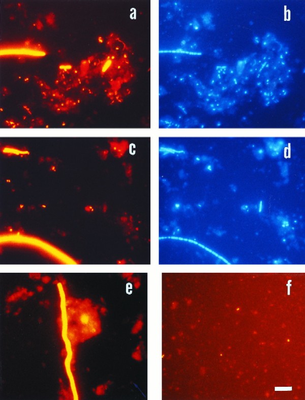
Epifluorescence micrographs of bacteria in sediment samples from the Jadebusen Bay of the German Wadden Sea. (a) Hybridization with probe EUB338, specific for Bacteria. (b) Same microscopic field as in panel a with UV excitation (DAPI staining). (c and d) Identical microscopic fields with probe SRB385 (c) and DAPI staining (d). (e) Hybridization with probe DNMA657, specific for Desulfonema. (f) Specific hybridization for Arcobacter with probe ARC94. Bar, 10 μm (applies to all panels).
References
-
- Cammen L M. Annual bacterial production in relation to benthic microalgal production and sediment oxygen uptake in an intertidal sandflat and an intertidal mudflat. Mar Ecol Prog Ser. 1991;71:13–25.
LinkOut - more resources
Full Text Sources
Other Literature Sources
