. 1998 Jul 1;12(13):1986-97.
doi: 10.1101/gad.12.13.1986.

Identification of Xenopus SMC protein complexes required for sister chromatid cohesion

Affiliations

Identification of Xenopus SMC protein complexes required for sister chromatid cohesion

A Losada et al. Genes Dev. .

Abstract

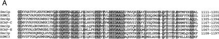
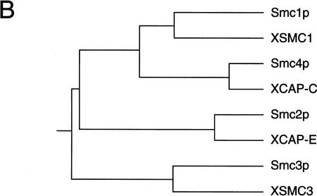
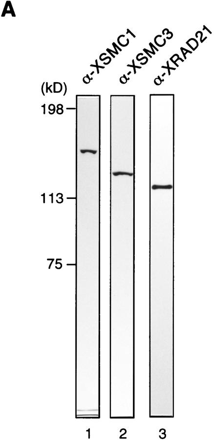
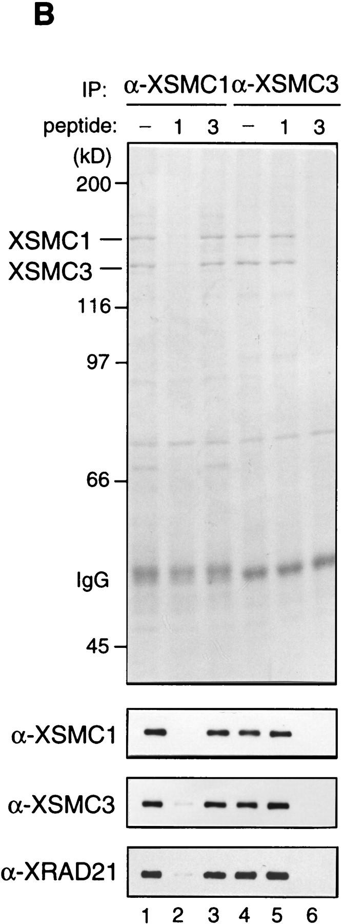
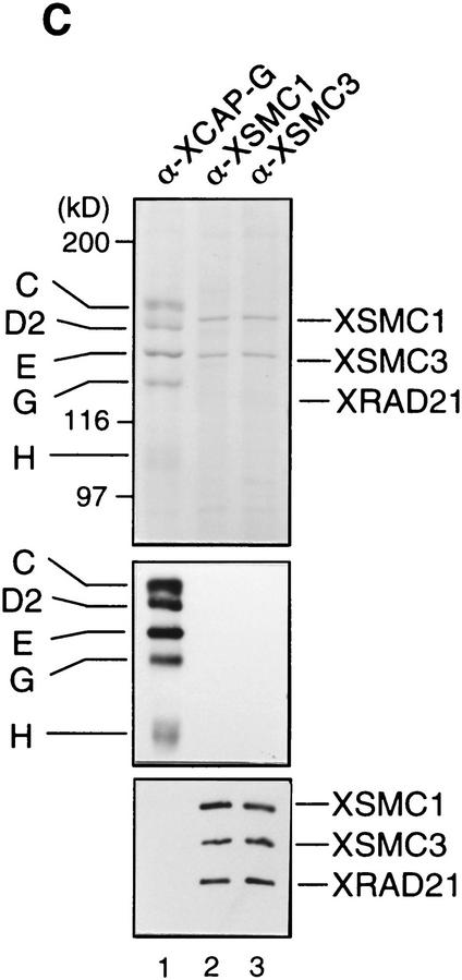
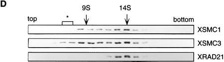
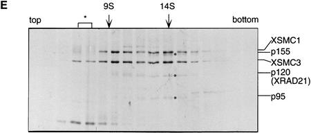
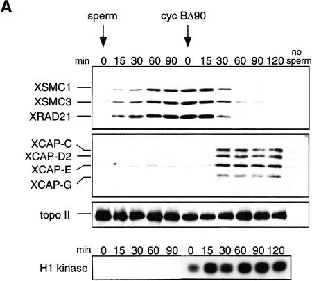
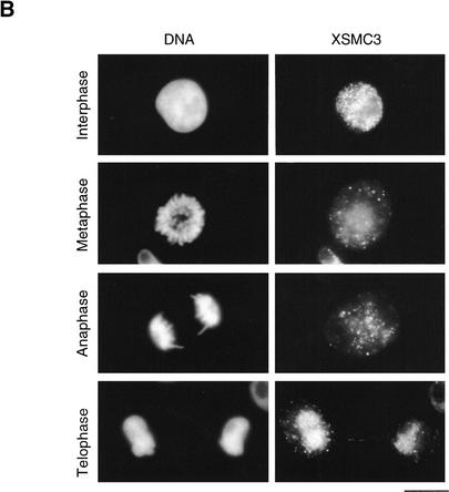
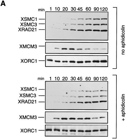
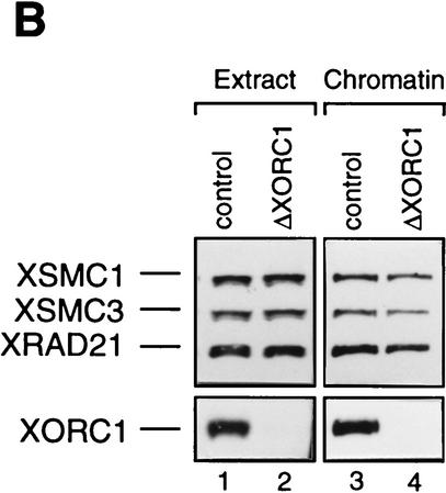
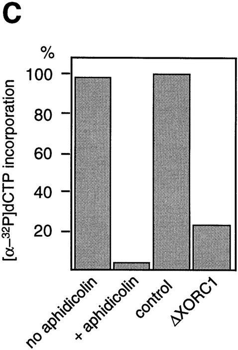
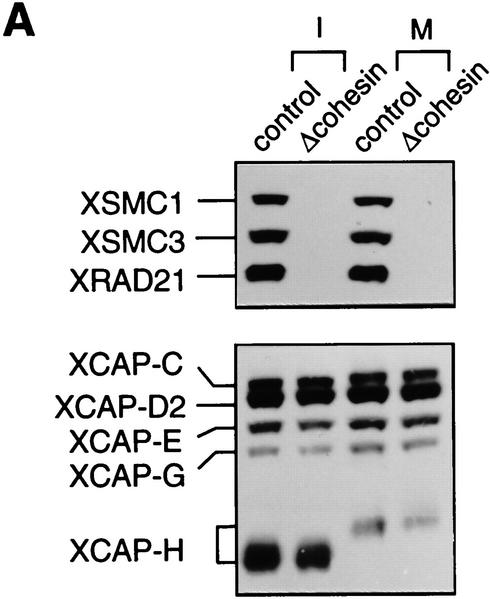
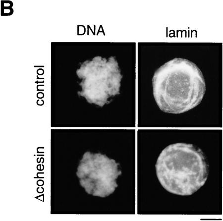
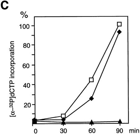
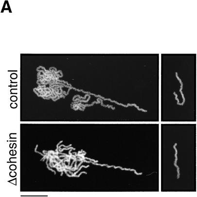
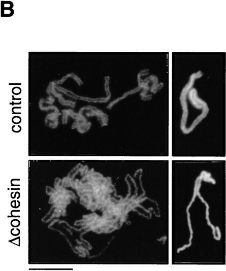
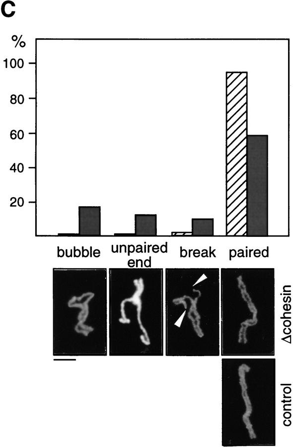
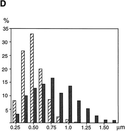
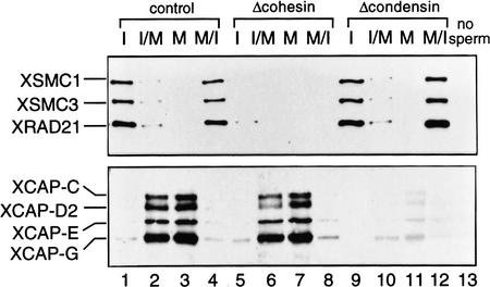
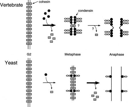
The structural maintenance of chromosomes (SMC) family is a growing family of chromosomal ATPases. The founding class of SMC protein complexes, condensins, plays a central role in mitotic chromosome condensation. We report here a new class of SMC protein complexes containing XSMC1 and XSMC3, Xenopus homologs of yeast Smc1p and Smc3p, respectively. The protein complexes (termed cohesins) exist as two major forms with sedimentation coefficients of 9S and 14S. 9S cohesin is a heterodimer of XSMC1 and XSMC3, whereas 14S cohesin contains three additional subunits. One of them has been identified as a Xenopus homolog of the Schizosaccharomyces pombe Rad21p implicated in DNA repair and the Saccharomyces cerevisiae Scc1p/Mcd1p implicated in sister chromatid cohesion. 14S cohesin binds to interphase chromatin independently of DNA replication and dissociates from it at the onset of mitosis. Immunodepletion of cohesins during interphase causes defects in sister chromatid cohesion in subsequent mitosis, whereas condensation is unaffected. These results suggest that proper assembly of mitotic chromosomes is regulated by two distinct classes of SMC protein complexes, cohesins and condensins.


Figures

Figure 1
Figure 1
Four SMC subtypes in yeast and Xenopus. (A) Sequence alignment of four SMC subtypes within the DA-box region. Yeast sequences, Smc1p, Smc2p, Smc3p, and Smc4p; Xenopus sequences, XSMC1, XCAP-E, XSMC3, and XCAP-C. (B) A phylogenetic tree prepared on the basis of the DA-box sequences. The program GeneWorks (IntelliGenetics) was used for sequence analysis.
Figure 1
Figure 1
Four SMC subtypes in yeast and Xenopus. (A) Sequence alignment of four SMC subtypes within the DA-box region. Yeast sequences, Smc1p, Smc2p, Smc3p, and Smc4p; Xenopus sequences, XSMC1, XCAP-E, XSMC3, and XCAP-C. (B) A phylogenetic tree prepared on the basis of the DA-box sequences. The program GeneWorks (IntelliGenetics) was used for sequence analysis.
Figure 2
Figure 2
Biochemical characterization of protein complexes containing XSMC1 and XSMC3. (A) Immunoblot analysis of an egg extract with affinity-purified antibodies against XSMC1 (lane 1), XSMC3 (lane 2), or XRAD21 (lane 3). (B) Coimmunoprecipitation of XSMC1 and XSMC3. Aliquots of an interphase high-speed supernatant were incubated with anti-XSMC1 (lanes 1–3) or anti-XSMC3 (lanes 4–6) antibodies raised against the carboxy-terminal peptide sequences. The antigen peptide for XSMC1 (lanes 2,5) or XSMC3 (lanes 3,6) was added (0.4 mg/ml) to demonstrate the specificity of immunoprecipitation reactions. Immunoprecipitates were separated by SDS-PAGE followed by Coomassie blue stain (top) or by immunoblotting (bottom). (C) Independent immunoprecipitation of condensin and cohesin subunits. Immunoprecipitates obtained with anti-XCAP-G (lane 1), anti-XSMC1 (lane 2), or anti-XSMC3 (lane 3) were analyzed by Coomassie blue stain (top) or by immunoblotting (middle and bottom). (D) Sucrose gradient centrifugation of a total extract. An interphase high-speed supernatant was fractionated in a 5%–20% sucrose gradient, and fractions were analyzed by immunoblotting with anti-XSMC1, anti-XSMC3, and anti-XRAD21 antibodies. The two major peaks of 9S and 14S are indicated. A minor population of free XSMC3 is indicated by the asterisk. (E) Sucrose gradient centrifugation of an affinity-purified fraction. The cohesin complexes were affinity purified with anti-XSMC3 antibody and fractionated in a 5%–20% sucrose gradient. Fractions were TCA-precipitated, separated by SDS-PAGE, and stained with silver. Asterisks indicate positions of the bands corresponding to p155, p120, and p95 that cofractionate with XSMC1 and XSMC3 in the 14S peak.
Figure 2
Figure 2
Biochemical characterization of protein complexes containing XSMC1 and XSMC3. (A) Immunoblot analysis of an egg extract with affinity-purified antibodies against XSMC1 (lane 1), XSMC3 (lane 2), or XRAD21 (lane 3). (B) Coimmunoprecipitation of XSMC1 and XSMC3. Aliquots of an interphase high-speed supernatant were incubated with anti-XSMC1 (lanes 1–3) or anti-XSMC3 (lanes 4–6) antibodies raised against the carboxy-terminal peptide sequences. The antigen peptide for XSMC1 (lanes 2,5) or XSMC3 (lanes 3,6) was added (0.4 mg/ml) to demonstrate the specificity of immunoprecipitation reactions. Immunoprecipitates were separated by SDS-PAGE followed by Coomassie blue stain (top) or by immunoblotting (bottom). (C) Independent immunoprecipitation of condensin and cohesin subunits. Immunoprecipitates obtained with anti-XCAP-G (lane 1), anti-XSMC1 (lane 2), or anti-XSMC3 (lane 3) were analyzed by Coomassie blue stain (top) or by immunoblotting (middle and bottom). (D) Sucrose gradient centrifugation of a total extract. An interphase high-speed supernatant was fractionated in a 5%–20% sucrose gradient, and fractions were analyzed by immunoblotting with anti-XSMC1, anti-XSMC3, and anti-XRAD21 antibodies. The two major peaks of 9S and 14S are indicated. A minor population of free XSMC3 is indicated by the asterisk. (E) Sucrose gradient centrifugation of an affinity-purified fraction. The cohesin complexes were affinity purified with anti-XSMC3 antibody and fractionated in a 5%–20% sucrose gradient. Fractions were TCA-precipitated, separated by SDS-PAGE, and stained with silver. Asterisks indicate positions of the bands corresponding to p155, p120, and p95 that cofractionate with XSMC1 and XSMC3 in the 14S peak.
Figure 3
Figure 3
Cell cycle-dependent chromosomal targeting of cohesins in vivo and in vitro. (A) Cell cycle-dependent chromosomal targeting of cohesins and condensins. Sperm chromatin was incubated with an interphase low-speed supernatant for 90 min and then a non-degradable form of sea urchin cyclin B (cyc BΔ90) was added to drive the cell cycle into mitosis. Aliquots were taken at the indicated time points, and chromatin-bound protein fractions were isolated and analyzed by immunoblotting with antibodies against cohesin subunits (XSMC1, XSMC3, and XRAD21), condensin subunits (XCAP-C, XCAP-D2, XCAP-E, and XCAP-G) or topoisomerase IIα. The level of H1 kinase activity in the extract was also assayed. (B) Immunolocalization of XSMC3 in tissue culture cells. Xenopus XL177 tissue culture cells fixed with paraformaldehyde were stained with DAPI (left) and anti-XSMC3 (right). Bar, 10 μm.
Figure 3
Figure 3
Cell cycle-dependent chromosomal targeting of cohesins in vivo and in vitro. (A) Cell cycle-dependent chromosomal targeting of cohesins and condensins. Sperm chromatin was incubated with an interphase low-speed supernatant for 90 min and then a non-degradable form of sea urchin cyclin B (cyc BΔ90) was added to drive the cell cycle into mitosis. Aliquots were taken at the indicated time points, and chromatin-bound protein fractions were isolated and analyzed by immunoblotting with antibodies against cohesin subunits (XSMC1, XSMC3, and XRAD21), condensin subunits (XCAP-C, XCAP-D2, XCAP-E, and XCAP-G) or topoisomerase IIα. The level of H1 kinase activity in the extract was also assayed. (B) Immunolocalization of XSMC3 in tissue culture cells. Xenopus XL177 tissue culture cells fixed with paraformaldehyde were stained with DAPI (left) and anti-XSMC3 (right). Bar, 10 μm.
Figure 4
Figure 4
Cohesin subunits associate with chromatin independently of DNA replication. (A) Effect of aphidicolin treatment on the association of cohesins with chromatin. Sperm chromatin was incubated with interphase low-speed supernatants in the absence (top) or presence of aphidicolin (bottom). At the indicated times after sperm addition, aliquots were removed from the extracts and chromatin-bound proteins were analyzed by immunoblotting with antibodies against the cohesin subunits, XMCM3 and XORC1. (B) Effect of XORC depletion on the association of cohesins with chromatin. Interphase extracts were immunodepleted with a control serum (lanes 1,3) or an anti-XORC1 serum (lanes 2,4). Extracts (lanes 1,2) or chromatin-bound fractions (lanes 3,4) were analyzed by immunoblotting with antibodies against the cohesin subunits or XORC1. (C) DNA replication in aphidicolin-treated or XORC-depleted extracts. DNA replication in the extracts was assayed by measuring the incorporation of [α-32P] dCTP into sperm chromatin. DNA synthesis is expressed as a percentage of the synthesis achieved in the control-depleted extract.
Figure 4
Figure 4
Cohesin subunits associate with chromatin independently of DNA replication. (A) Effect of aphidicolin treatment on the association of cohesins with chromatin. Sperm chromatin was incubated with interphase low-speed supernatants in the absence (top) or presence of aphidicolin (bottom). At the indicated times after sperm addition, aliquots were removed from the extracts and chromatin-bound proteins were analyzed by immunoblotting with antibodies against the cohesin subunits, XMCM3 and XORC1. (B) Effect of XORC depletion on the association of cohesins with chromatin. Interphase extracts were immunodepleted with a control serum (lanes 1,3) or an anti-XORC1 serum (lanes 2,4). Extracts (lanes 1,2) or chromatin-bound fractions (lanes 3,4) were analyzed by immunoblotting with antibodies against the cohesin subunits or XORC1. (C) DNA replication in aphidicolin-treated or XORC-depleted extracts. DNA replication in the extracts was assayed by measuring the incorporation of [α-32P] dCTP into sperm chromatin. DNA synthesis is expressed as a percentage of the synthesis achieved in the control-depleted extract.
Figure 5
Figure 5
Normal interphase nuclear functions in the absence of cohesins. (A) Immunodepletion of cohesins. Interphase (I) or CSF-arrested (M) extracts were immunodepleted with control IgG (control; lanes 1,3) or a mixture of anti-XSMC1 and anti-XSMC3 (Δcohesin; lanes 2,4). The extracts were analyzed by immunoblotting with antibodies against the cohesin subunits (XSMC1, XSMC3, and XRAD21) or the condensin subunits (XCAP-C, XCAP-D2, XCAP-E, XCAP-G, and XCAP-H). The CSF-arrested extracts were used in the chromosome assembly assays described in Fig. 6. (B) Nuclear assembly in the absence of cohesins. Sperm chromatin was incubated in the cohesin-depleted (Δcohesin) or control extracts at 22°C for 2 hr, fixed, and stained with DAPI (DNA) or antilamin LIII antibody (lamin). Bar, 10 μm. (C) DNA replication in the absence of cohesins. DNA replication in the cohesin-depleted (♦) or control (□) extracts was assayed as in Fig. 4C. Incorporation of [α-32P]dCTP was measured after a 2 hr-incubation of the sperm chromatin with the corresponding extract. DNA replication in an aphidicolin-treated extract (▴) is also shown as a control.
Figure 5
Figure 5
Normal interphase nuclear functions in the absence of cohesins. (A) Immunodepletion of cohesins. Interphase (I) or CSF-arrested (M) extracts were immunodepleted with control IgG (control; lanes 1,3) or a mixture of anti-XSMC1 and anti-XSMC3 (Δcohesin; lanes 2,4). The extracts were analyzed by immunoblotting with antibodies against the cohesin subunits (XSMC1, XSMC3, and XRAD21) or the condensin subunits (XCAP-C, XCAP-D2, XCAP-E, XCAP-G, and XCAP-H). The CSF-arrested extracts were used in the chromosome assembly assays described in Fig. 6. (B) Nuclear assembly in the absence of cohesins. Sperm chromatin was incubated in the cohesin-depleted (Δcohesin) or control extracts at 22°C for 2 hr, fixed, and stained with DAPI (DNA) or antilamin LIII antibody (lamin). Bar, 10 μm. (C) DNA replication in the absence of cohesins. DNA replication in the cohesin-depleted (♦) or control (□) extracts was assayed as in Fig. 4C. Incorporation of [α-32P]dCTP was measured after a 2 hr-incubation of the sperm chromatin with the corresponding extract. DNA replication in an aphidicolin-treated extract (▴) is also shown as a control.
Figure 6
Figure 6
Cohesins are required for sister chromatid cohesion but not for condensation. (A) Single chromatid assembly assay. Sperm chromatin was incubated with CSF-arrested high-speed supernatants (cohesin-depleted or control), fixed, and stained with anti-XCAP-E. Individual chromosomes are shown on the right. Bar, 10 μm. (B) Double-chromatid assembly assay. Sperm chromatin was incubated with interphase extracts (cohesin-depleted or control) to allow nuclear assembly and DNA replication. The cohesin-depleted or control nuclei were converted into mitotic chromosomes by the addition of cohesin-depleted or control CSF-arrested extracts, respectively. The chromosomes were stained as described in A. Bar, 10 μm. (C) Classification of defective phenotypes. Chromosome morphology was classified into four groups: bubble (an unpaired region somewhere along the chromosome); unpaired end (an unpaired region at one end of the chromosome); break (a double-strand break in one of the chromatids); paired (chromatids paired along the length of the chromosome). (Hatched bars) Control; (solid bars) Δcohesin. Bar, 5 μm. (D) Distance between sister chromatids in paired chromosomes. Chromosomes formed in control (hatched bars) or cohesin-depleted extracts (solid bars) that were classified in the paired group were randomly selected, and the distance between the two sister chromatids measured at regular intervals along the entire length of the chromosomes (see Materials andMethods). The results were plotted as number of times (expressed as a percentage of the total number of measurements made, y-axis) in which the distance between the sister chromatids corresponded to a given value (distance in micrometers, x-axis). The mean values and standard deviations for each case are indicated in the legend.
Figure 6
Figure 6
Cohesins are required for sister chromatid cohesion but not for condensation. (A) Single chromatid assembly assay. Sperm chromatin was incubated with CSF-arrested high-speed supernatants (cohesin-depleted or control), fixed, and stained with anti-XCAP-E. Individual chromosomes are shown on the right. Bar, 10 μm. (B) Double-chromatid assembly assay. Sperm chromatin was incubated with interphase extracts (cohesin-depleted or control) to allow nuclear assembly and DNA replication. The cohesin-depleted or control nuclei were converted into mitotic chromosomes by the addition of cohesin-depleted or control CSF-arrested extracts, respectively. The chromosomes were stained as described in A. Bar, 10 μm. (C) Classification of defective phenotypes. Chromosome morphology was classified into four groups: bubble (an unpaired region somewhere along the chromosome); unpaired end (an unpaired region at one end of the chromosome); break (a double-strand break in one of the chromatids); paired (chromatids paired along the length of the chromosome). (Hatched bars) Control; (solid bars) Δcohesin. Bar, 5 μm. (D) Distance between sister chromatids in paired chromosomes. Chromosomes formed in control (hatched bars) or cohesin-depleted extracts (solid bars) that were classified in the paired group were randomly selected, and the distance between the two sister chromatids measured at regular intervals along the entire length of the chromosomes (see Materials andMethods). The results were plotted as number of times (expressed as a percentage of the total number of measurements made, y-axis) in which the distance between the sister chromatids corresponded to a given value (distance in micrometers, x-axis). The mean values and standard deviations for each case are indicated in the legend.
Figure 7
Figure 7
Independent chromosomal association and dissociation of cohesins and condensins in Xenopus egg extracts. Sperm nuclei were incubated with interphase extracts (lanes 1,5,9), or with interphase extract followed by addition of a half volume of CSF-arrested extract to enter mitosis (lanes 2,6,10). Alternatively, sperm nuclei were incubated with CSF-arrested extracts (lanes 3,7,11), or with CSF-arrested extracts followed by addition of 0.4 mm CaCl2, which promotes exit from mitosis and entry into interphase (lanes 4,8,12). Chromatin fractions were isolated and analyzed by immunoblotting. Lanes 1–4, control; lanes 5–8, cohesin depletion; lanes 9–12, condensin depletion; lane 13, a control without sperm.
Figure 8
Figure 8
Sister chromatid cohesion and condensation in vertebrate and yeast chromosomes. Hypothetical chromosome architecture of vertebrate (top) and yeast (bottom) chromosome is shown. In vertebrates, the major transition occurs at the onset of mitosis (G2 to metaphase) when cohesion is largely reorganized and chromatids fully condense (top). In yeast, the structural reorganization of chromosomes takes place primarily at the metaphase–anaphase transition (bottom). Cohesins and condensins are shown by rectangles and circles, respectively. The single cohesion site drawn on the vertebrate metaphase chromosome by no means imply that cohesion is restricted to a centromeric region.

Similar articles

Cited by

References

    1. Benavente R, Krohne G, Franke WW. Cell type-specific expression of nuclear lamina proteins during development of Xenopus laevis. Cell. 1985;41:177–790. - PubMed
    1. Birkenbihl RP, Subramani S. Cloning and characterization of rad21, an essential gene of Schizosaccharomyces pombe involved in DNA double-strand-break repair. Nucleic Acids Res. 1992;20:6605–6611. - PMC - PubMed
    1. Blow JJ, Laskey RA. Initiation of DNA replication in nuclei and purified DNA by a cell-free extract of Xenopus eggs. Cell. 1986;47:577–587. - PubMed
    1. Castaño IB, Brzoska PM, Sadoff BU, Chen H, Christman MF. Mitotic chromosome condensation in the rDNA requires TRF4 and DNA topoisomerase I in Saccharomyces cerevisiae. Genes & Dev. 1996;10:2564–2576. - PubMed
    1. Coleman TR, Carpenter PB, Dunphy WG. The Xenopus cdc6 protein is essential for the initiation of a single round of DNA replication in cell-free extracts. Cell. 1996;87:53–63. - PubMed

Publication types

MeSH terms

Associated data