. 1998 Jul 1;12(13):2022-35.
doi: 10.1101/gad.12.13.2022.

Functional intertwining of Dpp and EGFR signaling during Drosophila endoderm induction

Affiliations

Functional intertwining of Dpp and EGFR signaling during Drosophila endoderm induction

D Szüts et al. Genes Dev. .

Abstract

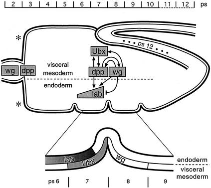
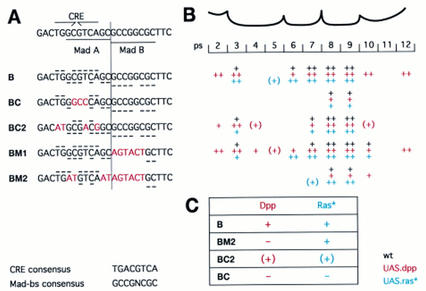
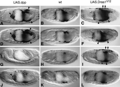
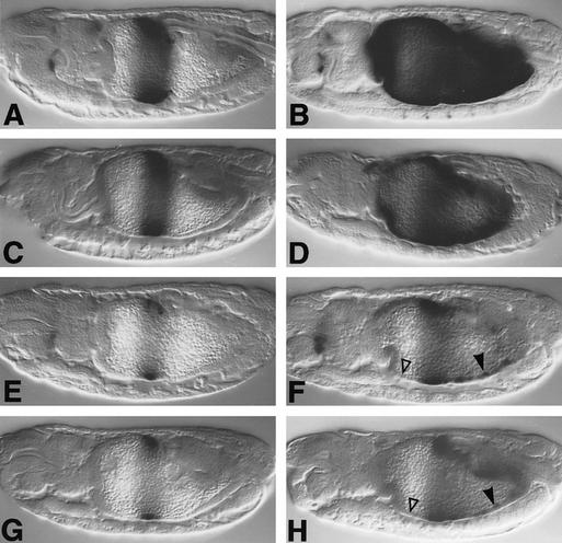
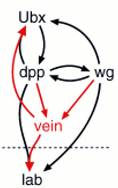
Endoderm induction in Drosophila is mediated by the extracellular signals Decapentaplegic (Dpp) and Wingless (Wg). We discovered a secondary signal with a permissive role in this process, namely Vein, a neuregulin-like ligand that stimulates the epidermal growth factor receptor (EGFR) and Ras signaling. Dpp and Wg up-regulate vein expression in the midgut mesoderm in two regions overlapping the Dpp sources. Experiments based on lack of function and ectopic stimulation of Dpp and EGFR signaling show that these two pathways are functionally interdependent and that they synergize with each other, revealing functional intertwining. The transcriptional response elements for the Dpp signal in midgut enhancers from homeotic target genes are bipartite, comprising CRE sites as well as binding sites for the Dpp signal-transducing protein Mad. Of these sites, the CRE seems to function primarily in the response to Ras, the secondary signal of Dpp. We discuss the potential significance of why an inductive process might use a secondary signal whose function is intertwined with that of the primary signal.


Figures

Figure 1
Figure 1
Endoderm induction. The embryonic midgut is drawn schematically in relation to parasegmental position (ps); outer cell layer, visceral mesoderm; inner cell layer, endoderm. The three constrictions are positioned at the junctions between ps5/ps6, ps7/ps8, and ps9/ps10; the proventriculus spans approximately ps2, and the gastric caeca are budding in ps3 [asterisks (*)]; the midgut ends posteriorly to ps12 (ps12 is stretched at this stage as indicated by dots). Within the midgut outline, the main genes mediating endoderm induction and their regulatory relationships are sketched out (note that D-Fos was omitted, for clarity; see Riese et al. 1997a). Underneath the drawing, the critical region of ps6-ps9 is blown up to highlight the spatial relationships of Ubx, Lab, and Wg expression (for references, see text).
Figure 2
Figure 2
Requirements of EGFR and vein during endoderm induction. Side views of 12- to 14-hr-old embryos stained with Lab (A–E) or Ubx antibody (F, G) or with lacZ antibody to visualize Ubx B expression (H,I). Wild type (A,F,H); flb1F26 homozygote (B); 48Y.GAL4/UAS.DN-DER (C); 24B.GAL4/UAS.DN-DER (G,I); veinγ4 homozygote (D); veinγ3/veinP1749 (E). Note the loss of Lab staining and the reduced Ubx staining (▵ in B–E, G) and the loss of Ubx B-mediated expression in ps6/ps7 and in ps3 (▵ in I), owing to lack of function of EGFR or vein. Anterior to the left, dorsal up (orientation the same in all figures).
Figure 3
Figure 3
Expression of vein transcripts in wild-type and mutant midguts. Side views of 12- to 15-hr-old embryos, after in situ hybridization to vein transcripts: Wild type (A), dpps4 homozygote (B), wgIL114 homozygote (C), 24B.GAL4/UAS.Dpp (D), 24B.GAL4/UAS.Wg (E); underneath each picture, a high-magnification view is shown of the corresponding genotype at a slightly older stage, to highlight the ventral ps3–ps10 region of the midgut. Maximal vein expression in the wild type is seen in posterior ps7, just anterior to the middle midgut constriction [indicated by asterisks (*) in A]. Note the reduction of vein expression in the ps7 and ps4 regions in dpp and wg mutants (▵ in B; in C, only the reduction in ps7 is indicated by ▵ as the reductive effect in ps4 of wg mutants is mild) and also the novel expression in the first constriction of dpp mutants (arrows in B). Additional vein expression is visible in the ps8–ps10 and ps12 regions after Dpp expression (arrowheads in D) and in ps2–ps7 after Wg expression throughout the midgut (arrowheads in E).
Figure 4
Figure 4
Interdependence and synergy between EGFR and Dpp signaling. Side views of 12- to 15-hr-old embryos stained with Lab antibody (A–D) or with LacZ antibody to visualize Ubx B expression (E–J): Wild type (A,E); 48Y.GAL4/UAS.Dpp (B); 48Y.GAL4/UAS.Dpp/UAS.DN-DER (C); 48Y.GAL4/UAS.Dpp/UAS.Dras1V12 (D); wild type (E); 24B.GAL4/UAS.DN-DER (F); 24B.GAL4/UAS.Dpp (G); 24B.GAL4/UAS.Dpp/UAS.DN-DER (H); 24B.GAL4/UAS.Dras1V12 (I); 24B.GAL4/UAS.Dpp/UAS.Dras1V12 (J). DN-DER antagonizes the stimulatory effects of Dpp in the anterior midgut (▵ in C,F,H; cf. with B,E,G, respectively), whereas Dras1V12 synergizes with Dpp [as judged by conspicuously strong and novel expression marked by arrowheads in D, J; cf. with the regions marked by asterisks (*) in B,G,I in which only low or no expression is seen in embryos expressing Dpp or Dras1V12 alone].
Figure 5
Figure 5
Targets for Vein/EGFR and Dpp signaling in the Ubx B enhancer. (A) Sequences of wild-type (B) and mutant enhancers (BC, BC2, BM1, BM2), underneath a sketch laying out the palindromic CRE and the two Mad binding sites (Mad A and Mad B); matches to the CRE or Mad binding site consensus sequences (given at bottom) are indicated with horizontal bars in wild-type and mutant enhancers (for CRE above, for Mad below sequences). (B) Activities of these enhancers in the various ps of the visceral mesoderm; black, wild type; red, 24B.GAL4/UAS.Dpp; blue, 24B.GAL4/UAS.Dras1V12. Levels of lacZ expression are estimated to be strong, ++, or weak, +; expression trailing into a ps is indicated by (+). Note that BM1 mediates additional expression in the wild type and in response to ectopic Dpp and Dras1V12, indicating a constitutive repressor binding to the Mad B sequence. (C) Summary of the results detailed in B, implicating the CRE as a response sequence for Ras signaling and Mad A as a response sequence for Dpp signaling (see also text); +, strong and consistent response; (+) disabled and, in the case of Ras, patchy response; − no response.
Figure 6
Figure 6
Responses of wild-type and mutant Ubx enhancers to Dpp and Dras1V12. Side views of 12- to 15-hr-old embryos, bearing Ubx B (A–C), BM1 (D–F), BM2 (G–I), or BC2 (J–L), stained with lacZ antibody; (left) 24B.GAL4/UAS.Dpp; (middle) wild type; (right) 24B.GAL4/UAS.Dras1V12. Additional staining is seen in response to ectopic Dpp in ps2, in ps9/ps10, and in ps12 (arrowheads in A,D; note that the staining in ps9/ps10 is partly attributable to derepression of endogenous Wg expression; see text); Dras1V12 produces increased staining mostly in ps6 but also in ps8/ps9 (arrowheads in C,F,I) and in the dorsal somatic mesoderm (arrows in C,I; dorsal mesoderm staining is also seen in BM1 embryos, but this is not visible in the focal plane of F).
Figure 7
Figure 7
Responses of wild-type and mutant lab enhancers to Dpp. Side views of 12- to 13-hr-old embryos, bearing lab HZ550 (A,B), 550M (C,D), 550C (E,F), or 550CM (G,H), stained with lacZ antibody; (left) wild type; (right) 48Y.GAL4/UAS.Dpp. Open triangles in F and H indicate complete loss of response to ectopic Dpp in the anterior midgut, whereas arrowheads point to residual response in ps8–ps10 (which may reflect a response to expanded Wg expression; see text).
Figure 8
Figure 8
Vein, a secondary signal in the inductive cascade: summary. Vein, the activating ligand of EGFR in the midgut, is a secondary signal whose expression is up-regulated by Dpp and Wg. Vein/EGFR signaling is required for lab induction in the endoderm (below broken line) and stimulates Ubx expression in the visceral mesoderm (above broken line). Its functional intertwining with Dpp signaling is indicated by merged arrows. Other stimulatory interactions between signaling pathways and responding genes are given by black arrows (most of these interactions are known to be direct, based on physical evidence; see text).

References

    1. Arora K, Dai H, Kazuko SG, Jamal J, O’Connor MB, Letsou A, Warrior R. The Drosophila schnurri gene acts in the Dpp/TGF-β signaling pathway and encodes a transcription factor homologous to the human MBP family. Cell. 1995;81:781–790. - PubMed
    1. Basler K, Struhl G. Compartment boundaries and the control of Drosophila limb pattern by Hedgehog protein. Nature. 1994;368:208–214. - PubMed
    1. Bienz M. Endoderm induction in Drosophila: The nuclear targets of the inducing signals. Curr Opin Genet Dev. 1997;7:683–688. - PubMed
    1. Bier E, Jan LY, Jan YN. rhomboid, a gene required for dorsoventral axis establishment and peripheral nervous system development in Drosophila melanogaster. Genes & Dev. 1990;4:190–203. - PubMed
    1. Brand AH, Perrimon N. Targeted gene expression as a means of altering cell fates and generating dominant phenotypes. Development. 1993;118:401–415. - PubMed

Publication types

MeSH terms

LinkOut - more resources