. 1998 Jul 1;12(13):2036-47.
doi: 10.1101/gad.12.13.2036.

Proneural gene self-stimulation in neural precursors: an essential mechanism for sense organ development that is regulated by Notch signaling

Affiliations

Proneural gene self-stimulation in neural precursors: an essential mechanism for sense organ development that is regulated by Notch signaling

J Culí et al. Genes Dev. .

Abstract

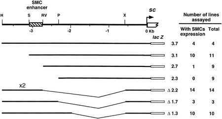
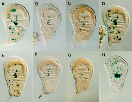
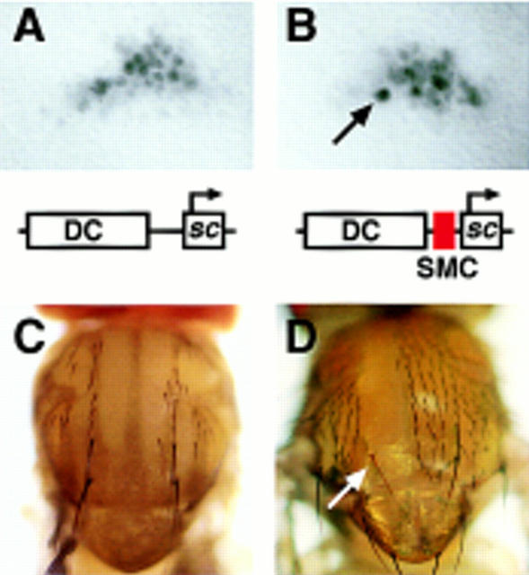
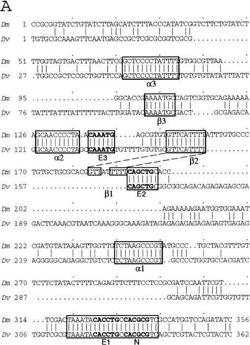
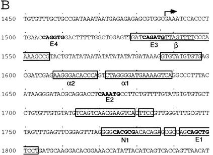
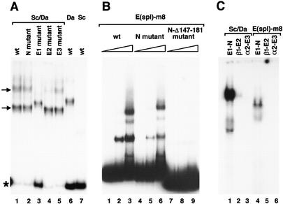
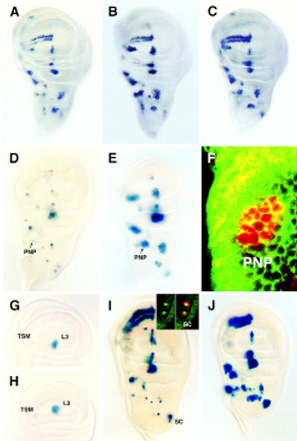
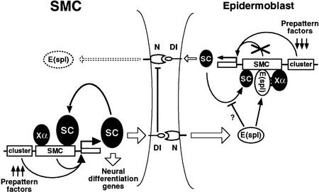
To learn about the acquisition of neural fate by ectodermal cells, we have analyzed a very early sign of neural commitment in Drosophila, namely the specific accumulation of achaete-scute complex (AS-C) proneural proteins in the cell that becomes a sensory organ mother cell (SMC). We have characterized an AS-C enhancer that directs expression specifically in SMCs. This enhancer promotes Scute protein accumulation in these cells, an event essential for sensory organ development in the absence of other AS-C genes. Interspecific sequence comparisons and site-directed mutagenesis show the presence of several conserved motifs necessary for enhancer action, some of them binding sites for proneural proteins. These and other data indicate that the enhancer mediates scute self-stimulation, although only in the presence of additional activating factors, which most likely interact with conserved motifs reminiscent of NF-kappaB-binding sites. Cells neighboring the SMC do not acquire the neural fate because the Notch signaling pathway effectors, the Enhancer of split bHLH proteins, block this proneural gene self-stimulatory loop, possibly by antagonizing the action on the enhancer of the NF-kappaB-like factors or the proneural proteins. These data suggest a mechanism for SMC committment.


Figures

Figure 1
Figure 1
Localization of the DNA harboring an SMC enhancer. (Top line) A map of the 3.7-kb sc upstream region. Only the restriction sites used to prepare deletions of the 3.7 sc fragment are shown. Restriction site nomenclature: (H) HindIII; (P) PstI; (RV) EcoRV; (S) SacII; (X) XbaI. In the following lines, the extent of the sc promoter fragments used to prepare the transgenes, the nomenclature of each transgene, the number of lines that showed expression in SMCs, and the total number of stained lines are indicated. Construct Δ2.2 contains a direct tandem repeat of the HindIII–EcoRV distal DNA fragment.
Figure 2
Figure 2
Expression of SRV–lacZ and several of its mutant forms in late third instar wing discs. (A) Expression of the unmodified SRV–lacZ transgene. (B–E) Expression of SRV–lacZ in which either the E1, E2, E3 or N box (see Fig. 4), respectively, have been mutated. (F,G) The α2 or the β2 box has been eliminated, respectively. Transgene mutated in the α3 was expressed as in F. Transgenes mutated in α1 and β3 had essentially wild-type expressions. Arrowheads in A, D, and E point at the posterior dorsocentral SMC. (H) Expression of SRV–lacZ in C-765; UAS–sc discs. The strong staining in the dorsal radius region is attributable to a large number of SMCs that differentiate in this region.
Figure 3
Figure 3
Expression of sc minigenes and their rescuing ability of the DC macrochaetae. A 5.8-kb DNA fragment containing the DC enhancer was linked to the sc structural gene, with and without the 356-bp sequence comprising the SMC enhancer, as schematically represented. (A,B) sc expression in the DC region of late third instar wing discs promoted by these minigenes, in the absence of the endogenous ac and sc genes [In(1)sc10.1 background], as detected by an anti-Sc antibody. Arrow points to a cell with high levels of Sc, most likely an SMC (Cubas et al. 1991; Skeath and Carroll 1991). No single cells with preferential Sc accumulation were detected with the minigene lacking the SMC enhancer. (C) Notum of an In(1)sc10.1 fly carrying the DC–sc minigene. The four DC macrochaetae and several microchaetae were normally rescued. The same rescuing activity was observed with the DC–SMC–sc minigene (2.06 and 1.95 chaetae/heminotum in 36 and 42 heminota examined for DC–SMC–sc and DC–sc, respectively). (D) Rescue of a y DC macrochaeta within a clone of cells homozygous for the Df(1)260-1, which removes the y gene and the entire AS-C, and carrying the DC–SMC–sc minigene. In these flies, 14 DC chaetae positions were found within homozygous Df(1)260-1 territories and 13 DC chaetae developed. In DC–sc flies, only 1 DC chaetae developed in 13 positions within the Df(1)260-1 homozygous territories.
Figure 4
Figure 4
Comparison of the D. melanogaster sc SMC enhancer and ase gene leader sequences with the corresponding genomic regions of D. virilis. (A) sc SMC enhancer sequences. The longest conserved regions are boxed. Within these three E boxes and one N box are marked in bold. (B) Sequences of the ase gene of D. melanogaster. Transcription starts at the arrow. Regions conserved in the homologous DNA of D. virilis are boxed. Note the presence of E, N, α, and β boxes within the conserved sequences. (C) Comparison of the α-like motifs from several genes of D. melanogaster (Dm) and D. virilis (Dv) with the consensus for NF-κB binding sites (Lenardo and Baltimore 1989). Nucleotides that fit the consensus are highlighted. (D) Similar comparison of β-like boxes in the sc and ase SMC enhancers.
Figure 4
Figure 4
Comparison of the D. melanogaster sc SMC enhancer and ase gene leader sequences with the corresponding genomic regions of D. virilis. (A) sc SMC enhancer sequences. The longest conserved regions are boxed. Within these three E boxes and one N box are marked in bold. (B) Sequences of the ase gene of D. melanogaster. Transcription starts at the arrow. Regions conserved in the homologous DNA of D. virilis are boxed. Note the presence of E, N, α, and β boxes within the conserved sequences. (C) Comparison of the α-like motifs from several genes of D. melanogaster (Dm) and D. virilis (Dv) with the consensus for NF-κB binding sites (Lenardo and Baltimore 1989). Nucleotides that fit the consensus are highlighted. (D) Similar comparison of β-like boxes in the sc and ase SMC enhancers.
Figure 5
Figure 5
Binding of Sc/Da heterodimers and E(spl)-m8 protein to SMC enhancer motifs. (A) Electrophoretic mobility-shift assays (EMSA) performed with a mixture of Sc and Da proteins (lanes 1–5) or with only Da (lane 6) or Sc (lane 7) proteins. DNA probe was the 356-bp enhancer in wild-type or mutant forms, as indicated. Arrows point at the complexes that probably contain one (lower band) and two Sc/Da heterodimers. Asterisk marks the free probe bands. (B) EMSA performed with 0, 1.3, and 6.6 μg of E(spl)-m8 protein and wild-type and mutant enhancer probes, as indicated. (C) EMSA carried out with mixtures of Sc and Da proteins (lanes 1–3) and with E(spl)-m8 protein (lanes 4–6). Oligonucleotide probes (see Materials and Methods) contained the boxes indicated.
Figure 6
Figure 6
Differential sensitivity of transcription directed by proneural cluster and SMC enhancers to N signaling. (A–C) Sc accumulation in wild-type, Nts, and da–Gal4/UAS–m8 imaginal wing discs, respectively. Note in B the presence of multiple cells with increased Sc accumulation, presumably SMCs, within proneural clusters. In C, overexpression of UAS–m8 did not modify appreciably Sc accumulation in proneural cluster cells, although it suppressed most SMCs. (D,E); SRV–lacZ expression in wild-type and Nts discs, respectively. Arrows point to postnotopleural (PNP) region. (F) Confocal section of a PNP cluster stained with anti-β-galactosidase antibody (red) and FITC–phalloidin (green) to visualize cell outlines. (G,H) lacZ expression driven by the L3/TSM enhancer in wild-type and Nts discs, respectively. Discs were understained (cf. with Fig. 5C of Gómez-Skarmeta et al. 1996) to better detect modifications in expression. (I,J) lacZ expression driven by minienhancers composed of four copies of an oligonucleotide containing E and α boxes or only E boxes, respectively. (Inset in I) Confocal image of the scutellar (SC) region of a disc carrying the complete minienhancer and showing that only single cells accumulate β-galactosidase (in cytoplasm, red). These cells are SMCs, as they also accumulate Ase protein (in nuclei, green).
Figure 7
Figure 7
Model for the regulation of sc in SMCs and neighboring epidermoblasts. In all proneural cluster cells, including the emerging SMC, sc transcription is activated by prepattern genes acting on proneural cluster-specific enhancers (Gómez-Skarmeta et al. 1996). This activation is largely independent of the N signaling pathway. Additionally in the SMC (left), the sc SMC-specific enhancer, by means of E boxes that bind Sc protein (as heterodimers with Da), promotes sc self-activation. The self-stimulatory loop also requires the unidentified activating factor Xα, which should bind to α boxes (NF-κB-like binding sites). The resulting increased Sc accumulation promotes strong activation of Dl in the SMC, which signals to the neighboring proneural cluster cells (right) and promotes in them the transcription of E(spl)-C genes. The E(spl)-C proteins prevent the sc self-stimulatory loop in these cells, which become epidermoblasts, by interacting with the Xα factor and, possibly, by complexing with Sc and Da proteins (Gigliani et al. 1996). (DNA sites capable of binding E(spl) proteins, although dispensable, may facilitate or stabilize their interaction with the Xα factor.) As a consequence, the Sc concentration remains low and the inhibitory signaling through Dl to the SMC will be weak. The fact that E(spl)-C proteins in SMCs are undetectable (Jennings et al. 1995) suggests the presence of a mechanism that reduces E(spl)-C transcription in SMCs. As proposed for dorsoventral boundary formation at the wing margin (de Celis et al. 1996b), sequestering of N molecules by the high concentration of active Dl in the SMC may block reception of signaling from the surrounding cells. In addition, low levels of N activation in the SMC may be compatible with lack of E(spl)-C transcription, as small amounts of Su(H) activator can be titrated by the constitutive levels of the Hairless antagonist (Bang et al. 1995).

References

    1. Alifragis P, Poortinga G, Parkhurst SM, Delidakis C. A network of interacting transcriptional regulators involved in Drosophila neural fate specification revealed by the yeast two-hybrid system. Proc Natl Acad Sci. 1997;94:13099–13104. - PMC - PubMed
    1. Arora K, Dai H, Kazuko SG, Jamal J, O’Connor MB, Letsou A, Warrior R. The Drosophila schnurri gene acts in the Dpp/TGFβ signalling pathway and encodes a transcription factor homologous to the human MBP family. Cell. 1995;81:781–790. - PubMed
    1. Artavanis-Tsakonas S, Simpson P. Choosing a cell fate: A view from the Notch locus. Trends Genet. 1991;7:403–408. - PubMed
    1. Bailey AM, Posakony JW. Suppressor of Hairless directly activates transcription of Enhancer of split complex genes in response to Notch receptor activity. Genes & Dev. 1995;9:2609–2622. - PubMed
    1. Bang AG, Bailey AM, Posakony JW. Hairless promotes stable commitment to the sensory organ precursor cell fate by negatively regulating the activity of the Notch signaling pathway. Dev Biol. 1995;172:479–494. - PubMed

Publication types

MeSH terms

Associated data

LinkOut - more resources