. 1998 Jul 1;12(13):2073-90.
doi: 10.1101/gad.12.13.2073.

New POU dimer configuration mediates antagonistic control of an osteopontin preimplantation enhancer by Oct-4 and Sox-2

Affiliations

New POU dimer configuration mediates antagonistic control of an osteopontin preimplantation enhancer by Oct-4 and Sox-2

V Botquin et al. Genes Dev. .

Abstract

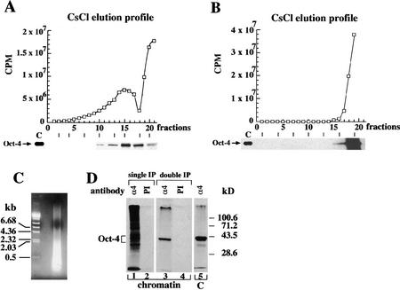
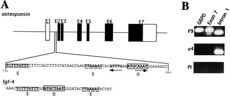
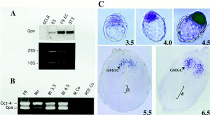
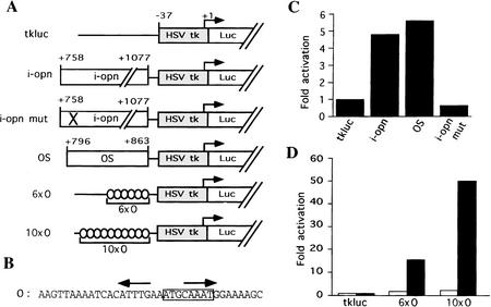
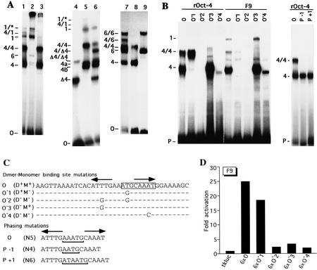
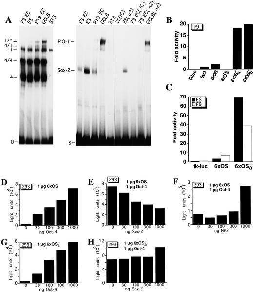
The POU transcription factor Oct-4 is expressed specifically in the germ line, pluripotent cells of the pregastrulation embryo and stem cell lines derived from the early embryo. Osteopontin (OPN) is a protein secreted by cells of the preimplantation embryo and contains a GRGDS motif that can bind to specific integrin subtypes and modulate cell adhesion/migration. We show that Oct-4 and OPN are coexpressed in the preimplantation mouse embryo and during differentiation of embryonal cell lines. Immunoprecipitation of the first intron of OPN (i-opn) from covalently fixed chromatin of embryonal stem cells by Oct-4-specific antibodies indicates that Oct-4 binds to this fragment in vivo. The i-opn fragment functions as an enhancer in cell lines that resemble cells of the preimplantation embryo. Furthermore, it contains a novel palindromic Oct factor recognition element (PORE) that is composed of an inverted pair of homeodomain-binding sites separated by exactly 5 bp (ATTTG +5 CAAAT). POU proteins can homo- and heterodimerize on the PORE in a configuration that has not been described previously. Strong transcriptional activation of the OPN element requires an intact PORE. In contrast, the canonical octamer overlapping with the downstream half of the PORE is not essential. Sox-2 is a transcription factor that contains an HMG box and is coexpressed with Oct-4 in the early mouse embryo. Sox-2 represses Oct-4 mediated activation of i-opn by way of a canonical Sox element that is located close to the PORE. Repression depends on a carboxy-terminal region of Sox-2 that is outside of the HMG box. Expression, DNA binding, and transactivation data are consistent with the hypothesis that OPN expression is regulated by Oct-4 and Sox-2 in preimplantation development.


Figures

Figure 1
Figure 1
Immunoprecipitation of Oct-4-containing chromatin of embryonal cells. (A) Protein elution profiles of isopyknic CsCl gradients of sheared cross-linked chromatin of P19 EC cells. (B) In parallel to equilibrium ultracentrifugation in CsCl the chromatin that was decross-linked before fractionation. In A and B CsCl density gradients were fractionated and the 35S levels (□) determined in each fraction. Fraction 1 is bottom of the gradient. The Oct-4 protein elution profile was obtained from aliquots of gradient fractions by hydrolyzing cross-links, and detected by 35S on SDS-PAGE. Lane C contains a control Oct-4 immunoprecipitate of 35S-labeled P19 EC protein extracts. Exposure of the Oct-4 profile in B is three times longer than in A to show that Oct-4 was only detected at the top of the gradient. (C) Cross-linked chromatin was physically fragmented to an average DNA size of 400 bp as determined by an agarose gel stained with ethidium bromide. (D) Immunoprecipitation of cross-linked chromatin with Oct-4-specific antibodies. All immunoprecipitations were performed with aliquots of fraction 15. 35S-Labeled proteins in precipitates were analyzed on 10% SDS-PAGE. (Lane 1) Immunoprecipitation with Oct-4 antibodies (α4); (lane 2) immunoprecipitation with preimmune (PI) antibodies; (lane 3) Oct-4 immunoprecipitated chromatin was hydrolyzed and subjected to a second round of immunoprecipitation with α4 antibodies; (lane 4) reverse cross-linked PI immunoprecipitated chromatin was hydrolyzed and subjected to a second round of immunoprecipitation with PI antibodies; (lane 5) α4-immunoprecipitates of 35S-labeled P19 EC protein extract. All immunoprecipitations were done with affinity-purified Oct-4 polyclonal antibodies.
Figure 2
Figure 2
OPN first intron is enriched in EC chromatin αOct-4 immunoprecipitates. (A) Two elements identified by a computer search for genes containing combination of potential binding sites for Oct-4 (O, boxed) and Sox-2 (S, underlined). An engrailed-like factor-binding site is represented by E, dashed line. Schematic organization of OPN gene and localization of the first intron. Solid boxes are coding regions; open boxes are untranslated regions. PORE sequence is represented by inverted arrows. (B) Enrichment of OPN first intron in αOct-4 EC chromatin immunoprecipitates. PCR was performed on F9 chromatin immunoprecipitated with αOct-4 antibodies (α4) or with preimmune antibodies (PI). PCR on purified F9 genomic DNA (F9) serves as a control for the PCR reactions. Primer pairs were selected to amplify a promoter region of G6PD and exon 7 and intron 1 fragments of OPN.
Figure 3
Figure 3
OPN expression in embryonal cell lines and pregastrulation embryos. (A) Northern blot analyses of OPN mRNA (Opn) expression in F9 EC, GCLB, MBL-1 ES, and fibroblast (3T3) cell lines. Each lane contains 1 μg of poly(A)+ RNA. mRNA levels were normalized by comparison of corresponding ethidium bromide-stained 28S and 18S rRNA bands. (B) RT–PCR of OPN and Oct-4 mRNA in different stages of preimplantation embryos. Multiplex PCR of Oct-4 and Opn was performed on reverse transcripts obtained from F9 cells, morulae (Mo), blastocysts of 3.5 dpc (Bl 3.5) and 4.5 dpc (Bl 4.5). PCR negative controls were done with H2O (PCR Co) and M2 medium treated with RNA extraction buffers and reverse transcription buffers (M Co). (C) In situ hybridization of 3.5–6.5 dpc embryos with OPN DIG-labeled mRNA. Blastocyst staining is observed in ICM and forming hypoblast, but not in trophectoderm and 4.5 dpc hypoblast. In sections of 5.5 and 6.5 dpc embryos only granulated metrial gland cells (GMGC) are stained. (E) embryo. Hybridization with sense probes did not generate staining (not shown).
Figure 4
Figure 4
OPN first intron functions as an EC-specific enhancer. (A) Reporter constructs. HSV tk minimal promoter cloned upstream of the luciferase gene (Luc); i-opn and i-opn mut are PCR fragments of the OPN first intron from +758 to +1077 wild-type and point mutated octamer motif, respectively; OS denotes OPN first intron from +796 to +863. 6 × O and 10 × O denote 6 or 10 copies of the O oligonucleotide, respectively. All fragments were cloned upstream of −37tkluc. (B) Oligonucleotide O represents a short fragment of i-opn that binds to Oct-4. The octamer motif is boxed and the PORE is represented by two inverted arrows. (C,D) F9 (solid bars) and 3T3 (open bars) cells were transfected transiently with 10 μg of respective reporter and 2 μg of human β-actin–LacZ used as an internal standard to normalize luciferase activity. Fold activation refers to the quotient of luciferase activities in the test and control (tkluc) construct.
Figure 5
Figure 5
Oct-4 binding to and transactivation by the PORE (A) EMSA demonstrating Oct-4 homo- and heterodimerization on a PORE-containing oligonucleotide (O). (Lane 1) F9 cell extracts; (lane 2) F9 cell extracts incubated with Oct-4 antibodies; (lane 3) F9 cell extracts incubated with Oct-1 antibodies; (lane 4) COS extract containing an Oct-4 protein lacking 98 amino acids at the amino terminus; (lane 5) F9 EC cell extract; (lane 6) mixture of extracts used in lanes 4 and 5; (lane 7) mixture of purified recombinant Oct-4 and Oct-6; (lane 8) recombinant Oct-4; (lane 9) recombinant Oct-6. (B) EMSA of recombinant Oct-4 (rOct-4) and F9 extracts (F9) with the O oligonucleotide and mutated versions thereof (see O) O−1, O−2, O−3, O−4, P−1, and P+1 in C). (P) Free probe. Complexes in A and B are as follows: (1) Oct-1 monomer; (4) Oct-4 monomer; the complexes labeled 4a and 4b correspond to Oct-4 monomer in mobility and are considered to represent two differentially phosphorylated forms of Oct-4 (V. Botquin, unpubl.). These bands are not clearly resolved in lanes 1 and 3 of A, lane O with F9 extracts of B and therefore, are merely labeled 4. (1/*) Oct-1 homo- or heterodimer; (4/4) Oct-4 homodimer; (Δ4/Δ4) homodimer of truncated Oct-4; (4/Δ4) heterodimer of full-length and truncated Oct-4; (6/6) Oct-6 homodimer; (4/1) heterodimer of Oct-4 and Oct-1; (4/6) heterodimer of Oct-4 and Oct-6. (C) Specific point and phasing mutations in O oligonucleotide to analyze binding and transactivation requirements in B and D. Point mutations were introduced at base pairs predicted to make central contacts with the POUS domain (O−1), the POUHD (O−3, O−4), or both (O−2). Oligonucleotides capable of binding Oct-4 as a dimer or a monomer are denoted by D+ and M+, respectively. (N) Number of nucleotides between the two half sites of the PORE. The octamer motif is boxed and the PORE is labeled by two inverted arrows. Dots represent nucleotides identical to those in oligonucleotide O. (D) Transactivation by mutated versions of oligonucleotide O. 6 × O, 6 × O−1, 6 × O−2, 6 × O−3, and 6 × O−4 are six copies of O, O−1, O−2, O−3, and O−4, respectively, inserted upstream of tkluc. All constructs contain a TATA box at similar distances from the insert. F9 EC cells were transfected transiently with 10 μg of respective reporter and 2 μg of human β-actin LacZ, which served as an internal standard to normalize luciferase activities for transfection efficiency. Fold activation refers to the quotient of luciferase activities in test and control (tkluc) constructs.
Figure 6
Figure 6
Sox-2 binds i-opn and represses Oct-4-mediated transactivation. (A) EMSA of EC cell (P19 and F9), ES cell, GCLB, and fibroblast (3T3) cell extracts on O and S oligonucleotides. Sox-2 binding was affected with either poly[d(I-C)] (IC) or Sox-2 antibody (α2). Complexes are as follows: (4) Oct-4 monomer; (4/4) Oct-4 homodimer; (4/1) heterodimer of Oct-1 and Oct-4; (1) Oct-1 monomer; (1/*) Oct-1 homo- or heterodimer; (PIO-1) protein binding to intron 1 of osteopontin). (B,C) Transient transfection assays in F9 EC and ES cells with reporters carrying octamer and Sox-binding sites. 6 × O represents a hexamer of oligonucleotide O (PORE only), 6 × OS a hexamer of OS (PORE and Sox-binding sites), 6 × OS a hexamer of OS (mutated PORE, intact Sox-2-binding site), 6 × OSa and 6 × OSb hexamers of OSa and OSb, respectively (intact PORE, mutated Sox-binding site). All hexamers were cloned upstream of tkluc. F9 EC and ES cells were transfected transiently with 10 μg of respective reporter and 2 μg of human β-actin–LacZ as an internal standard. Fold activation refers to the quotient of luciferase activities in test and control (6 × O) constructs. (D) Cotransfection assay in 293 cells of 1 μg of 6 × OS reporter and increasing amount of CMVOct-4 effector. (E) Cotransfection assay in 293 cells of 1 μg of 6 × OS reporter, constant amount of CMVOct-4 and increasing amounts of CMVSox-2 effector. (F) Cotransfection assay in 293 cells of 1 μg of 6 × OS reporter, constant amount of CMVOct-4 and increasing amount of CMVNP2 effector. (G) Cotransfection assay in 293 cells of 1 μg of 6 × OSa reporter and increasing amount of CMVOct-4 effector. (H) Cotransfection assay in 293 cells of 1 μg of 6 × OSa reporter, constant amount of CMVOct-4 and increasing amounts of CMVSox-2 effector. All luciferase activities were normalized for β-galactosidase expression. Light units refers to the units of luciferase counts.
Figure 7
Figure 7
Correlation of OPN, Oct-4, and Sox-2 expression during F9 and P19 differentiation. (A) F9 EC cells were induced to differentiate into parietal endoderm by exposing adherent cells to 10−1 μm all-trans retinoic acid (RA) (nonaggregates) or into visceral endoderm by exposing cells grown in suspension to 10−1 μm RA (aggregates). P19 were treated with 1 μm all-trans RA. Total RNA was extracted at day 0 (untreated F9 and P19 cells), day 1 and 2 of RA treatment. Northern blot analyses were performed for OPN, Oct-4, and 28S rRNA probes. Each lane contains 10 and 30 μg of total mRNA in F9 and P19 EC cells, respectively. The same filter was sequentially hybridized to OPN, Oct-4, and 28S rRNA probes. (B) EMSA of differentiating F9 and P19 EC cell extracts. Whole cell extracts at day 0 (untreated F9 and P19 EC cells), day 1 and 2 of RA treatment were incubated with the O and S oligonucleotide used as probe. Complexes are denoted as follows: (4) Oct-4 monomer; (4/4) Oct-4 homodimer; (4/1) heterodimer of Oct-1 and Oct-4; (1) Oct-1 monomer; (1/*) Oct-1 homo- or heterodimer. PIO-1 complex in P19 EC cells could be observed only after longer exposure.
Figure 7
Figure 7
Correlation of OPN, Oct-4, and Sox-2 expression during F9 and P19 differentiation. (A) F9 EC cells were induced to differentiate into parietal endoderm by exposing adherent cells to 10−1 μm all-trans retinoic acid (RA) (nonaggregates) or into visceral endoderm by exposing cells grown in suspension to 10−1 μm RA (aggregates). P19 were treated with 1 μm all-trans RA. Total RNA was extracted at day 0 (untreated F9 and P19 cells), day 1 and 2 of RA treatment. Northern blot analyses were performed for OPN, Oct-4, and 28S rRNA probes. Each lane contains 10 and 30 μg of total mRNA in F9 and P19 EC cells, respectively. The same filter was sequentially hybridized to OPN, Oct-4, and 28S rRNA probes. (B) EMSA of differentiating F9 and P19 EC cell extracts. Whole cell extracts at day 0 (untreated F9 and P19 EC cells), day 1 and 2 of RA treatment were incubated with the O and S oligonucleotide used as probe. Complexes are denoted as follows: (4) Oct-4 monomer; (4/4) Oct-4 homodimer; (4/1) heterodimer of Oct-1 and Oct-4; (1) Oct-1 monomer; (1/*) Oct-1 homo- or heterodimer. PIO-1 complex in P19 EC cells could be observed only after longer exposure.
Figure 8
Figure 8
Computer modeling of Oct-4 POU domain dimer conformation on the PORE binding sequence. (A,B) Lateral and frontal view of Oct-4 POU domain dimer conformation on the PORE binding site. Oct-4 POU domains were modeled into the coordinates of Oct-1. Each Oct-4 molecule has one color. The POUS domain has four α-helices (labeled 1–4) and the POUHD has three α-helices (labeled 1–3). DNA structure is represented by purple for phosphate, red for oxygen, green for carbon, and blue for nitrogen. A and B generated with WHAT IF software. (C) Amino acid sequence alignment of POU specific, linker, and POU homeodomain of Oct-1 and Oct-4. Amino acids numeration are taken from Herr and Cleary (1995). Dots represents identical amino acids found in Oct-4. α-Helices are indicated by boxes. (D) Schematic representation of Oct-4 and Pit-1 homodimers. Each molecule is given one color.

References

    1. Adamson ED. Cell-lineage-specific gene expression in development. In: Rossant J, Pederson RA, editors. Experimental approaches to mammalian embryonic development. Cambridge, UK: Cambridge University Press; 1986. pp. 321–364.
    1. Ambrosetti DC, Basilico C, Dailey L. Synergistic activation of the fibroblast growth factor 4 enhancer by Sox2 and Oct-3 depends on protein-protein interactions facilitated by a specific spatial arrangement of factor binding sites. Mol Cell Biol. 1997;17:6321–6329. - PMC - PubMed
    1. Behrendtsen O, Alexander CM, Werb Z. Cooperative interactions between extracellular matrix, integrins and parathyroid hormone-related peptide regulate parietal endoderm differentiation in mouse embryos. Development. 1995;121:4137–4148. - PubMed
    1. Caelles C, Hennemann H, Karin M. M-phase-specific phosphorylation of the POU transcription factor GHF-1 by a cell cycle-regulated protein kinase inhibits DNA binding. Mol Cell Biol. 1995;15:6694–6701. - PMC - PubMed
    1. Chinea G, Padron G, Hooft RW, Sander C, Vriend G. The use of position-specific rotamers in model building by homology. Proteins. 1995;23:415–421. - PubMed

MeSH terms