. 1998 Jul 15;18(14):5112-23.
doi: 10.1523/JNEUROSCI.18-14-05112.1998.

Neuroprotective actions of dipyridamole on cultured CNS neurons

Affiliations

Neuroprotective actions of dipyridamole on cultured CNS neurons

S E Farinelli et al. J Neurosci. .

Abstract

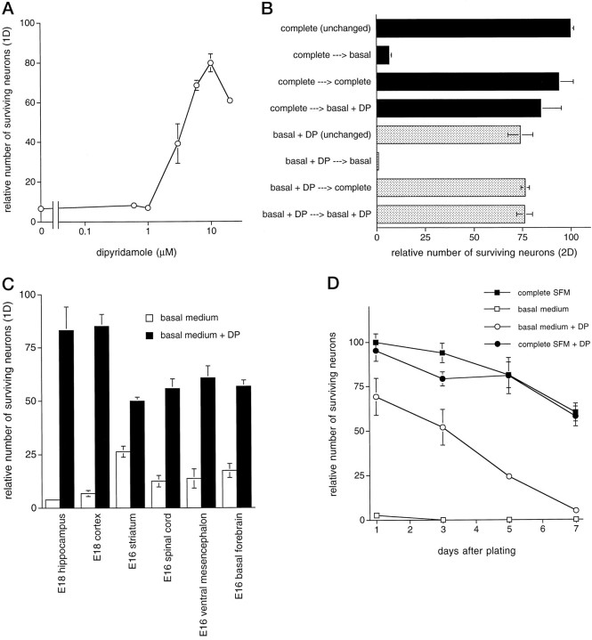
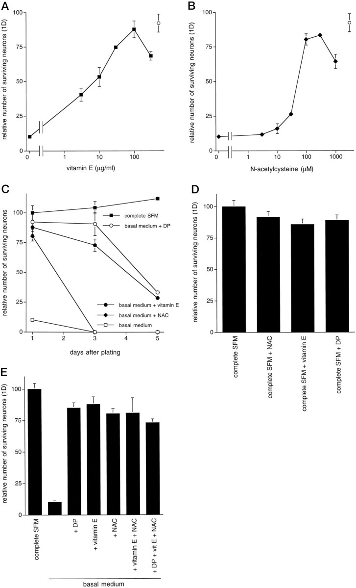
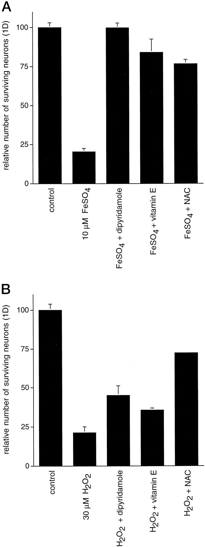
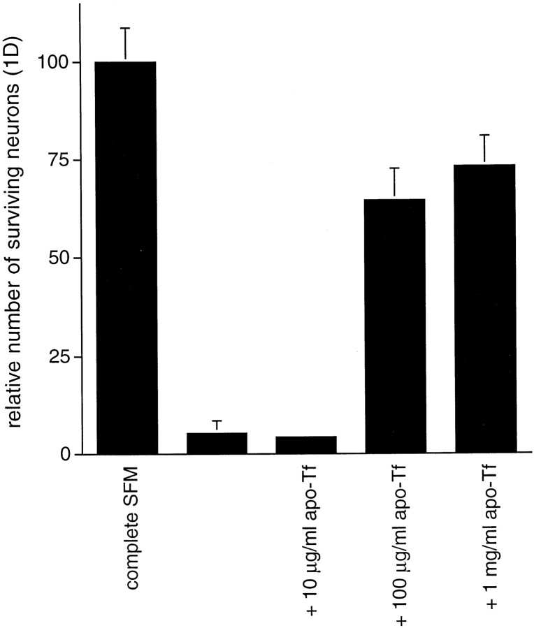
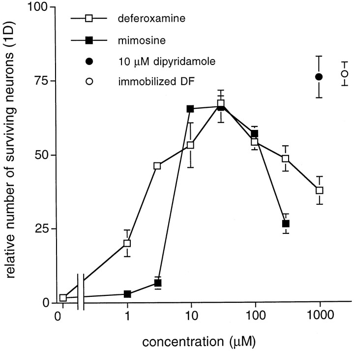
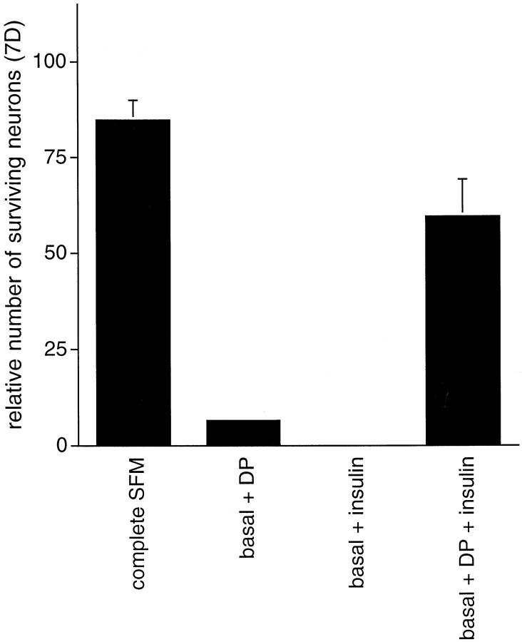
We report that dipyridamole is neuroprotective for a variety of rat embryonic CNS neurons cultured in serum-free basal medium lacking trophic factors or other additives. We also describe the mechanism underlying this action. Neurons died rapidly in basal medium but were rescued in large measure by 10 microM dipyridamole. The protective action of dipyridamole seems to be attributable to its antioxidant property. Vitamin E and N-acetylcysteine provided comparable neuroprotection in basal medium, whereas an array of compounds that mimic other actions of dipyridamole (inhibition of phosphodiesterases, blockade of nucleoside and chloride transport, interference with the multidrug resistance protein, and enhancement of prostacyclin synthesis) failed to promote survival. Thus, a major cause of neuronal death in this system seems to be oxidative stress that is relieved by dipyridamole. Iron plays a significant role in generation of such stress, as indicated by the observations that addition of apotransferrin or iron chelators to basal medium or use of iron-free medium also afforded protection. Although oxidative stress was a major determinant of neuronal death, it was not the only factor. Dipyridamole or other antioxidant measures did not provide sustained neuroprotection. However, provision of insulin, which was not protective alone in basal medium, along with dipyridamole significantly enhanced long-term neuronal survival. Hence, optimal protection requires both trophic support and relief from oxidative stress. These findings lend credence to the potential use of dipyridamole or its derivatives in prevention and/or treatment of CNS injuries and degenerative disorders in which oxidative stress is a significant component.


Figures

Fig. 1.
Fig. 1.
Phase contrast micrographs of E18 hippocampal neurons maintained in complete SFM for 24 hr (A), no additives (B), 10 μmdipyridamole (C), or 100 μg/ml (230 μm) vitamin E (D).
Fig. 2.
Fig. 2.
Dipyridamole promotes survival of primary CNS neurons in basal medium. A, Dose–response relationship for the effect of dipyridamole on survival of E18 hippocampal neurons in basal medium (1 d). Survival data are expressed relative to the number of neurons alive in complete SFM 1 d after plating (arbitrarily set at 100). B, Viability of dipyridamole-supported cells confirmed by responsiveness to complete SFM. Cells were cultured for 24 hr in basal medium with or without 10 μm dipyridamole or complete SFM, and the culture medium was then replaced as indicated. Cells maintained continuously in complete SFM or in basal medium containing 10 μmdipyridamole are shown for reference. Cell numbers were quantified 2 d after plating and expressed relative to the number of neurons in replicate cultures maintained continuously in complete SFM.C, Protection of neurons from several different brain regions by dipyridamole. Survival data are expressed relative to the number of neurons from the indicated brain region alive in complete SFM 1 d after plating. D, Time course. E18 hippocampal neurons were plated in the indicated media, and replicate cultures were assessed for surviving neurons at various times. No subsequent addition of dipyridamole was given. Data are the mean ± SEM of three samples. In this and subsequent figures, the apparent absence of error bars indicates that the error was smaller than thesymbol used. DP, Dipyridamole.
Fig. 3.
Fig. 3.
The antioxidants vitamin E and NAC promote survival of primary CNS neurons in basal medium. A,B, Dose–response relationships for the effect of vitamin E (A) and NAC (B) on promotion of survival of E18 hippocampal neurons in basal medium (1 d) are shown. The open circle represents survival mediated by 10 μm dipyridamole. Survival data are expressed relative to the number of neurons alive in complete SFM 1 d after plating (arbitrarily set at 100). C, Time course is shown. E18 hippocampal neurons were plated in either complete SFM or basal medium in the presence of the indicated additives. No subsequent addition of these agents was given. D, Antioxidants do not afford additional protection beyond that provided by complete SFM alone. E, Antioxidant effects on survival in basal medium are not additive and fail to increase dipyridamole-promoted survival. All survival data are expressed relative to the number of neurons alive in complete SFM 1 d after plating.
Fig. 4.
Fig. 4.
Dipyridamole, vitamin E, and NAC provide protection to E18 hippocampal neurons under conditions of oxidative stress. Neurons were plated in complete SFM and allowed to become established for 3 d. On the third day, cultures were treated with 10 μm FeSO4 (A) or 30 μm H2O2 (B) in complete SFM with the indicated additions. Cultures were not pretreated with the agents before addition of FeSO4 or H2O2. Cultures were assessed for survival 24 hr later. All survival data are expressed relative to the number of neurons in untreated control cultures.
Fig. 5.
Fig. 5.
Phase contrast micrographs of E18 hippocampal neurons maintained in complete SFM for 3 d followed by a 1 d treatment with no additives (complete SFM control) (A), 10 μm FeSO4(B), or 10 μm FeSO4plus 10 μm dipyridamole (C).
Fig. 6.
Fig. 6.
Transferrin promotes survival of primary hippocampal neurons in basal medium. Survival data are expressed relative to the number of neurons alive in complete SFM 1 d after plating (arbitrarily set at 100). Tf, Transferrin.
Fig. 7.
Fig. 7.
The iron chelators deferoxamine and mimosine promote survival of primary CNS neurons in basal medium. Dose–response relationships for the effects of deferoxamine and mimosine on survival of E18 hippocampal neurons in basal medium (1 d) are shown. Theclosed circle represents survival mediated by 10 μm dipyridamole. The open circle indicates survival in basal medium depleted of iron by immobilized deferoxamine (DF). All survival data are expressed relative to the number of neurons in complete SFM 1 d after plating.
Fig. 8.
Fig. 8.
Death of neurons is enhanced in basal medium (MEM/F12), compared with MEM, a culture medium lacking FeSO4. E18 hippocampal neurons were plated in the indicated medium and assessed for survival at daily intervals. A, Survival is enhanced in iron-free MEM compared with that in iron-containing basal medium and is diminished by the addition of
Fig. 9.
Fig. 9.
Trophic factors contribute to longer-term neuronal survival. E16 hippocampal neurons were plated in basal medium supplemented with either dipyridamole, insulin, or dipyridamole plus insulin and assessed for survival at 1 week. The survival data are expressed relative to the number of neurons alive in complete SFM 1 d after plating.

Similar articles

Cited by

References

    1. Aizenman Y, Weichsel ME, Jr, de Vellis J. Changes in insulin and transferrin requirements of pure brain neuronal cultures during embryonic development. Proc Natl Acad Sci USA. 1986;83:2263–2266. - PMC - PubMed
    1. Ayesh S, Shao YM, Stein WD. Co-operative, competitive and non-competitive interactions between modulators of p-glycoprotein. Biochim Biophys Acta. 1996;1316:8–18. - PubMed
    1. Barbin G, Selak I, Manthorpe M, Varon S. Use of central neuronal cultures for the detection of neuronotrophic agents. Neuroscience. 1984;12:33–43. - PubMed
    1. Batistatou A, Greene LA. Aurintricarboxylic acid rescues PC12 cells and sympathetic neurons from cell death caused by nerve growth factor deprivation: correlation with suppression of endonuclease activity. J Cell Biol. 1991;115:461–471. - PMC - PubMed
    1. Bottenstein JE, Sato GH. Growth of a rat neuroblastoma cell line in serum-free supplemented medium. Proc Natl Acad Sci USA. 1979;76:514–517. - PMC - PubMed

Publication types

LinkOut - more resources