. 1998 Jul 15;18(14):5136-50.
doi: 10.1523/JNEUROSCI.18-14-05136.1998.

Electrophysiological characterization of a putative supporting cell isolated from the frog taste disk

Affiliations

Electrophysiological characterization of a putative supporting cell isolated from the frog taste disk

A Bigiani et al. J Neurosci. .

Abstract

Chemosensory cells in vertebrate taste organs have two obvious specializations: an apical membrane with access to the tastants occurring in food, and synapses with sensory axons. In many species, however, certain differentiated taste cells have access to the tastants but lack any synaptic contacts with axons, and a supportive rather than chemosensory function has been attributed to them. Until now, no functional data are available for these taste cells. To begin to understand their role in taste organ physiology, we have characterized with patch-clamp recording techniques the electrophysiological properties of a putative supporting cell-the so-called wing cell-isolated from frog taste disks. Wing cells were distinguished from chemosensory elements by the presence of a typical, sheet-like apical process. Their resting potential was approximately -52 mV, and the average input resistance was 4.8 GOmega. Wing cells possessed voltage-gated Na+ currents sensitive to TTX, and an inactivating, voltage-gated K+ current sensitive to TEA. Current injections elicited single action potentials but not repetitive firing. We found no evidence for voltage-gated Ca2+ currents under various experimental conditions. Amiloride-sensitive Na+ channels, thought to be involved in Na+ chemotransduction, were present in wing cells. Many of the membrane properties of wing cells have been also reported for chemosensory taste cells. The presence of ion channels in wing cells might be suggestive of a role in controlling the microenvironment inside the taste organs or the functioning of chemosensory cells or both. In addition, they might participate directly in the sensory transduction events by allowing loop currents to flow inside the taste organs during chemostimulation.


Figures

Fig. 1.
Fig. 1.
Differential interference contrast photomicrographs of living taste cells isolated from the frog taste disk. A, A wing (supporting) cell characterized by its wide, sheet-like apical process. B, A chemosensory cell characterized by the rod-like apical process. Scale bar, 5 μm.
Fig. 2.
Fig. 2.
Whole-cell currents recorded from the wing cell shown in Figure 1A. The cell membrane was held at −85 mV and stepped in 10 mV increments from −75 to +45 mV. Capacitative and leakage currents were not subtracted from the records. Bath perfusion was APS; pipette solution was standard 105 mm KCl. After recording and adequate processing, this cell was observed at the scanning electron microscope to confirm its identity. From the photomicrograph on the right it is possible to recognize readily the typical wide process that characterizes wing cells. Scale bar, 5 μm.
Fig. 3.
Fig. 3.
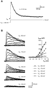
Voltage-gated Na+ currents in wing cells. A, Whole-cell currents elicited in an isolated wing cell by a series of depolarizing pulses between −75 and +35 mV, in 10 mV increments, from a holding potential of −85 mV. During bath perfusion with 0.5 μm TTX, the transient inward current was completely blocked, indicating that it was a Na+ current. Im, Membrane currents. Capacitative and leakage currents were not subtracted from the records. Pipette solution was standard 105 mm KCl. B, A family of Na+ currents recorded from an isolated wing cell. Currents (Im) were elicited by stepping the membrane held at −85 mV from −75 to +85 mV in 10 mV increments. Capacitative and leakage currents were subtracted from the records. Bath, APS; pipette solution, standard 105 mm CsCl. Current–voltage relationship for the peak transient inward current reveals that voltage-gated Na+ current activated at approximately −40 mV and reached a maximum value at approximately −15 mV in this wing cell.
Fig. 4.
Fig. 4.
Properties of the inactivation of voltage-gated Na+ currents in wing cells. A, Voltage-dependence of the steady-state inactivation. Maximal peak inward Na+ currents recorded after holding the membrane for 10 sec at −105, −95, −85, −75, and −65 mV (Vh). Note that the Na+ currents decreased at less negative potentials (inactivation). Capacitative and leakage currents were subtracted from the records. Bath solution, APS; pipette solution, standard 105 mm CsCl. In the steady-state inactivation–voltage relationship (bottom), the maximal peak Na+ currents (I) obtained for holding potentials between −105 and −45 mV were normalized with respect to that obtained withVh = −105 mV (Imax), and then plotted against the holding potential. Each point represents the mean ± SD of two to seven values from seven wing cells. Data were fitted by a sigmoid curve. The calculated half-maximal voltage (V0.5) was −70.7 mV, and the slope was 6.8 mV. B, Recovery from inactivation for voltage-gated Na+ currents at −85 mV. Na+ currents were elicited with a two-pulse protocol. The first 20 msec pulse moved the voltage from a holding potential of −85 to −15 mV. After a variable delay spent at −85 mV, a second 20 msec pulse of the same amplitude was applied to the cell membrane and the current was recorded. Current elicited by the second pulse was measured and normalized to the first preceding pulse and plotted against the interpulse interval. Data points could be fitted approximately by a single exponential function with a time constant of 18.6 msec for the cell shown here.
Fig. 5.
Fig. 5.
Voltage-gated K+ currents in wing cells. A, TEA sensitivity of the outward currents recorded from an isolated wing cell. Voltage-gated Na+ currents were blocked by adding 0.5 μm TTX to all solutions. Membrane currents (Im) were elicited by a series of depolarizing pulses between −75 and +35 mV, in 10 mV increments, from a holding potential of −85 mV. Capacitative and leakage currents were subtracted from the records. Outward currents recorded in regular APS (control, top) were totally abolished by 5 mm TEA (middle). The effect was reversible, as indicated by the recovery of the currents during washout (bottom). Pipette solution was standard 105 mm KCl. B, Voltage-dependence of K+ currents recorded from an isolated wing cell. A family of currents (Im) was elicited by stepping the cell membrane, held at −85 mV, from −75 to +45 mV in 10 mV increments. Capacitative and leakage currents were subtracted from the records. Bath perfusion was APS + 0.2 μm TTX to block voltage-gated Na+ currents. Pipette solution was standard 105 mm KCl. In theIV plot, both the peak value (•) and the value measured at the end of the voltage pulse (○) are shown. It can be noted that the peak current after reaching a maximal value between approximately 20 and 40 mV decreases with further depolarization.
Fig. 6.
Fig. 6.
Effect of Co2+ on the voltage-gated K+ currents recorded from an isolated wing cell. A, Sample currents elicited by a series of depolarizing voltage pulses from a holding potential of −85 mV when the cell was bathed in regular APS (top) and in the presence of 10 mm Co2+(bottom). TTX (0.5 μm) was added to all bath solutions to block voltage-gated Na+ channels. Pipette solution was standard 105 mm KCl. Capacitative and leakage currents were not subtracted from the records.B, IV plots for the peak value (circles) and the value at the end of the voltage pulses (squares) of the outward current elicited in regular APS (filled symbols) and during Co2+ application (open symbols). Co2+ caused the IVrelationship to shift in a positive direction.
Fig. 7.
Fig. 7.
Properties of the inactivation of voltage-gated K+ currents in wing cells. A, Time course of inactivation of the K+ current recorded from an isolated wing cell. The membrane was held at −85 mV and stepped for 200 msec to +15 mV. The decay of the current was fit (solid line) with a single exponential function with a time constant of 21.8 msec in this cell.Im, Membrane current.B, The effect of holding potential (Vh) on the inactivation of K+ currents in another wing cell. Currents were elicited by a series of depolarizing voltage pulses in 10 mV increments from different holding potentials (Vh: −85, −75, −55, −45, and −35 mV). All records are from the same cell. Bath solution was APS + 0.5 μm TTX to block voltage-gated Na+ channels. Pipette solution was standard 105 mm KCl. Capacitative and leakage currents were subtracted from the records. The corresponding IVplots for the peak K+ currents elicited at different holding potentials are shown on the right.
Fig. 8.
Fig. 8.
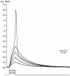
Action potential recorded from an isolated wing cell under current-clamp conditions. Cell membrane was held at approximately −80 mV, and brief depolarizing current pulses in 2 pA increments were injected. The first four pulses were subthreshold and failed to elicit action potentials. In this cell, the firing threshold was between −30 and −40 mV. Pipette solution was standard 105 mm KCl. Vm, Membrane potential.
Fig. 9.
Fig. 9.
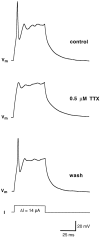
Contribution of sodium currents to action potential in wing cells. The cell was held at approximately −80 mV, and a 14 pA pulse of current was injected. In control conditions (bath solution: APS) an action potential was fired (top). In the presence of 0.5 μm TTX, the same stimulation protocol failed to elicit a spike-like action potential (middle). In voltage-clamp configuration, the inward Na+currents were completely abolished (data not shown). After the drug was washed out, wing cell membrane recovered the capability of firing action potential (bottom). Pipette solution was standard 105 mm KCl. Vm, Membrane potential.
Fig. 10.
Fig. 10.
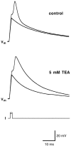
Contribution of potassium currents to the repolarization phase of the action potential in wing cells. Cell membrane was held at approximately −80 mV, and two current pulses were injected. The first pulse (70 pA) was subthreshold, whereas the second one (80 pA) elicited an action potential (top,control). When 5 mm TEA was applied, the action potential broadened considerably (bottom). In voltage-clamp configuration, the inactivating K+current was completely abolished (data not shown). Pipette solution was standard 105 mm KCl. Vm, Membrane potential.
Fig. 11.
Fig. 11.
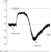
Response of whole-cell current to amiloride in an isolated wing cell. Cell membrane was held at −75 mV. Bath application of 40 μm amiloride in regular APS caused a rapid decrease in the stationary inward current (holding current) and in current noise. Note the undershooting transient (undershoot) on washout of amiloride. Pipette solution was standard 105 mm CsCl.Im, Membrane current.
Fig. 12.
Fig. 12.
Change in the input resistance of a wing cell during response to amiloride (30 μm). Input resistance was monitored by application of −20 mV steps from a holding potential (Vh) of −75 mV. Input resistance increased from 1.6 GΩ before amiloride application to 3.9 GΩ during the amiloride response, suggesting that membrane conductance was reduced. Note that during washout, cell input resistance recovered to the control value. Im, Membrane current.
Fig. 13.
Fig. 13.
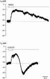
Effect of Na+-free saline and amiloride on the stationary inward current (Im) recorded from an isolated wing cell held at −60 mV. Replacing extracellular Na+with NMDG, a large impermeant cation, induced a decrease in the stationary inward current (top trace) similar to that elicited by bath-applying 30 μm amiloride to the same cell (bottom trace). However, time courses of the current recovery during washout were markedly different.
Fig. 14.
Fig. 14.
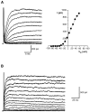
Whole-cell recordings from isolated rod cells lacking voltage-gated Na+ currents.A, Outward currents (left) elicited in regular APS by a series of depolarizing pulses between −75 and +55 mV, in 10 mV increments, from a holding potential of −85 mV. The corresponding IV plot (right) indicated that the outward current in this cell activated at approximately −40 mV. These recordings were obtained from the cell shown in Figure 1B. B, Outward currents recorded in another rod cell. Voltage protocol was the same as for the cell in A, but the pulse duration was 200 msec. It can be noted that in this cell the outward current did not inactivate significantly during the stimulation period (compare with the time course of inactivation shown in Fig. 7A). Capacitative and leakage currents were not subtracted in these recordings. Pipette solution was standard 105 mmKCl.
Fig. 15.
Fig. 15.
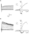
Whole-cell recording from other rod cells showing various membrane ionic currents. A, Transient inward Na+ current and sustained outward K+ currents recorded from an isolated rod cell (left). The IV plots for these currents are shown on the right. Voltage-clamp protocol: holding potential, −85 mV; depolarizing pulses in 10 mV increments from −75 to +85 mV. Capacitative and leakage currents were not subtracted in these recordings. B, Voltage-gated currents recorded from another isolated rod cell (left) with their corresponding IV plots (right). Note that the currents were similar to those recorded in the wing cells (for example, compare with Fig. 2). Voltage-clamp protocol was the same as in A. Capacitative and leakage currents were subtracted in these recordings. Pipette solution was standard 105 mm KCl.

Similar articles

Cited by

References

    1. Akabas MH, Dodd J, Al-Awqati Q. Identification of electrophysiologically distinct subpopulations of rat taste cells. J Membr Biol. 1990;114:71–78. - PubMed
    1. Avenet P, Lindemann B. Patch-clamp study of isolated taste receptor cells of the frog. J Membr Biol. 1987;97:223–240. - PubMed
    1. Avenet P, Lindemann B. Amiloride-blockable sodium currents in isolated taste receptor cells. J Membr Biol. 1988;105:245–255. - PubMed
    1. Barres BA, Chun LLY, Corey DP. Ion channels in vertebrate glia. Annu Rev Neurosci. 1990;13:441–474. - PubMed
    1. Barry PH, Lynch JW. Liquid junction potentials and small cell effects in patch clamp analysis. J Membr Biol. 1991;121:101–117. - PubMed

Publication types

LinkOut - more resources