. 1998 Jul 15;18(14):5180-90.
doi: 10.1523/JNEUROSCI.18-14-05180.1998.

Dopamine D1-like receptor activation excites rat striatal large aspiny neurons in vitro

Affiliations

Dopamine D1-like receptor activation excites rat striatal large aspiny neurons in vitro

T Aosaki et al. J Neurosci. .

Abstract

The aim of this study was to elucidate electrophysiologically the actions of dopamine and SKF38393, a D1-like dopamine receptor agonist, on the membrane excitability of striatal large aspiny neurons (cholinergic interneurons). Whole-cell and perforated patch-clamp recordings were made of striatal cholinergic neurons in rat brain slice preparations. Bath application of dopamine (1-100 microM) evoked a depolarization/inward current with an increase, a decrease, or no change in membrane conductance in a dose-dependent manner. This effect was antagonized by SCH23390, a D1-like dopamine receptor antagonist. The current-voltage relationships of the dopamine-induced current determined in 23 cells suggested two conductances. In 10 cells the current reversed at -94 mV, approximately equal to the K+ equilibrium potential (EK); in three cells the I-V curves remained parallel, whereas in 10 cells the current reversed at -42 mV, which suggested an involvement of a cation permeable channel. Change in external K+ concentration shifted the reversal potential as expected for Ek in low Na+ solution. The current observed in 2 mM Ba2+-containing solution reversed at -28 mV. These actions of dopamine were mimicked by application of SKF38393 (1-50 microM) or forskolin (10 microM), an adenylyl cyclase activator, and were blocked by SCH23390 (10 microM) or SQ22536 (300 microM), an inhibitor of adenylyl cyclase. These data indicate, first, that dopamine depolarizes the striatal large aspiny neurons by a D1-mediated suppression of resting K+ conductance and an opening of a nonselective cation channel and, second, that both mechanisms are mediated by an adenylyl cyclase-dependent pathway.


Figures

Fig. 1.
Fig. 1.
Effects of DA on neostriatal large aspiny neurons.A, Membrane properties in response to constant-current pulses applied intracellularly. The resting membrane potentials (∼ −60 mV) and input resistances (∼430 MΩ), as well as the long-duration, large-amplitude afterhyperpolarization and the prominent sag during hyperpolarizing current pulses all fit well with the physiological properties of large aspiny neurons (LA cells).B, Dose–response relation for DA in neostriatal large aspiny cells. The holding potential was −60 mV. DA was bath-applied with 50 μm sodium metabisulfite or 0.1% ascorbic acid to prevent its oxidative degradation. Vertical linesrepresent SD values. Numbers in parentheses refer to numbers of tested cells. C, DA depolarization of a striatal large aspiny neuron. The external solution containedl(+)-ascorbic acid (0.1%). DA (50 μm), bath-applied at 3 ml/min, caused a transient depolarization and a train of action potentials. Haloperidol (100 μm) completely blocked the DA response. D, The effects of DA were analyzed under a whole-cell voltage clamp (holding potential −60 mV).Da, Suppression of the 100 μm DA-induced current by pretreatment with SCH23390 (10 μm), an antagonist of the D1-like DA receptor. TTX was present at 0.5 μm in the external solution. Note that the amplitude of DA-induced current gradually increased during washout of the antagonist. Db, SCH23390 (10 μm)) alone evoked an outward shift of the holding current. Application of DA (100 μm) elicited a small inward current. TTX was 0.5 μm. Membrane conductances were monitored periodically with hyperpolarizing voltage steps of 10 mV. Holding potential was −60 mV. Calibration bars apply to both a andb.
Fig. 2.
Fig. 2.
Two conductances mediate the DA-induced inward current. Shown are typical examples expressing a reversal potential close to the potassium equilibrium potential (EK, −100.6 mV) (A), or a reversal close to −40 mV (B), or no reversal potential within the tested voltage range (C). Voltage ramps (10 mV/sec) were used to construct IV curves forA and B. Data forIV plots in C were taken at the end of the voltage pulse. C2, open circles indicate before DA application; filled circles indicate after DA application. There was no significant difference between measurements taken at the beginning and at the end of the voltage pulse. A3, B3, and C3 indicate the net DA-induced currents, calculated as differences between IVplots obtained before and during the peak of the DA responses.
Fig. 3.
Fig. 3.
One of the inward currents caused by DA is K+ dependent. A Nernst plot of reversal potentials against three K+ concentrations. Na+ concentration in the external solutions was reduced to 27 mm to maximize the K+component. TTX was present at 0.5 μm. Thecontinuous line plots the mean values of the reversal potentials obtained at each concentration. The dashed line was calculated from the Nernst equation for a K+-selective electrode. Numbers in parentheses refer to numbers of tested cells.
Fig. 4.
Fig. 4.
Lowering external Na+ ion concentration reduces the amplitude of the DA-induced inward current in a cell. Inward currents caused by DA (100 μm) in 27 mm (A1) and 151 mm(B1) [Na+]o, respectively. Voltage ramps (−125 to −60 mV, 9.3 mV/sec) were applied before (1, 2) and during (3, 4) the inward shift of the holding current. A2,B2, Two superimposedIV plots, one before (1, 3) and the other during (2, 4) the peak DA response. A3, B3,IV plots of the net DA-induced currents. Note that the reversal potential was close toEK in 27 mm[Na+]o, but no reversal potential was obtained within the voltage range tested in 151 mm [Na+]o. In a small population of the cells, DA elicited an early outward current followed by an inward current as observed in B1.
Fig. 5.
Fig. 5.
The Ba2+-resistant component of the DA-induced current was evoked by DA (100 μm) in a solution containing 2 mm Ba2+(A) and a solution containing Ba2+ (2 mm), Cs+ (2 mm), and Cd2+ (200 μm) (B). A2, Ba2, Bb2, Two superimposed IV plots, one before and the other during the peak DA response.A3, The net DA-induced current showed an atypical IV relation. Addition of Cd2+ did not eliminate the net DA inward current seen at more depolarized potentials than −35 mV (Ba3). In another cell the current reversed the sign at −28 mV (Bb3).
Fig. 6.
Fig. 6.
Summary histograms representing mean ± SD of the amplitude of DA-induced inward currents. SDs are shown withbars. Numbers in parentheses refer to numbers of tested cells. From the left, the current induced by DA (100 μm) as a control (open bar), that with DA plus SCH 23390 (10 μm) (filled bar) in saline with TTX (0.5 μm), that with DA alone in a solution containing 27 mm Na+ (gray bar), that in a solution containing Ba2+, that in a solution containing Ba2+, Cs+, Cd2+, and nifedipine (5 μm), that recorded with a Cs+-filled pipette in a saline, and that in a low Na+ (27 mm) solution. Comparisons were made with the Student’s t test against the control group of neurons tested in the saline solution (*p < 0.05; ** p < 0.01).
Fig. 7.
Fig. 7.
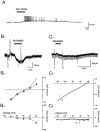
Effects of the D1-like agonist, SKF38393, on striatal LA neurons. A, A whole-cell current-clamp recording with a resting membrane potential of −65 mV illustrates a slowly rising, prolonged, and reversible membrane depolarization with action potentials occurring during the peak of the response. B1, C1, Voltage-clamp traces (holding potential −60 mV) recorded from another LA cell in saline containing TTX (0.5 μm) illustrate a slow inward current induced by SKF38393. Two superimposedIV plots before (open circles in B2 and 1 inC2) and during the peak of the response (filled circles in B2 and2 in C2) show a reversal potential close to EK(B2) and no reversal potential within the tested voltage range (C2). B3,C3, IV plots of the net SKF38393-induced currents (filled triangles in B3 and 2–1in C3).
Fig. 8.
Fig. 8.
Forskolin, an activator of adenylyl cyclase, mimics and occludes the effects of SKF38393 on the striatal LA cells.A, Dideoxyforskolin (10 μm,stippled bar), an inactive forskolin analog, evoked no response, whereas forskolin (10 μm, filled bar) caused a slow and reversible inward current. Holding potential was −60 mV. B, Application of SKF38393 (filled bars) resulted in a slow inward current (left trace). Fifteen minutes after washout of SKF38393 (10 μm), treatment with forskolin (10 μm,stippled bar) elicited a large slow inward current and occluded the effect of the second application of SKF38393.
Fig. 9.
Fig. 9.
Slow inward currents evoked by SKF38393 (10 μm), substance P (0.5 μm), and forskolin (10 μm) are unaffected by intracellular application of BAPTA (20 mm), a potent Ca2+chelator.

References

    1. Abercrombie ED, DeBoer P. Substantia nigra D1 receptors and stimulation of striatal cholinergic interneurons by dopamine: a proposed circuit mechanism. J Neurosci. 1997;17:8498–8505. - PMC - PubMed
    1. Ajima A, Yamaguchi T, Kato T. Modulation of acetylcholine release by D-1, D-2 dopamine receptors in rat striatum under freely moving conditions. Brain Res. 1990;518:193–198. - PubMed
    1. Anderson JJ, Kuo S, Chase TN, Engber TM. Dopamine D1 receptor-stimulated release of acetylcholine in rat striatum is mediated indirectly by activation of striatal neurokinin-1 receptors. J Pharmacol Exp Ther. 1994;269:1144–1151. - PubMed
    1. Aosaki T, Kawaguchi Y. Actions of substance P on rat neostriatal neurons in vitro. J Neurosci. 1996;16:5141–5153. - PMC - PubMed
    1. Arenas E, Alberch J, Perez-Navarro E, Solsona C, Marsal J. Neurokinin receptors differentially mediate endogenous acetylcholine release evoked by tachykinins in the neostriatum. J Neurosci. 1991;11:2332–2338. - PMC - PubMed

Publication types

MeSH terms