. 1998 Jul 15;18(14):5253-63.
doi: 10.1523/JNEUROSCI.18-14-05253.1998.

Membrane current induced by protein kinase C activators in rhabdomeric photoreceptors: implications for visual excitation

Affiliations

Membrane current induced by protein kinase C activators in rhabdomeric photoreceptors: implications for visual excitation

M del Pilar Gomez et al. J Neurosci. .

Abstract

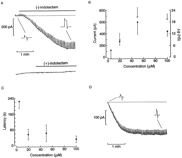
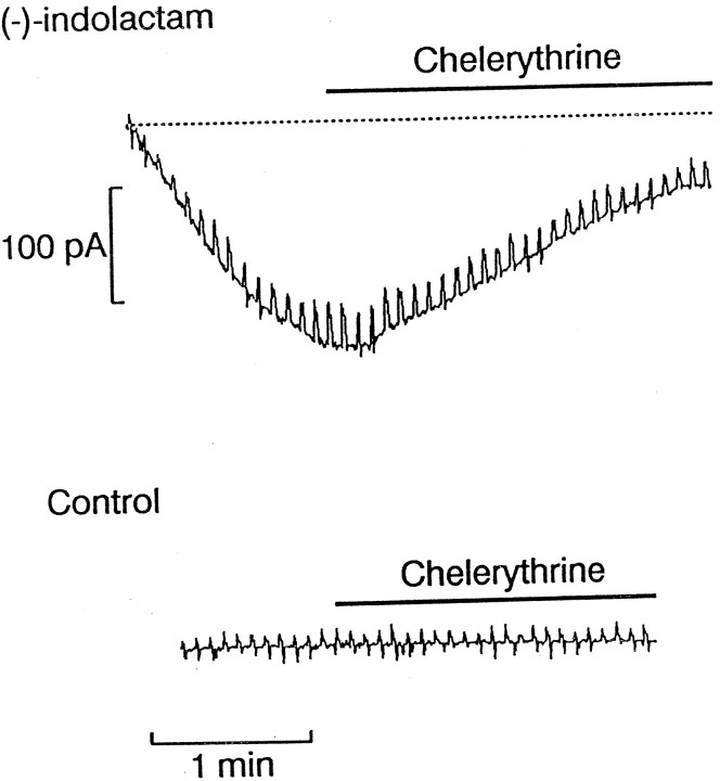
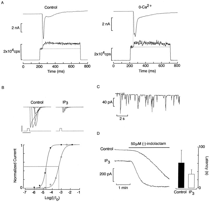
Visual excitation in rhabdomeric photoreceptors is thought to be mediated by activation of a light-regulated phospholipase C (PLC) and the consequent hydrolysis of phosphatidylinositol bisphosphate. Whereas much attention has been devoted to inositol trisphosphate (IP3) production and intracellular Ca2+ release, little is known about the possible role of the DAG branch in the generation of the light response. We have tested the effect of chemically distinct surrogates of DAG on isolated Lima photoreceptors. Application of the phorbol ester PMA (0.5-10 microM) or the alkaloid (-)-indolactam (20-100 microM) from a holding potential of -50 mV elicited an inward current, several hundred picoamperes in amplitude, accompanied by a pronounced increase in membrane conductance. The stereoisomers 4alpha-PMA and (+)-indolactam were both inactive, arguing for the specificity of the effects. Elevation of cytosolic Ca2+ by intracellular dialysis accelerated this current, whereas chelerythrine antagonized it, suggesting the involvement of PKC. The reversal potential of the membrane current induced by PKC activators was approximately +10 mV; replacement of extracellular Na with impermeant N-methyl-D-glucamine decreased its amplitude and shifted the reversal potential in the negative direction. Stimulation by PMA and (-)-indolactam was accompanied by a pronounced depression of light responsiveness; conversely, steady illumination reduced the size of the current elicited by these PKC activators. Taken together, these results support the notion that the DAG branch of the PLC cascade, in addition to its suggested participation in visual adaptation, may play a role in the activation of the photoresponse or a component thereof, probably in synergy with IP3-mediated Ca2+ release.


Figures

Fig. 1.
Fig. 1.
Activation of a membrane current by PMA.A, A photoreceptor cell was voltage clamped at −50 mV, with a repetitive 3 mV rectangular pulse superimposed on the holding potential. A puffer pipette containing 1 μm PMA in ASW (final DMSO concentration 0.1%) was positioned ∼50 μm away from the cell. The thick bar indicates pressure ejection. A large inward current was elicited. The change in the current steps (insets) indicates that an increase of membrane conductance occurred. B, Lack of effects of dialysis with normal internal solution (top trace), pressure-application of ASW, ASW containing 5% DMSO, and 1 μm 4α-PMA. In the last trace, no voltage command steps were superimposed on the holding potential; the downward deflections are quantum bumps. Calibration: 1 min. C, Inward current elicited by internal dialysis with 0.5 μmPMA via the patch pipette. A similar inward current, as inA, was elicited, but with a reduced latency.Insets illustrate the increase in conductance during the development of the inward current.
Fig. 2.
Fig. 2.
Effect of indolactam V on membrane current.A, Local superfusion with 100 μm(−)-indolactam of a rhabdomeric cell held at −50 mV in the dark caused a large inward current accompanied by an increase in membrane conductance. Administration of the stereoisomer (+)-indolactam at the same concentration was ineffective (bottom trace).B, Effects of different concentrations of indolactam on the peak amplitude of the inward current elicited at −50 mV (filled squares) and membrane conductance (open squares). Error bars indicate SD. The response appears to saturate at <50 μm. C, Time required to reach 10% of the maximum amplitude of the current, plotted as a function of concentration. D, Inward current obtained by administering 50 μm (−)-indolactam intracellularly. Notice the dramatic reduction in the latency of the inward current.
Fig. 3.
Fig. 3.
DAG surrogates act via PKC activation. To ascertain whether the current induced by administration of indolactam is mediated by its ability to activate PKC, photoreceptors were first stimulated with 50 μm (−)-indolactam V applied by internal dialysis. Once a conspicuous inward current developed at −50 mV, a puffer pipette was used to pressure eject 50 μm of the PKC inhibitor chelerythrine. After several seconds of application of the antagonist, the inward current and the associated increase in membrane conductance were substantially reversed. Bottom trace shows that application of chelerythrine to a control cell dialyzed with the normal internal solution was ineffective.
Fig. 4.
Fig. 4.
Facilitation of the effects of PKC activators by elevation of intracellular Ca2+. A, Recordings were obtained from two different photoreceptor cells internally dialyzed with Ca-free solution (−) and with 1 μm free calcium (+), respectively. The inward current evoked by local application of 5 μm indolactam (top thick bar) had a much more rapid onset and a larger amplitude when an internal solution with elevated calcium was used.B, Responses elicited by 50 μm indolactam in four different cells dialyzed with high Ca2+. In all cases the inward current attained its peak amplitude within 1 min.Bottom traces are from four control cells dialyzed with 1 μm free calcium without indolactam application.C, Mean latency (t10%) of the inward current evoked by the two concentrations of indolactam in the presence or absence of Ca2+ in the internal solution. Error bars indicate the SD.
Fig. 5.
Fig. 5.
Light-induced Ca2+ release and effects of IP3. A, Left, Simultaneous recording of membrane current (top trace) and fluorescence (bottom trace) in a photoreceptor loaded with 100 μm Ca Green 2 and voltage-clamped at −50 mV. At the onset of the excitation light step (500 msec, 480 nm), the optical signal jumped abruptly to a baseline level, and ∼30 msec later a secondary increase in fluorescence was observed, reflecting the rise in cytosolic calcium. Right, A similar result was obtained in a cell bathed for many minutes in a Ca-free solution.B, Light-intensity series obtained with the standard intracellular solution or one containing 10 μmIP3, respectively; access to the cell interior was through the somatic lobe. The normalized peak amplitude of the photocurrent plotted as a function of light intensity (bottom) reveals a marked desensitization induced by IP3. Calibration: 800 pA, 400 msec. C, Oscillatory inward current evoked by dialysis with IP3directly into the rhabdomeric lobe of a photoreceptor. The recording started immediately on rupturing the patch of membrane to gain access to the cell interior. D, Left, Comparison of the current elicited in the dark by puffer application of 50 μm (−)-indolactam (horizontal bar) in a cell internally dialyzed with the standard intracellular solution and one dialyzed with 10 μm IP3.Right, Histogram comparing the mean latency (t10%) of the indolactam-evoked current in control and IP3-treated photoreceptors (n = 7 and 9, respectively).
Fig. 6.
Fig. 6.
Interactions between light and PKC activators.A, Depression of the photocurrents by application of (−)-indolactam (50 μm). A photoreceptor cell was voltage clamped at −50 mV and stimulated with 100 msec flashes of light (3.7 × 10¹⁴photons · sec−1 · cm−2); subsequently, indolactam was applied by a puffer pipette, evoking a sustained inward current (data not shown). Delivery of another identical flash (top trace) elicited a dramatically reduced photocurrent. B, Effect of steady light on the amplitude and latency of the response to indolactam. The recordings at the top illustrate the responses evoked by 50 μm indolactam (thick lines) in two cells maintained in the dark (left) and two cells continuously illuminated (3.7 × 10¹⁴photons · sec−1 · cm−2).Bottom shows the mean steady-state current amplitude (filled bars) and the mean latency (t10%) (open bars) pooled for six cells in each of the two conditions. Error bars indicate SD.
Fig. 7.
Fig. 7.
Effect of external sodium removal on the current induced by PKC activators. A, Top, Replacement of Na with NMDG reduced the amplitude of the inward current evoked by application of 50 μm indolactam (thick horizontal lines). Membrane potential: −50 mV. Calibration: 100 pA, 1 min. Bottom, pooled data for several cells stimulated in the presence and absence of Na (n = 3 in each condition). Error bars indicate SD. B,Top, Reversal of the indolactam-evoked current in ASW. The membrane potential was clamped at the levels indicated; the traces were recorded from different cells. Bottom, Shift in the reversal voltage during superfusion with Na-free solution (replaced with NMDG).

Similar articles

Cited by

References

    1. Acharya JK, Jalink K, Hardy RW, Hartenstein V, Zuker CS. InsP3 receptor is essential for growth and differentiation but not for vision in Drosophila. Neuron. 1997;18:881–887. - PubMed
    1. Baer KM, Saibil HR. Light- and GTP-activated hydrolysis of phosphatidylinositol bisphosphate in squid photoreceptor membranes. J Biol Chem. 1988;263:17–20. - PubMed
    1. Bloomquist BT, Shortridge RD, Schneuwly S, Perdew M, Montell C, Steller H, Rubin G, Pak WL. Isolation of a putative phospholipase C gene of Drosophila, norpA, and its role in phototransduction. Cell. 1988;54:723–733. - PubMed
    1. Bode H-P, Göke B. Protein kinase C activates capacitative calcium entry in the insulin secreting cell line RINm5F. FEBS Lett. 1994;339:307–311. - PubMed
    1. Brown JE, Blinks JR. Changes in intracellular free calcium concentration during illumination of invertebrate photoreceptors. J Gen Physiol. 1974;64:643–665. - PMC - PubMed

Publication types

LinkOut - more resources