. 1998 Jul 15;18(14):5285-93.
doi: 10.1523/JNEUROSCI.18-14-05285.1998.

Vasoactive intestinal peptide enhances its own expression in sympathetic neurons after injury

Affiliations

Vasoactive intestinal peptide enhances its own expression in sympathetic neurons after injury

R P Mohney et al. J Neurosci. .

Abstract

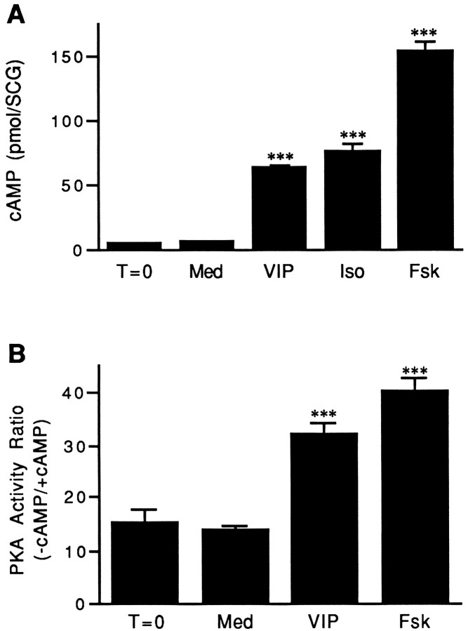
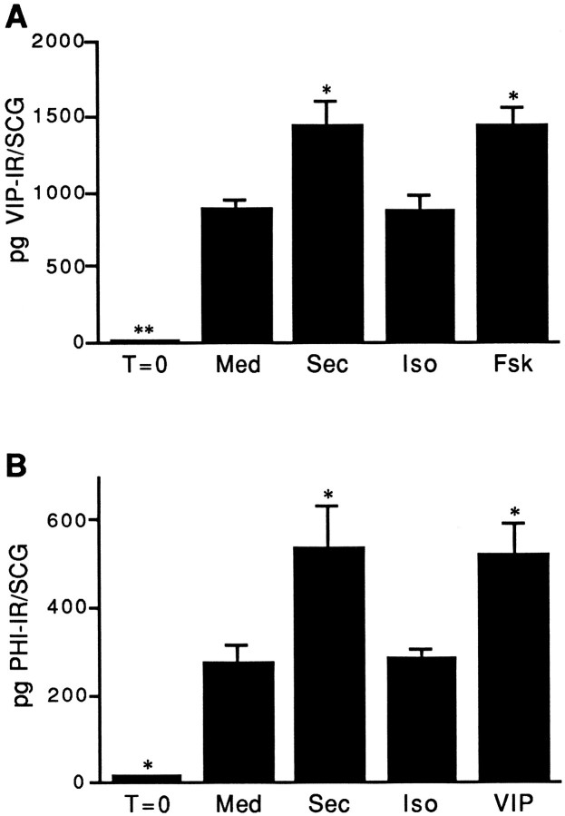
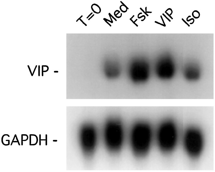
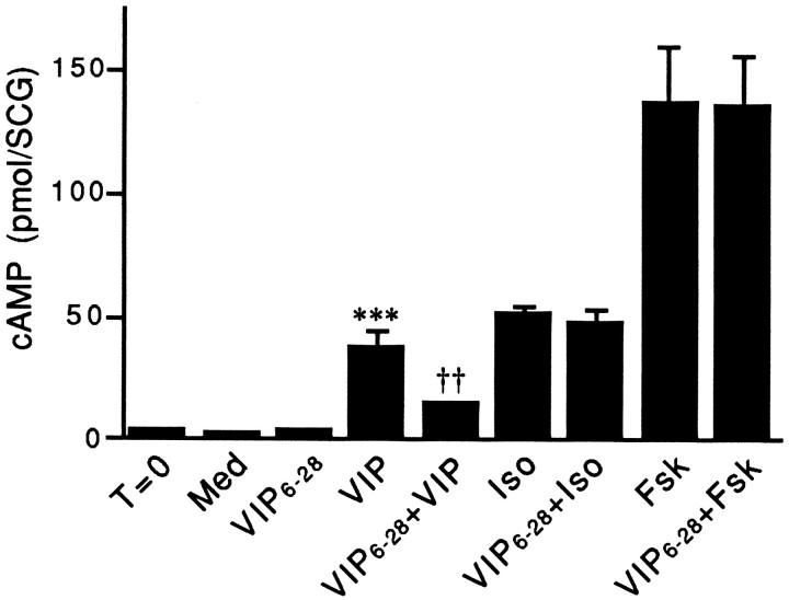
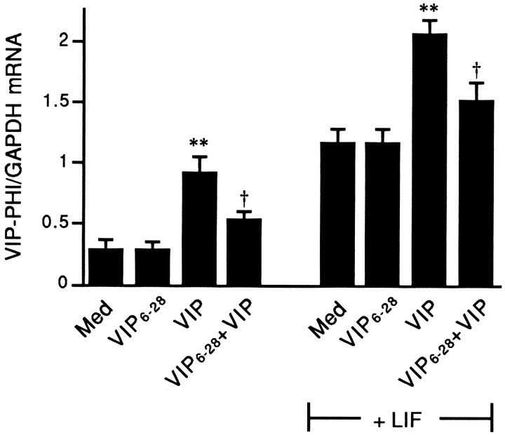
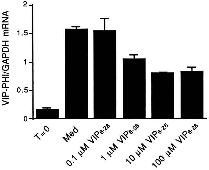
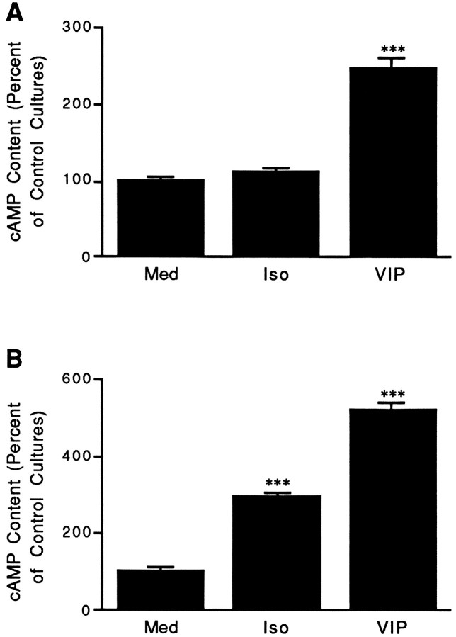
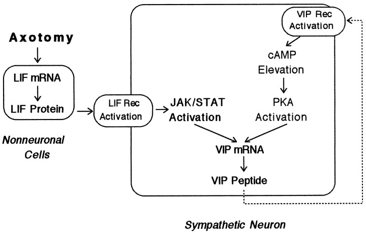
Neurons in the adult rat superior cervical sympathetic ganglion (SCG) dramatically increase their content of vasoactive intestinal peptide (VIP) and its mRNA after axotomy in vivo and after explantation. Because the VIP gene contains a functional cAMP response element, the effects of cAMP-elevating agents on VIP expression were examined. VIP, forskolin, or isoproterenol increased cAMP accumulation in explanted ganglia. Secretin, a peptide chemically related to VIP, or forskolin increased VIP levels above those seen in ganglia cultured in control medium, whereas treatment with VIP or secretin increased the level of peptide histidine isoleucine (PHI), a peptide coded for by the same mRNA that encodes VIP. VIP or forskolin also increased VIP-PHI mRNA. In contrast, isoproterenol did not alter levels of VIP, PHI, or VIP-PHI mRNA. Although VIP or forskolin increased cAMP levels in both dissociated neurons and in non-neuronal cells, isoproterenol significantly stimulated cAMP accumulation only in the latter. VIP6-28 was an effective antagonist of the actions of exogenous VIP on cAMP and VIP-PHI mRNA in neuron-enriched cultures. When adult SCG explants were cultured in defined medium, endogenous VIP immunoreactivity was released. When VIP6-28 was added to such cultures, it significantly inhibited the increase in VIP-PHI mRNA that normally occurs. These data indicate that VIP, or a closely related molecule, produced by adult neurons after injury can enhance the expression of VIP. Such a mechanism may prolong the period during which VIP is elevated after axonal damage. The possibility is also discussed that, because VIP is present in preganglionic neurons in normal animals, its release during periods of increased sympathetic nerve activity could alter VIP expression in the SCG.


Figures

Fig. 1.
Fig. 1.
Stimulation of cAMP levels and PKA activity in adult rat SCG. A, VIP, isoproterenol, or forskolin increases cAMP levels in SCG in short-term organ culture. Control ganglia (T = 0) were removed from animals and processed immediately as described in Materials and Methods. Cultured ganglia were incubated for 30 min in medium containing IBMX alone (Med) or IBMX together with 10 μm VIP (VIP), isoproterenol (Iso), or forskolin (Fsk). Data represent the mean ± SEM of at least 12 SCGs. B, VIP or forskolin increases PKA activity in SCG in short-term organ culture. Control ganglia were removed from animals and frozen until assayed (T = 0). Other SCGs were cultured for 1 hr in defined medium containing IBMX alone (Med) or IBMX together with 10 μm VIP (VIP) or forskolin (Fsk). The data are expressed as the ratio of PKA activity measured in the absence of added cAMP relative to that in its presence. Data represent the mean ± SEM of 10 SCGs. ***p < 0.001 compared with Med or T = 0.
Fig. 2.
Fig. 2.
Stimulation of VIP or PHI-IR in cultured adult rat SCG. A, Secretin or forskolin, but not isoproterenol, significantly increases VIP-IR in SCG explants. SCGs were removed from animals and either frozen immediately (T = 0) or cultured for 48 hr in defined medium containing IBMX alone (Med) or IBMX together with 10 μmsecretin (Sec), isoproterenol (Iso), or forskolin (Fsk). VIP-IR was determined by radioimmunoassay. B, Secretin or VIP, but not isoproterenol, increases PHI-IR in SCG explants. SCGs were treated as described in part A except that the agonists tested were 10 μm secretin (Sec), isoproterenol (Iso), or VIP, and PHI-IR was measured by radioimmunoassay. For both parts A and B, data represent the mean ± SEM of four ganglia. *p < 0.05 and **p < 0.01 compared with Med.
Fig. 3.
Fig. 3.
VIP or forskolin, but not isoproterenol, increases the steady-state levels of VIP–PHI mRNA in adult SCG explants. SCGs were removed from the animal and either frozen directly (T = 0) or maintained in organ culture for 24 hr in defined medium containing IBMX (Med) or in medium containing IBMX plus 10 μm forskolin (Fsk), VIP, or isoproterenol (Iso). Total RNA was extracted from pairs of ganglia, and Northern blot analyses were performed using radiolabeled cDNA probes for VIP–PHI or GAPDH mRNA.
Fig. 4.
Fig. 4.
VIP6–28 reduces the increase in cAMP levels measured in adult SCG stimulated with exogenous VIP. Control ganglia were removed from animals and processed immediately as described in Materials and Methods (T = 0). After a 30 min preincubation in the presence of IBMX, cultured ganglia were incubated for an additional 30 min in medium containing IBMX alone (Med) or together with 10 μm VIP (VIP), isoproterenol (Iso), or forskolin (Fsk). To examine the blocking ability of VIP6–28, ganglia were preincubated for 25 min in medium containing IBMX, transferred to medium containing IBMX and 30 μm VIP6–28 for 5 min, and then incubated for an additional 30 min in medium containing 30 μmVIP6–28 alone or together with 10 μm VIP (VIP6–28 + VIP), isoproterenol (VIP6–28 + Iso), or forskolin (VIP6–28 + Fsk). Data represent the mean ± SEM of four SCGs. ***p < 0.001 compared with T = 0, Med, or Ant, and ††p < 0.01 compared with VIP.
Fig. 5.
Fig. 5.
VIP6–28 reduces the effect of VIP but not LIF on VIP–PHI mRNA expression in neuron-enriched cultures. Neuron-enriched cultures were maintained for 48 hr in medium alone (Med) or stimulated with 100 μmVIP6–28 (VIP6–28), 10 μm VIP (VIP), or both. Other cultures were stimulated with VIP, VIP6–28, or both, and also exposed to 10 ng/ml LIF. Total RNA was isolated and Northern blot analyses were performed. Data represent the mean ± SEM of five cultures. **p < 0.01 compared with Med (± LIF, as appropriate); †p < 0.05 compared with VIP (± LIF, as appropriate).
Fig. 6.
Fig. 6.
VIP6–28 reduces the elevation in steady-state levels of VIP–PHI mRNA that result from placement of adult rat SCG into organ culture for 48 hr in defined medium. Ganglia were removed from the animal and frozen (T = 0) or cultured in defined medium alone (Med) or in medium supplemented with 0.1, 1, 10, or 100 μmVIP6–28 (VIP6–28) for 48 hr. Total RNA was isolated and examined by Northern blot analyses. Data represent the mean ± range of two lanes that each contained RNA from two ganglia.
Fig. 7.
Fig. 7.
The action of isoproterenol is cell-type specific.A, VIP, but not isoproterenol, increases cAMP levels in neuron-enriched cultures. Neonatal SCGs were dissociated and cultured under conditions that favor the survival of neurons but not non-neuronal cells. After 48 hr in culture, neuron-enriched cultures were incubated for 30 min in medium containing IBMX alone (Med) or IBMX together with 10 μmisoproterenol (Iso) or VIP. The content of cAMP is expressed relative to levels measured in cells cultured in control medium alone (Med). Data from several independent experiments were pooled together and represent the mean ± SEM (n = 27). B, VIP or isoproterenol increases cAMP levels in non-neuronal cell-enriched cultures. Neonatal SCGs were dissociated and cultured under conditions that favor the survival and proliferation of non-neuronal cells. After 48 hr in culture, non-neuronal cell-enriched cultures were incubated for 30 min in medium containing IBMX alone (Med) or IBMX together with 10 μm isoproterenol (Iso) orVIP. The content of cAMP is expressed relative to levels measured in cells cultured in control medium alone (Med). Data from several independent experiments were pooled together and represent the mean ± SEM (n = 12). ***p < 0.001 compared with Med.
Fig. 8.
Fig. 8.
Hypothesized mechanism of feedback stimulation of VIP expression in cultured sympathetic ganglia. Under normal conditions, ganglia express only low levels of VIP–PHI mRNA and peptide. When ganglia are placed into explant culture, LIF mRNA (and subsequently, LIF protein) increases in non-neuronal cells as a result of injury-induced factors (Sun et al., 1996). The release of LIF and its binding to receptors on neurons cause the activation of Janus kinases (JAK) and the phosphorylation of signal transducers and activators of transcription (STAT) proteins. Phosphorylated STAT proteins then translocate to the nucleus and bind to and increase the rate of transcription of the VIP gene (Symes et al., 1994, 1995). VIP is released by neurons in culture and binds to VIP6–28-sensitive, G-protein-coupled receptors in neurons to elevate cAMP levels. Elevations in cAMP levels activate PKA and lead to increased VIP gene transcription.

Similar articles

Cited by

References

    1. Aloisi F, Rosa S, Test U, Bonsi P, Russo G, Peschle C, Levi G. Regulation of leukemia inhibitory factor synthesis in cultured human astrocytes. J Immunol. 1994;152:5022–5031. - PubMed
    1. Ariano MA, Briggs CA, McAfee DA. Cellular localization of cyclic nucleotide changes in rat superior cervical ganglion. Cell Mol Neurobiol. 1982;2:143–156.
    1. Audigier S, Barberis C, Jard S. Vasoactive intestinal polypeptide increases inositol phospholipid breakdown in the rat superior cervical ganglion. Brain Res. 1986;376:363–367. - PubMed
    1. Baldwin C, Sasek CA, Zigmond RE. Evidence that some preganglionic sympathetic neurons in the rat contain vasoactive intestinal peptide- or peptide histidine isoleucine amide-like immunoreactivities. Neuroscience. 1991;40:175–184. - PubMed
    1. Brenneman DE, Eiden LE. Vasoactive intestinal peptide and electrical activity influence neuronal survival. Proc Natl Acad Sci USA. 1986;83:1159–1162. - PMC - PubMed

Publication types

MeSH terms

LinkOut - more resources