

Comparative Study
doi: 10.1523/JNEUROSCI.18-14-05294.1998.
Comparison of hippocampal dendritic spines in culture and in brain
Affiliations
- PMID: 9651212
- PMCID: PMC6793498
- DOI: 10.1523/JNEUROSCI.18-14-05294.1998
Item in Clipboard
Comparative Study
Comparison of hippocampal dendritic spines in culture and in brain
J Neurosci.
.
Abstract
We have quantified hippocampal spine structure at the light and ultrastructural levels in cell cultures approximately 1- 3 weeks old and in the brains of rodents 5 and 21 d old. The number of spines bearing synapses increases with age in cultures and in brain, but the structures are similar in both. In culture, about half of the synapses are formed on spines and the remainder are formed on dendritic shafts. In the 5-d-old brain, about half of the synapses occur on dendritic shafts, by 3 weeks of age only approximately 20% of synapses are found on dendritic shafts, and in the adult shaft synapses are very rare.
Figures
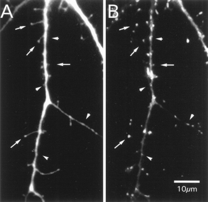
Original fluorescence staining of the same dendrite of a hippocampal cell in culture. A, Dendrite after filling with Lucifer yellow. B, The corresponding immunocytochemical staining against synaptophysin. Thearrows in both images point to the sites of spines, and the arrowheads point to the site shaft synapses.
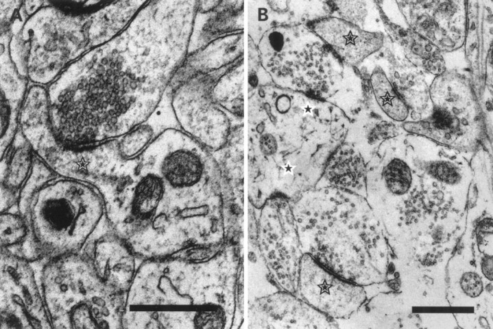
Ultrastructure of developing synapses.A, The largest spine (star) we encountered in our sample of synapses at P21. All typical structural features are present at that age; the postsynaptic density is aligned with the active zone. The spine had the typical grainy dense appearance, and the spine apparatus is visible. B, Spines from hippocampal neurons in culture after 14 DIV. Spines (stars) in culture show no qualitative difference from their counterparts in situ. Shaft synapses are marked with a star on a white background. Scale bars, 0.5 μm.
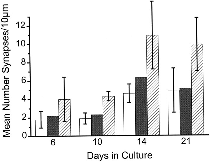
Comparison of synaptic density in culture as a function of time. The different bars depict the number of spine synapses (white bars), the number of shaft synapses (gray bar, calculated as the differences from total synapses minus spine synapses), and the number of all synapses (striped bars). Between 10 and 14 DIV there is a significant increase of synaptic density. Before and after this age the density of spine and shaft synapses is constant. Note that the relationship between shaft and spine synapses is equal for all ages.
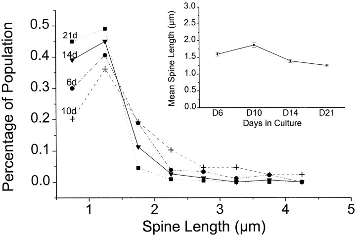
Spine length distribution in culture at different ages. Most of the spines have a length of 1–1.5 μm. Note that the occurrence of longer spines in young cultures is significantly increased. The inset depicts the decreasing mean spine length and SE over time.
References
-
- Banker G, Goslin K. Culturing nerve cells. MIT; Cambridge, MA: 1991.
-
- Cooper MW, Smith SJ. A real-time analysis of growth cone–target cell interactions during the formation of stable contacts between hippocampal neurons in culture. J Neurobiol. 1992;23:814–828. - PubMed
-
- Crick F. Do dendritic spines twitch? Trends Neurosci. 1982;5:44–46.
-
- Deisseroth K, Bito H, Tsien RW. Signaling from synapse to nucleus: postsynaptic CREB phosphorylation during multiple forms of hippocampal synaptic plasticity. Neuron. 1996;16:89–101. - PubMed
Publication types
MeSH terms
Grants and funding
LinkOut - more resources
Full Text Sources