. 1998 Jul 15;18(14):5301-10.
doi: 10.1523/JNEUROSCI.18-14-05301.1998.

A complex program of striatal gene expression induced by dopaminergic stimulation

Affiliations

A complex program of striatal gene expression induced by dopaminergic stimulation

J D Berke et al. J Neurosci. .

Abstract

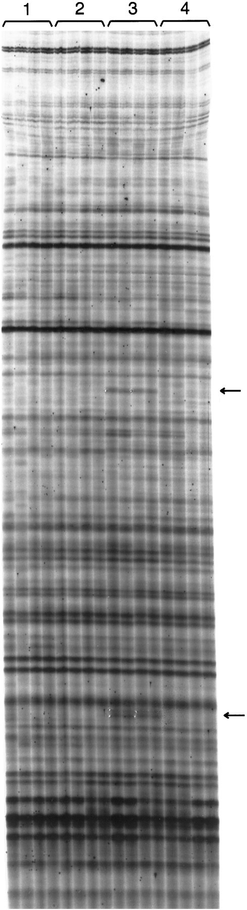
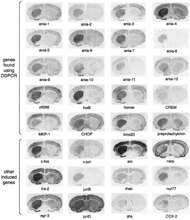
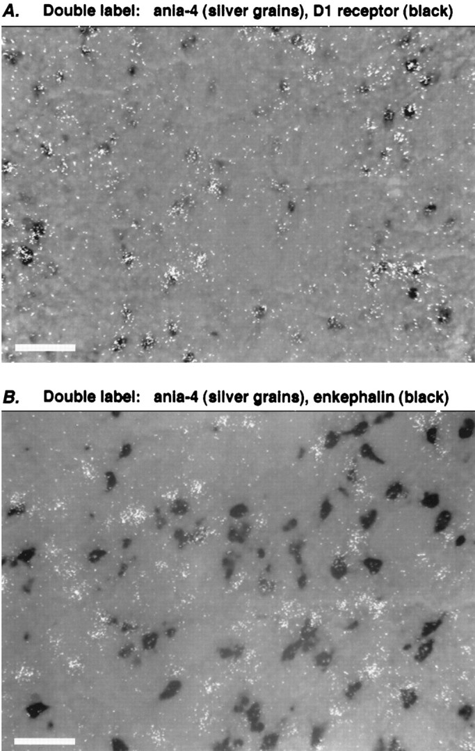
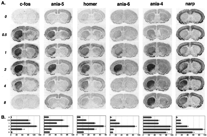
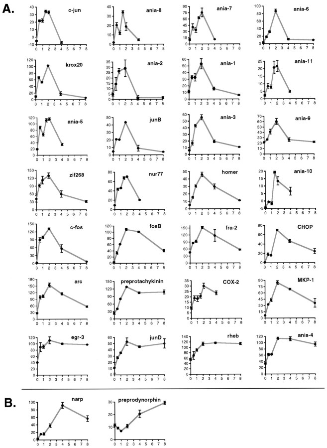
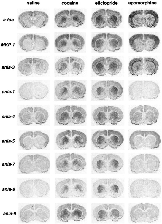
Dopamine acting in the striatum is necessary for normal movement and motivation. Drugs that change striatal dopamine neurotransmission can have long-term effects on striatal physiology and behavior; these effects are thought to involve alterations in gene expression. Using the 6-hydroxydopamine lesion model of Parkinson's disease and differential display PCR, we have identified a set of more than 30 genes whose expression rapidly increases in response to stimulation of striatal dopamine D1 receptors. The induced mRNAs include both novel and previously described genes, with diverse time courses of expression. Some genes are expressed at near-maximal levels within 30 min, whereas others show no substantial induction until 2 hr or more after stimulation. Some of the induced genes, such as CREM, CHOP, and MAP kinase phosphatase-1, may be components of a homeostatic response to excessive stimulation. Others may be part of a genetic program involved in cellular and synaptic plasticity. A very similar set of genes is induced in unlesioned animals by administration of the psychostimulant cocaine or the antipsychotic eticlopride, although in distinct striatal cell populations. In contrast to some previously described early genes, most of the novel genes are not induced in cortex by apomorphine, indicating specificity of induction. Thus we have identified novel components of a complex, coordinated genetic program that is induced in striatal cells in response to various dopaminergic manipulations.


Figures

Fig. 1.
Fig. 1.
A fragment of a typical differential display film. This portion of the film has an approximate size range of 600–1500 bp and is 30 cm long. The conditions used in the screen are as follows. Conditions 1 and 2 are the unlesioned (left) and lesioned (right) striata, respectively, from animals given saline injections and killed 1 hr later. Conditions 3 and 4 are the lesioned striata from animals given SKF38393 (5 mg/kg) and killed after 1 and 24 hr, respectively. For each condition we used RNA from two different animals for reverse transcription, and each reverse transcription reaction was used for two duplicate PCR reactions, for a total of four lanes per condition for each primer combination. Arrows mark examples of differentially expressed bands; the white spots at the edge of these bands are pinholes used to orient the film over the dried gel for band excision. The upper of the two arrowsindicates an 850 bp fragment of ania-6 mRNA.
Fig. 2.
Fig. 2.
Examples of genes acutely induced by SKF38393. Each image is of a coronal rat brain section through the middle striatum, after in situ hybridization with a probe to the gene indicated. All of the brain sections are from animals given unilateral 6-hydroxydopamine lesions and then injected with SKF38393 (5 mg/kg) and killed 2 hr later (except tPA, shown at 4 hr after SKF38393). The lesioned side is shown on the leftin each case. The genes designated “ania-1,” “ania-2,” etc., have no named matches in GenBank. Genes were designated as previously known on the basis of matches to the following accession numbers: homer U92079; CHOP (= GADD153) U36994; krox20 (= egr-2) U78102; preprotachykinin M34184; CREM M60285; fosBX14897; zif268 (= NGFIA/egr-1) M18416; MKP-1 (= CL100)S81478.
Fig. 3.
Fig. 3.
Selective gene induction in D1-receptor-bearing striatal neurons. Double-labelin situ hybridization was performed with probes to the gene ania-4 (silver grains) and either D1receptor (A) or enkephalin (B). The field in each case is taken from a brain section treated as in Figure 2 (i.e., 2 hr after SKF38393, 5 mg/kg), with additional hybridizations using digoxygenin-labeled probes, visualized by a dark reaction product. A, Induced ania-4 mRNA colocalizes with D1 receptor mRNA. B, Induced ania-4 mRNA does not colocalize with mRNA for enkephalin, a marker of D2-receptor-bearing striatopallidal neurons. Scale bars, ∼50 μm.
Fig. 4.
Fig. 4.
Five examples of distinct temporal patterns of gene expression after SKF38393 (5 mg/kg) given to 6-OHDA lesioned animals. Animals were killed at either 0, 0.5, 1, 2, 4, or 8 hr after injection of SKF38393, as indicated. A, Representativein situ hybridization images from each time point. Lesioned side is on the left as in Figure 2.B, Bar charts of average gray value measurements from the dorsal striatum. For each gene, sections from the same four animals were measured at each time point. The x-axis indicates average gray value, in arbitrary units. Error bars indicate SEM.
Fig. 5.
Fig. 5.
A, Time course examples for genes reaching peak induction within 2 hr. B, Preprodynorphin and narp take longer to reach peak induction. Graphs are quantitations from tissue sections at the same time points as in Figure 4, except for a few genes with rapid decay rates where data are shown from an experiment in which animals were killed at 0, 0.5, 1, 1.5, 2, and 4 hr after SKF38393 (5 mg/kg). The y-axis in each case is average gray value in dorsal striatum (in arbitrary units), and thex-axis is hours after SKF38393. In most casesn = 4 animals per time point; for a few casesn = 2 or 3. Error bars indicate SEM. Where no error bar is visible, the error was smaller than the graph symbol.
Fig. 6.
Fig. 6.
The striatal response to cocaine and eticlopride uses a similar set of genes. All brain sections shown are from normal (i.e., unlesioned) animals given the indicated drug and killed 1 hr later. All drugs were given intraperitoneally at the following doses: cocaine 25 mg/kg, apomorphine 2 mg/kg, eticlopride 1 mg/kg.

References

    1. Alberini CM, Ghirardi M, Metz R, Kandel ER. C/EBP is an immediate-early gene required for the consolidation of long-term facilitation in Aplysia. Cell. 1994;76:1099–1114. - PubMed
    1. Albin RL, Young AB, Penney JB. The functional anatomy of basal ganglia disorders. Trends Neurosci. 1989;12:366–375. - PubMed
    1. Brakeman PR, Lanahan AA, O’Brien R, Roche K, Barnes CA, Huganir RL, Worley PF. Homer: a protein that selectively binds metabotropic glutamate receptors. Nature. 1997;386:284–288. - PubMed
    1. Chase TN, Mouradian MM, Engber TM. Motor response complications and the function of striatal efferent systems. Neurology. 1993;43:S23–27. - PubMed
    1. Cole AJ, Bhat RV, Patt C, Worley PF, Baraban JM. D1 dopamine receptor activation of multiple transcription factor genes in rat striatum. J Neurochem. 1992;58:1420–1426. - PubMed

Publication types

Substances

LinkOut - more resources