. 1998 Jul 15;18(14):5311-21.
doi: 10.1523/JNEUROSCI.18-14-05311.1998.

The neural cell adhesion molecule L1 interacts with the AP-2 adaptor and is endocytosed via the clathrin-mediated pathway

Affiliations

The neural cell adhesion molecule L1 interacts with the AP-2 adaptor and is endocytosed via the clathrin-mediated pathway

H Kamiguchi et al. J Neurosci. .

Abstract

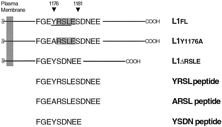
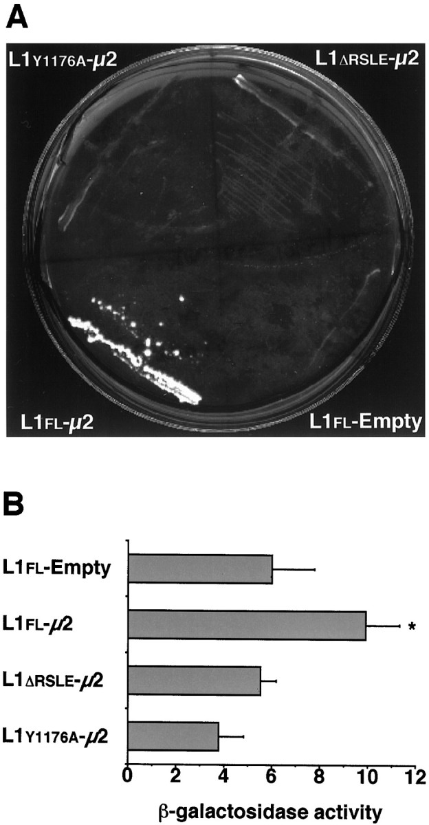
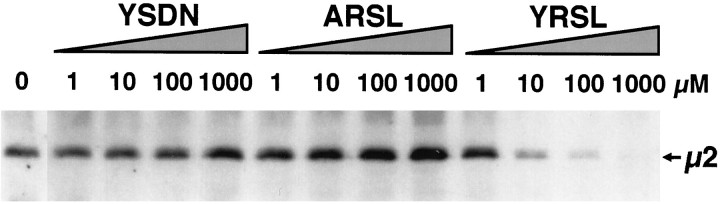
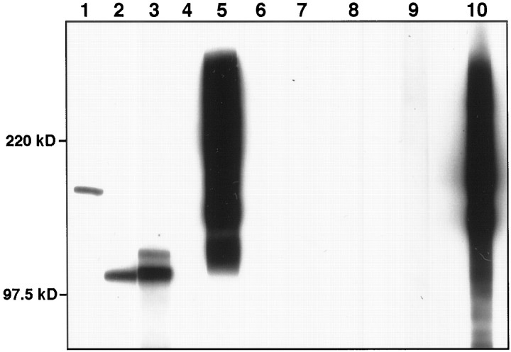
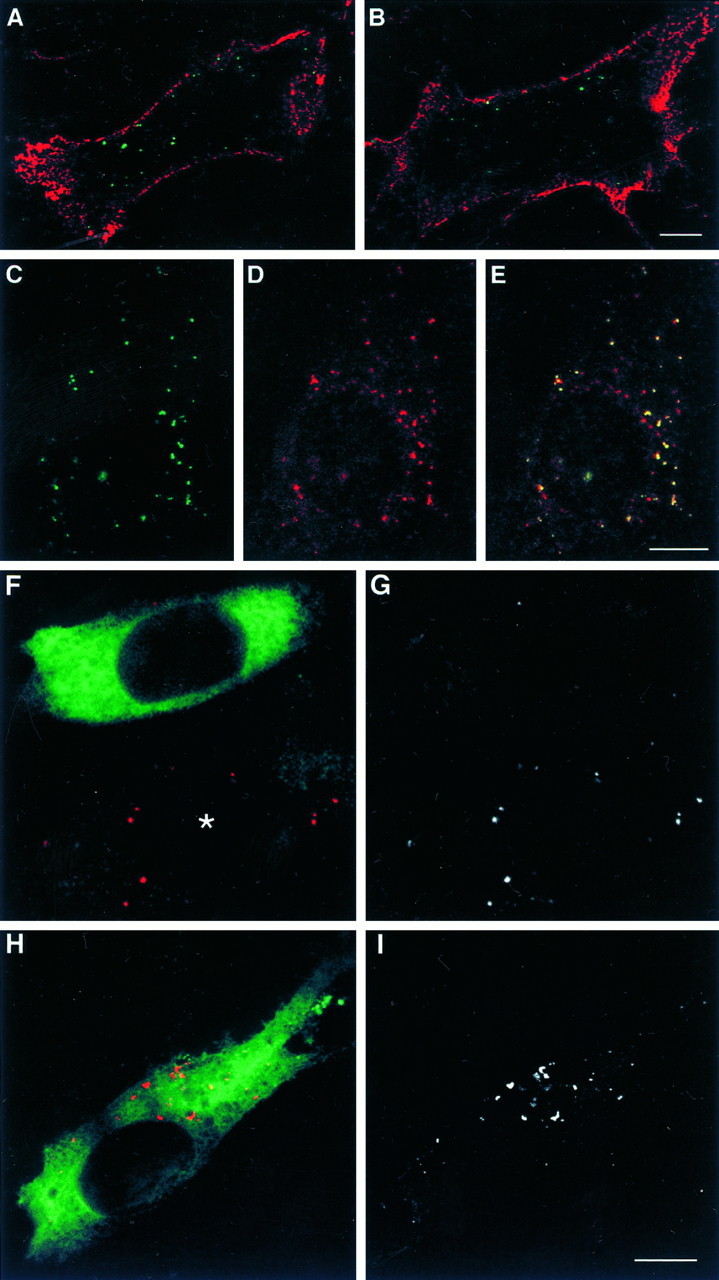
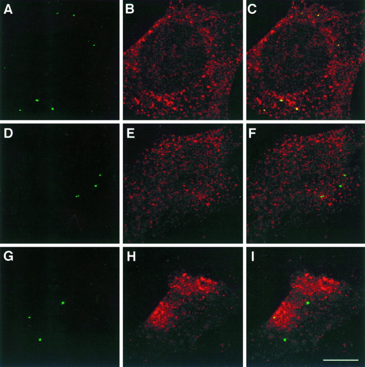
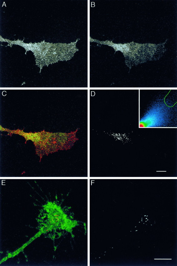
Cell-cell interactions mediated via cell adhesion molecules (CAMs) are dynamically regulated during nervous system development. One mechanism to control the amount of cell surface CAMs is to regulate their recycling from the plasma membrane. The L1 subfamily of CAMs has a highly conserved cytoplasmic domain that contains a tyrosine, followed by the alternatively spliced RSLE (Arg-Ser-Leu-Glu) sequence. The resulting sequence of YRSL conforms to a tyrosine-based sorting signal that mediates clathrin-dependent endocytosis of signal-bearing proteins. The present study shows that L1 associates in rat brain with AP-2, a clathrin adaptor that captures plasma membrane proteins with tyrosine-based signals for endocytosis by coated pits. In vitro assays demonstrate that this interaction occurs via the YRSL sequence of L1 and the mu 2 chain of AP-2. In L1-transfected 3T3 cells, L1 endocytosis is blocked by dominant-negative dynamin that specifically disrupts clathrin-mediated internalization. Furthermore, endocytosed L1 colocalizes with the transferrin receptor (TfR), a marker for clathrin-mediated internalization. Mutant forms of L1 that lack the YRSL do not colocalize with TfR, indicating that the YRSL mediates endocytosis of L1. In neurons, L1 is endocytosed preferentially at the rear of axonal growth cones, colocalizing with Eps15, another marker for the clathrin endocytic pathway. These results establish a mechanism by which L1 can be internalized from the cell surface and suggest that an active region of L1 endocytosis at the rear of growth cones is important in L1-dependent axon growth.


Figures

Fig. 1.
Fig. 1.
A portion of the L1 cytoplasmic domain is shown in the single-letter amino acid code. In the wild-type full-length L1 (L1fl), the cytoplasmic domain consists of 114 amino acids (1144–1257) and contains the RSLE sequence (shaded region), which is adjacent to Tyr1176. The resulting sequence of YRSL conforms to a tyrosine-based sorting motif, YxxL (underlined region). L1y1176ahas a single amino acid substitution (Y1176A) that mutates a critical tyrosine residue in the motif. In the non-neuronal form of L1 (L1Δrsle) that lacks the RSLE sequence, a hydrophobic amino acid leucine at position Y+3 is replaced by a polar amino acid asparagine. Therefore, the tyrosine-based motif YRSL is disrupted in L1y1176a and L1Δrsle. A cDNA that codes for the whole cytoplasmic domain of these forms of L1 (L1flCD, L1ΔrsleCD, or L1y1176aCD) was used in the yeast two-hybrid system. The synthetic peptides (YRSL,ARSL, and YSDN), which correspond to a part of L1flCD, L1ΔrsleCD, or L1y1176aCD, respectively, were used as a competitor in the UV-induced cross-linking experiments.
Fig. 2.
Fig. 2.
The yeast two-hybrid assay demonstrates the interaction between the YRSL sequence of the L1CD and the μ2 chain of AP-2. A, Yeast cells, which were cotransformed with L1flCD and μ2 sequences (L1fl-μ2), were able to grow and form colonies on histidine-deficient plates in the presence of 5 mm 3-AT between 3 and 5 d after plating. However, cotransformants containing either L1ΔrsleCD or L1y1176aCD and μ2 sequences (L1Δrsle-μ2 orL1y1176a-μ2, respectively) did not form colonies after 10 d under the same conditions.B, L1fl-μ2 expressed a significantly higher level of β-galactosidase activity than a background level (L1fl-Empty). However, bothL1Δrsle-μ2 andL1y1176a-μ2expressed comparable levels of β-galactosidase activity to the background level. Values represent the mean ± SD of five determinations. *p < 0.0005; one-way ANOVA, followed by post hoc Fisher’s PLSD as compared withL1fl-Empty.
Fig. 3.
Fig. 3.
The μ2 chain of AP-2 was labeled by the *YQRL peptide containing photoreactive cross-linker in the absence or presence of the competitor peptides (YRSL,ARSL, or YSDN; see Fig. 1). The concentrations of each competitor peptide are indicated in the figure. The YRSL peptide that carries the tyrosine-based motif interfered with the cross-linking reaction between the *YQRL peptide and the μ2 chain when the YRSL peptide was presented at a concentration as low as 10 μm. Both the ARSL and YSDNpeptides lacking the motif failed to interfere with the cross-linking reaction at concentrations up to 1 mm.
Fig. 4.
Fig. 4.
L1 immunoprecipitates from rat brain membrane extracts were probed with anti-AP180 antibody (lane 1), anti-β1- and β2-adaptin antibody (lane 2) or anti-α-adaptin antibody (lane 3), and no primary antibody control (lane 6), showing that AP-2 and AP180 coimmunoprecipitated with L1. As a control, the L1 immunoprecipitates (lane 4) and rat brain membrane extracts (lane 5) were probed with anti-rat NCAM antibody. As another control, NCAM immunoprecipitates from rat brain membrane extracts were probed with anti-AP180 antibody (lane 7), anti-β1- and β2-adaptin antibody (lane 8), anti-α-adaptin antibody (lane 9), or anti-NCAM antibody (lane 10).
Fig. 5.
Fig. 5.
A, B, Shown are confocal sections (0.83 μm in thickness) of L1fl-transfected NIH-3T3 cells in which cell surface L1 and endocytosed L1 were differentially labeled. Living cells were incubated with bivalent anti-L1 antibody (A) or anti-L1 Fab (B) for 1 hr to allow for L1 endocytosis. A superimposed image in which cell surface L1 is colored inred and endocytosed L1 in green is shown. Scale bar, 10 μm. CE, Confocal sections (0.83 μm in thickness) of a L1fl-transfected NIH-3T3 cell that was incubated with anti-L1 antibody and TxR-conjugated Tf for 1 hr to allow for the endocytosis of both probes. Shown are endocytosed L1 (C), endocytosed Tf (D), and a superimposed image showing colocalization of L1 and Tf as evidenced by yellow(E). Scale bar, 10 μm.FI, Confocal sections (0.71 μm in thickness) of L1fl-expressing NIH-3T3 cells that were transfected with K44A dynamin (F, G) or wild-type dynamin (H, I). Endocytosed L1 is colored in red and transfected dynamin in green (F, H). To facilitate the visualization of endocytosed L1, we have shown the red channel only in black and white(G, I). An asteriskindicates a cell that was not transfected with K44A dynamin. Scale bar, 10 μm.
Fig. 6.
Fig. 6.
Confocal sections (0.83 μm in thickness) of NIH-3T3 cells expressing L1fl(AC), L1y1176a(DF), or L1Δrsle(GI). Living cells were incubated with anti-L1 Fab for 1 hr to label endocytosed L1, and the cells were double-labeled with anti-TfR antibody. Endocytosed L1 (A, D, G), TfR (B, E, H), and superimposed images (C, F,I) are shown. Yellow indicates colocalization of endocytosed L1 and TfR. Scale bar, 10 μm.
Fig. 7.
Fig. 7.
AD, Confocal sections (0.71 μm in thickness) of a DRG growth cone growing on an L1 substrate. The growth cone was double-labeled with anti-L1CD antibody (74-5H7) (A) and anti-Eps15 antibody (B). Superimposition of the images with L1 colored in red and Eps15 in greenexhibits colocalization (yellow) at the rear of the growth cone (C). A black andwhite mask shows the colocalized distribution of L1 and Eps15 (D). High intensity pixels falling within the green tracing on the scatterplot (inset) were chosen to generate the mask (see Materials and Methods for details). Scale bar, 10 μm. E,F, Confocal sections (0.83 μm in thickness) of a DRG growth cone growing on an L1 substrate. The living neuron was incubated with anti-L1 antibody for 15 min to label endocytosed L1. The cell was double-labeled with anti-NCAM antibody to visualize the outline of the growth cone. Endocytosed L1 is colored in red and NCAM in green (E). To facilitate visualization of endocytosed L1, we have shown the red channel only inblack and white(F). Scale bar, 10 μm.

References

    1. Aberle H, Schwartz H, Kemler R. Cadherin–catenin complex: protein interactions and their implications for cadherin function. J Cell Biochem. 1996;61:514–523. - PubMed
    1. Ahle S, Ungewickell E. Purification and properties of a new clathrin assembly protein. EMBO J. 1986;5:3143–3149. - PMC - PubMed
    1. Ahle S, Ungewickell E. Auxilin, a newly identified clathrin-associated protein in coated vesicles from bovine brain. J Cell Biol. 1990;111:19–29. - PMC - PubMed
    1. Alconada A, Bauer U, Hoflack B. A tyrosine-based motif and a casein kinase II phosphorylation site regulate the intracellular trafficking of the varicella-zoster virus glycoprotein I, a protein localized in the trans-Golgi network. EMBO J. 1996;15:6096–6110. - PMC - PubMed
    1. Bailey CH, Chen M, Keller F, Kandel ER. Serotonin-mediated endocytosis of apCAM: an early step of learning-related synaptic growth in Aplysia. Science. 1992;256:645–649. - PubMed

Publication types