. 1998 Jul 10;273(28):17917-25.
doi: 10.1074/jbc.273.28.17917.

The gut-enriched Krüppel-like factor suppresses the activity of the CYP1A1 promoter in an Sp1-dependent fashion

Affiliations

The gut-enriched Krüppel-like factor suppresses the activity of the CYP1A1 promoter in an Sp1-dependent fashion

W Zhang et al. J Biol Chem. .

Abstract

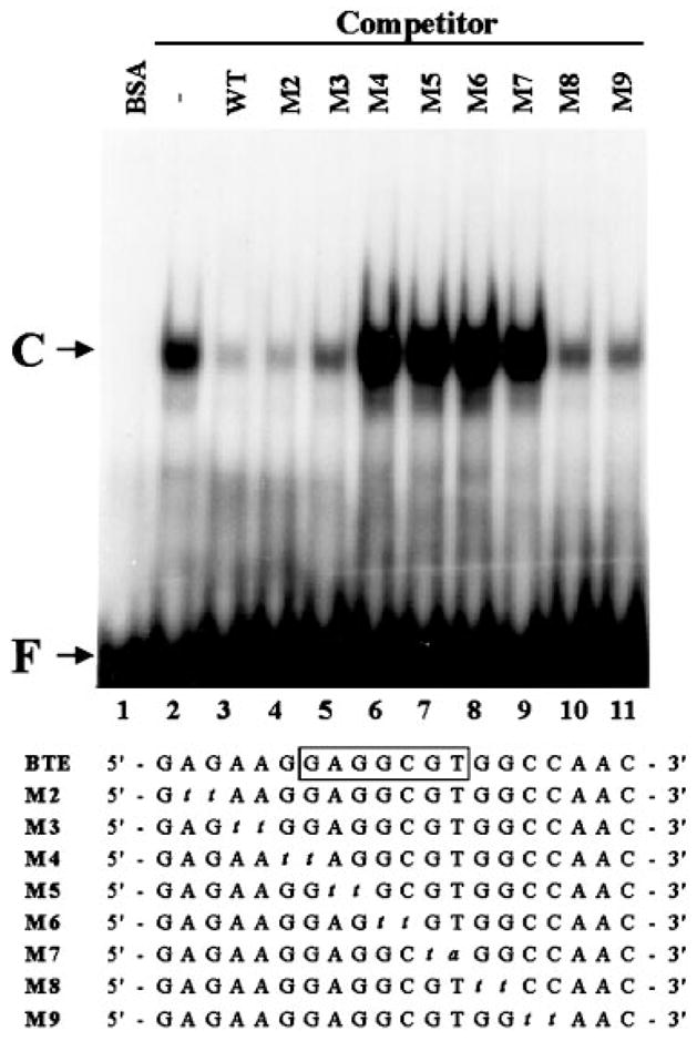
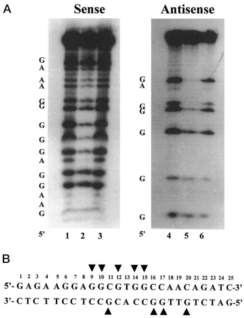
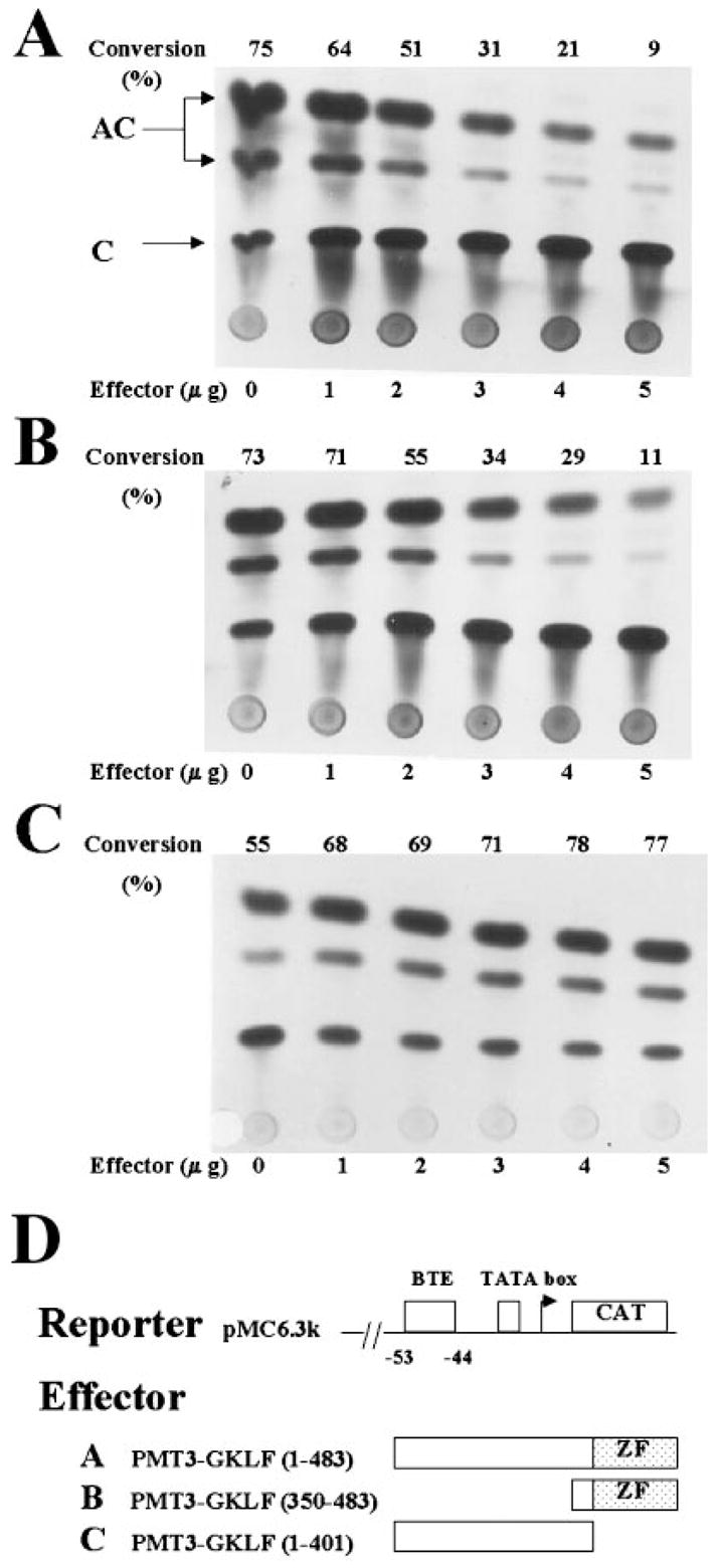
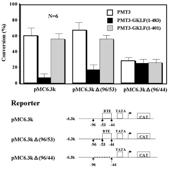
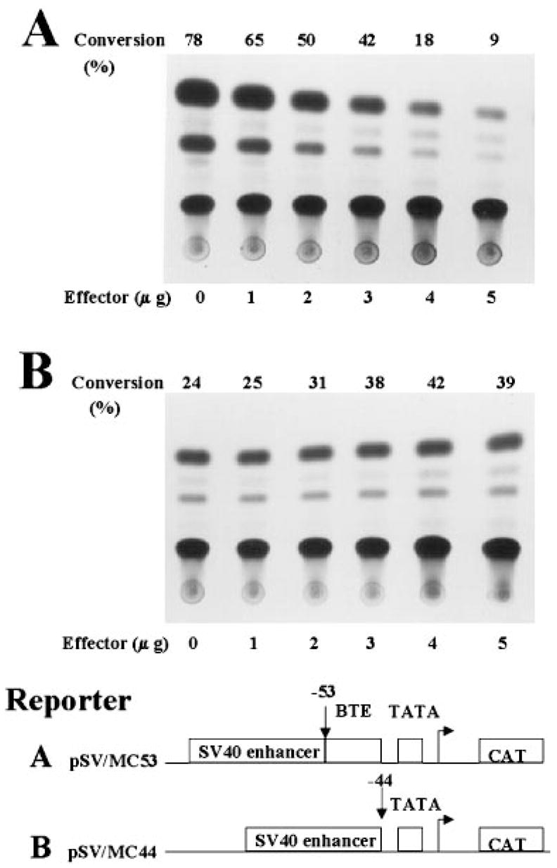
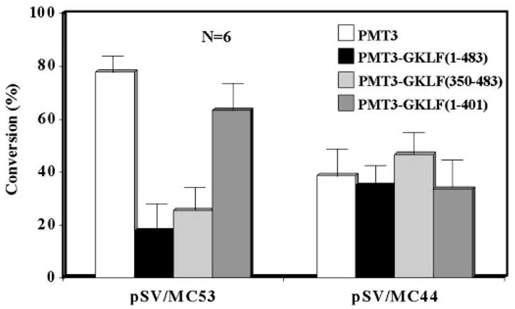
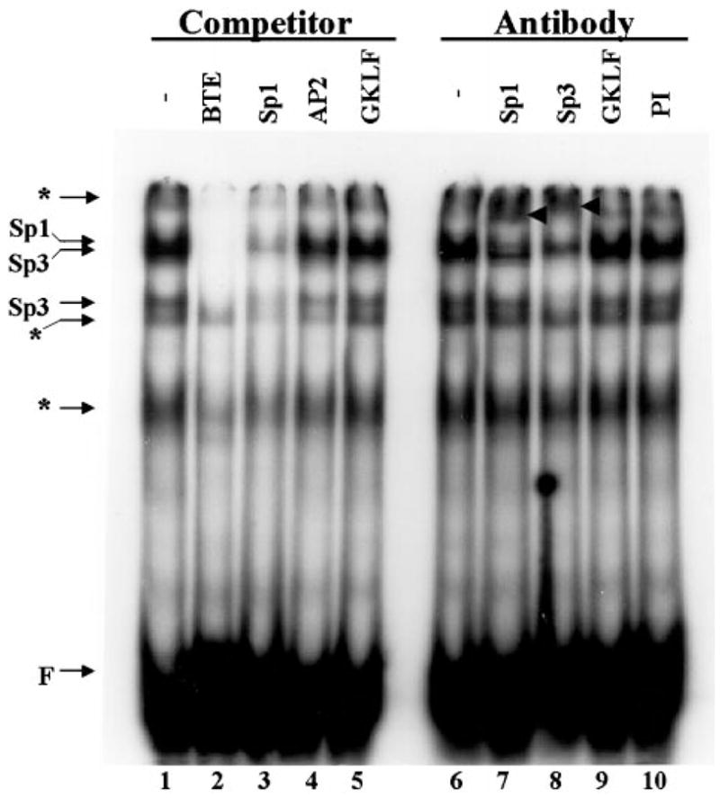
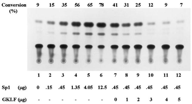
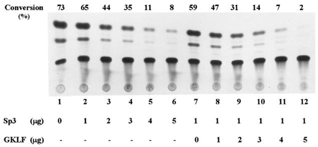
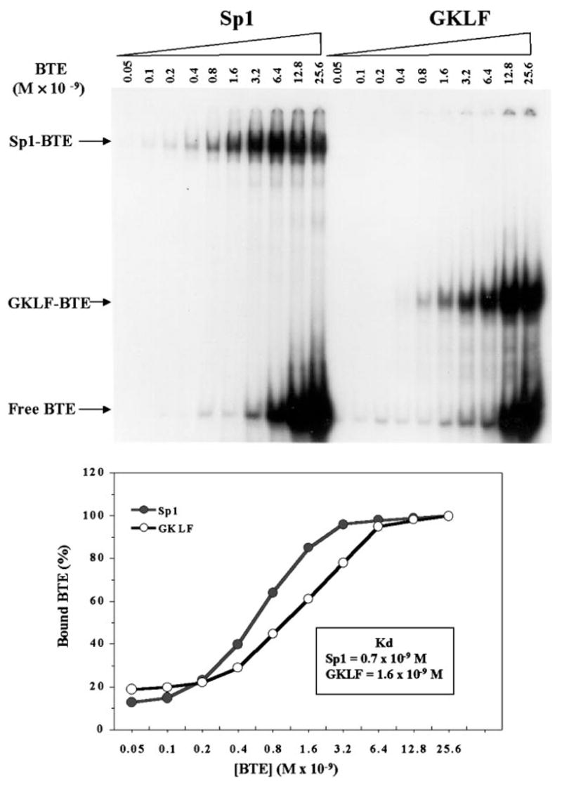
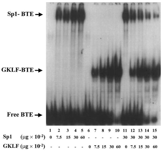
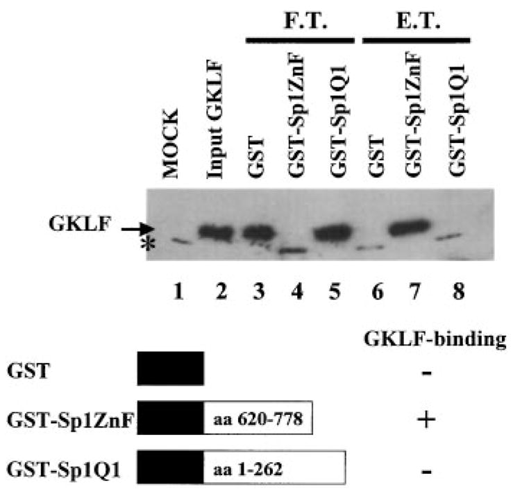
The gut-enriched Krüppel-like factor (GKLF) is a newly identified zinc finger-containing transcription factor. Recent studies indicate that GKLF binds to a core DNA sequence of 5'-(G/A)(G/A)GG(C/T)G(C/T)-3', which is found in an endogenous cis element, the basic transcription element (BTE) of the cytochrome P-450IA1 (CYP1A1) promoter. The present study characterizes the ability of GKLF to regulate CYP1A1 expression. By electrophoretic mobility gel shift assay (EMSA) and methylation interference assay, GKLF was found to bind BTE in a manner similar to several other transcription factors known to interact with BTE including Sp1 and BTEB. Cotransfection studies in Chinese hamster ovary cells showed that GKLF inhibited the CYP1A1 promoter in a dose- and BTE-dependent manner. The same experiments also revealed that BTE was responsible for a significant portion of the CYP1A1 promoter activity. EMSA of nuclear extracts from Chinese hamster ovary cells showed that Sp1 and Sp3 were two major proteins that interacted with BTE. Additional cotransfection studies showed that GKLF inhibited Sp1-mediated activation of the CYP1A1 promoter. In contrast, GKLF enhanced Sp3-dependent suppression of the same promoter. Moreover, the ability of GKLF to inhibit Sp1-dependent transactivation was in part due to physical interaction of the two proteins. These findings indicate that GKLF is a negative regulator of the CYP1A1 promoter in a BTE-dependent fashion and that this inhibitory effect is in part mediated by physical interaction with Sp1.


Figures

Fig. 1
Fig. 1. EMSA of wild-type and mutant BTE oligonucleotides with recombinant GKLF
EMSA was performed as described under “Experimental Procedures.” 0.1 pmol of labeled BTE oligonucleotide and 100 ng of purified recombinant GKLF were used in each reaction except for lane 1 which contained 100 ng of bovine serum albumin (BSA) as control. Unlabeled competitor oligonucleotides were added in 10-fold molar excess over the probe. The sequences of the wild-type and the individual mutant oligonucleotides are shown. The boxed sequence in the wild-type BTE indicates the established minimal essential binding site of GKLF (23). The mutated bases are indicated by italic lower-case. WT is wild-type BTE. C denotes DNA-protein complex, and F denotes free DNA probe.
Fig. 2
Fig. 2. Methylation interference assay of BTE by recombinant GKLF
Methylation interference assay was performed as described under “Experimental Procedures.” The double-stranded BTE oligonucleotide was labeled at the 5′ end of either the sense or antisense strand. A, is the result of the interference assay. The 5′ end of each strand is shown. The DNA included in lanes 1, 3, 4, and 6 was derived from the unbound free probe, whereas the DNA included in lanes 2 and 5 was from the bound probe. Methylated guanine and adenine residues are identified. B, shows the guanine residues (arrowheads) within the BTE, which when methylated resulted in an interference of binding to GKLF.
Fig. 3
Fig. 3. Cotransfection of CHO cells with a CYP1A1 promoter-linked CAT reporter and GKLF expression constructs
Transient transfections of CHO cells were performed with 5 μg/10-cm dish of pMC6.3k that contains 6.3 kilobase pairs of the rat CYP1A1 promoter linked to the CAT reporter (24) and increasing amounts of the mammalian expression plasmid PMT3 (30) containing either the full-length (effector A) or two truncated forms of GKLF (effectors B and C). The amount of extracts used to determine the CAT activity was first standardized to the β-galactosidase activity derived from the internal control, pCMV-SPORT-β-galactosidase. C denotes the substrate chloramphenicol, and AC denotes acetylated forms of chloramphenicol. The number at the top of each panel represents the percentage of substrate conversion ((AC/AC + C) × 100). ZF denotes the zinc fingers of GKLF.
Fig. 4
Fig. 4. Cotransfection of CHO cells with various CYP1A1 reporter and GKLF effector constructs
Transfection conditions were essentially the same as those described in Fig. 3. The amount of reporter and effector plasmids used was 5 μg/10-cm dish each. A description of the reporter constructs can be found under “Experimental Procedures.” PMT3 indicates the vector alone. Shown are the means ± S.E. of six independent experiments, each of which was carried out in duplicate.
Fig. 5
Fig. 5. Cotransfection of CHO cells with minimal CYP1A1 promoter-reporters and the full-length GKLF-expressing construct
A description of the two reporter constructs (A and B) was provided under “Experimental Procedures” and “Results.” Five μg/10-cm dish of the reporter and increasing amounts of the full-length GKLF construct, PMT3-GKLF-(1–483), were used in the experiments.
Fig. 6
Fig. 6. Cotransfection of CHO cells with minimal CYP1A1 promoter-reporters and expression constructs containing various forms of GKLF
The experimental conditions were similar to those described in Fig. 5. The amount of DNA used for all constructs was 5 μg/10-cm dish. Shown are the means ± S.E. of substrate conversion from six independent experiments. Each experiment was carried out in duplicate.
Fig. 7
Fig. 7. EMSA of BTE oligonucleotides using CHO cell nuclear extracts
EMSA was performed using 0.1 pmol of labeled BTE oligonucleotides and 10 μg of nuclear extracts obtained from CHO cells. Where indicated, competitor oligonucleotides (see “Experimental Procedures”) were added at a 10-fold molar excess of the labeled probe. In experiments involving antibodies, 2 μg of the stated antibody were added to the reaction. PI denotes pre-immune serum for GKLF. Super-shifted bands in lanes 7 and 8 are labeled with arrowheads. The positions of Sp1 and Sp3 in the DNA-protein complexes are indicated. The asterisks indicate either nonspecific complexes (since they competed poorly) or complexes containing as yet unidentified proteins.
Fig. 8
Fig. 8. Contransfection of CHO cells with a CYP1A1 promoter-reporter construct and expression plasmids containing Sp1 and GKLF
CHO cells were transiently transfected with 5μg/10-cm dish of pMC6.3k and increasing amounts of CMV-Sp1 (lanes 1–6) or increasing amounts PMT3-GKLF in the presence of 0.45 μg/10-cm dish of CMV-Sp1. Quantification of substrate conversion was performed as in previous figures. The amount of cell extracts used for the CAT assay was intentionally lowered by 5-flod to demonstrate the inductive effect of the promoter by Sp1. Shown is a representative result from three independent experiments.
Fig. 9
Fig. 9. Cotransfection of CHO cells with a CYP1A1 promoter-reporter and expression plasmids containing Sp3 and GKLF
Cotransfection experiments were performed as described in Fig. 8 with the exception that CMV-Sp3 was substituted for CMV-Sp1. The amount of cell extracts used for the CAT assay was similar to those used in Figs. 4 and 6. Shown is a representative result from two independent experiments.
Fig. 10
Fig. 10. Kinetics of binding between Sp1 and GKLF to BTE
EMSA was performed using increasing amounts of labeled BTE probe and 200 ng of purified human Sp1 or 60 ng of recombinant GKLF (top panel). Quantification of the intensity of the shifted bands was performed by densitometric measurement. One hundred percent bound was defined as the band that had the highest intensity for each protein (bottom panel).
Fig. 11
Fig. 11. Competition of binding of BTE to Sp1 by GKLF
EMSA was performed as before. 0.1 pmol of the labeled BTE oligonucleotide was incubated with increasing amounts of purified human Sp1 or recombinant GKLF (lanes 1–5 and lanes 6–10, respectively). In addition, the probe was incubated with 30 ng of purified Sp1 and increasing amounts of recombinant GKLF (lanes 11–15).
Fig. 12
Fig. 12. GST pull-down of GKLF by Sp1
The GST pull-down experiments are described under “Experimental Procedures.” Western blot analysis was performed on various protein fractions using a polyclonal rabbit GKLF antibody (13). Lanes 1 and 2 contained input lysates from mock- (vector alone-) transformed and GKLF-transformed bacteria, respectively. Lanes 3–5 contained the flow-through (F.T.) fractions of GKLF that did not bind to the glutathione-Sepharose 4B beads containing GST, GST-Sp1ZnF, and GST-Sp1Q1, respectively. Lanes 6–8 contained the eluant (E.T.) fractions of GKLF that bound to the beads and were eluted with 1 mm reduced glutathione. The location of GKLF is indicated and has a molecular mass of 24 kDa. The asterisk denotes a nonspecific cross-reacting material that was present in the lysates of mock-transformed bacteria.

References

    1. Klug A, Rhodes D. Cold Spring Harb Symp Quant Biol. 1987;52:473–482. - PubMed
    1. Berg JM. J Biol Chem. 1990;265:6513–6516. - PubMed
    1. Berg JM. Annu Rev Biophys Biophys Chem. 1990;19:405–421. - PubMed
    1. Klug A, Schwabe JW. FASEB J. 1995;9:597–604. - PubMed
    1. Berg JM, Shi Y. Science. 1996;271:1081–1085. - PubMed

Publication types

MeSH terms

Associated data

LinkOut - more resources