. 1998 Jul 6;188(1):5-16.
doi: 10.1084/jem.188.1.5.

CD4 T cell tolerance to human C-reactive protein, an inducible serum protein, is mediated by medullary thymic epithelium

Affiliations

CD4 T cell tolerance to human C-reactive protein, an inducible serum protein, is mediated by medullary thymic epithelium

L Klein et al. J Exp Med. .

Abstract

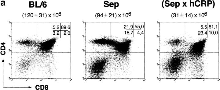
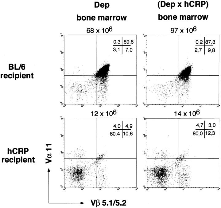
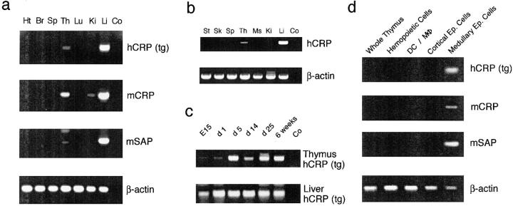
Inducible serum proteins whose concentrations oscillate between nontolerogenic and tolerogenic levels pose a particular challenge to the maintenance of self-tolerance. Temporal restrictions of intrathymic antigen supply should prevent continuous central tolerization of T cells, in analogy to the spatial limitation imposed by tissue-restricted antigen expression. Major acute-phase proteins such as human C-reactive protein (hCRP) are typical examples for such inducible self-antigens. The circulating concentration of hCRP, which is secreted by hepatocytes, is induced up to 1,000-fold during an acute-phase reaction. We have analyzed tolerance to hCRP expressed in transgenic mice under its autologous regulatory regions. Physiological regulation of basal levels (<10(-9) M) and inducibility (>500-fold) are preserved in female transgenics, whereas male transgenics constitutively display induced levels. Surprisingly, crossing of hCRP transgenic mice to two lines of T cell receptor transgenic mice (specific for either a dominant or a subdominant epitope) showed that tolerance is mediated by intrathymic deletion of immature thymocytes, irrespective of widely differing serum levels. In the absence of induction, hCRP expressed by thymic medullary epithelial cells rather than liver-derived hCRP is necessary and sufficient to induce tolerance. Importantly, medullary epithelial cells also express two homologous mouse acute-phase proteins. These results support a physiological role of "ectopic" thymic expression in tolerance induction to acute-phase proteins and possibly other inducible self-antigens and have implications for delineating the relative contributions of central versus peripheral tolerance.


Figures

Figure 1
Figure 1
Positive and negative selection of CD4 thymocytes specific for the dominant epitope of hCRP. Thymocytes of BL/6, TCR single transgenic Dep, and Dep × hCRP female mice were stained for coexpression of CD4 and CD8 (a) and the transgenic TCR chains Vα11 and Vβ5.1 (b), and were analyzed by four-color fluorescence. Note the efficient positive selection of TCR transgenic CD4 T cells in single transgenic mice and their deletion at the DP stage in noninduced Dep × hCRP mice. The total number of thymocytes (mean ± SD of at least three animals) and the percentages of subsets per quadrant are indicated. Male mice displayed an identical phenotype. Fluorescence intensity is shown on a four-decade logarithmic scale. 6–10-wk-old mice were analyzed.
Figure 1
Figure 1
Positive and negative selection of CD4 thymocytes specific for the dominant epitope of hCRP. Thymocytes of BL/6, TCR single transgenic Dep, and Dep × hCRP female mice were stained for coexpression of CD4 and CD8 (a) and the transgenic TCR chains Vα11 and Vβ5.1 (b), and were analyzed by four-color fluorescence. Note the efficient positive selection of TCR transgenic CD4 T cells in single transgenic mice and their deletion at the DP stage in noninduced Dep × hCRP mice. The total number of thymocytes (mean ± SD of at least three animals) and the percentages of subsets per quadrant are indicated. Male mice displayed an identical phenotype. Fluorescence intensity is shown on a four-decade logarithmic scale. 6–10-wk-old mice were analyzed.
Figure 2
Figure 2
Positive and negative selection of CD4 thymocytes specific for the subdominant epitope of hCRP. Thymocytes of BL/6, TCR single transgenic Sep, and Sep × hCRP female mice were stained for coexpression of the transgenic TCR chain Vβ8.3, CD4, and CD8, and were analyzed by three-color fluorescence. CD4 versus CD8 expression (a) and Vβ8.3 expression gated on the CD4 SP subset (b) are shown. Note the strong bias towards CD4 SP T cells in TCR single transgenic mice and its reversion in noninduced Sep × hCRP mice. The total number of thymocytes (mean ± SD of at least three animals) and the percentages of subsets per quadrant are indicated. Male mice displayed an identical phenotype. 6–10-wk-old mice were analyzed.
Figure 2
Figure 2
Positive and negative selection of CD4 thymocytes specific for the subdominant epitope of hCRP. Thymocytes of BL/6, TCR single transgenic Sep, and Sep × hCRP female mice were stained for coexpression of the transgenic TCR chain Vβ8.3, CD4, and CD8, and were analyzed by three-color fluorescence. CD4 versus CD8 expression (a) and Vβ8.3 expression gated on the CD4 SP subset (b) are shown. Note the strong bias towards CD4 SP T cells in TCR single transgenic mice and its reversion in noninduced Sep × hCRP mice. The total number of thymocytes (mean ± SD of at least three animals) and the percentages of subsets per quadrant are indicated. Male mice displayed an identical phenotype. 6–10-wk-old mice were analyzed.
Figure 3
Figure 3
Epitope-specific T cell tolerance in hCRP and hCRP × TCR transgenic mice. (a) BL/6, hCRP single transgenic, and Dep × hCRP male and female mice were immunized with a peptide corresponding to the dominant epitope of hCRP. (b) BL/6, hCRP single transgenic, and Sep × hCRP female mice were immunized with a peptide corresponding to the subdominant epitope of hCRP. 8–10 d later, draining lymph node cells were assessed for their proliferative response to the respective peptide. Irrespective of gender, hCRP single and Dep × hCRP mice are tolerant to the dominant epitope, whereas hCRP single and Sep × hCRP mice respond to the subdominant epitope.
Figure 3
Figure 3
Epitope-specific T cell tolerance in hCRP and hCRP × TCR transgenic mice. (a) BL/6, hCRP single transgenic, and Dep × hCRP male and female mice were immunized with a peptide corresponding to the dominant epitope of hCRP. (b) BL/6, hCRP single transgenic, and Sep × hCRP female mice were immunized with a peptide corresponding to the subdominant epitope of hCRP. 8–10 d later, draining lymph node cells were assessed for their proliferative response to the respective peptide. Irrespective of gender, hCRP single and Dep × hCRP mice are tolerant to the dominant epitope, whereas hCRP single and Sep × hCRP mice respond to the subdominant epitope.
Figure 4
Figure 4
Thymic deletion is not due to hemopoietic cell–derived hCRP. BL/6 and hCRP transgenic mice were lethally irradiated and reconstituted either with bone marrow cells from Dep or Dep × hCRP donors. 8 wk later, intrathymic selection of Dep T cells was analyzed by four-color fluorescence (see Fig. 1). Coexpression of the transgenic TCR chains Vα11 and Vβ5.1 on CD4 SP thymocytes is shown. Deletion is independent of the presence of the hCRP transgene in the donor bone marrow.
Figure 5
Figure 5
Thymus-derived hCRP is necessary and sufficient for tolerance in female hCRP transgenic mice. Thymectomized male and female hCRP transgenic mice were grafted with fetal BL/6 thymi, and thymectomized female BL/6 mice were grafted with hCRP transgenic thymi. 4 wk later, animals were lethally irradiated and reconstituted with Dep transgenic bone marrow. After another 6 wk intrathymic selection of Dep T cells was analyzed by four-color fluorescence (see Fig. 1). Coexpression of the transgenic TCR chains Vα11 and Vβ5.1 on CD4 SP thymocytes is shown (a). The same type of animals were immunized with peptide hCRP 89–100 (see Fig. 3) to assess their tolerance status (b). Thymus-derived hCRP is necessary in female, but not in male, hCRP transgenic mice for central deletion of hCRP-specific T cells and tolerance.
Figure 5
Figure 5
Thymus-derived hCRP is necessary and sufficient for tolerance in female hCRP transgenic mice. Thymectomized male and female hCRP transgenic mice were grafted with fetal BL/6 thymi, and thymectomized female BL/6 mice were grafted with hCRP transgenic thymi. 4 wk later, animals were lethally irradiated and reconstituted with Dep transgenic bone marrow. After another 6 wk intrathymic selection of Dep T cells was analyzed by four-color fluorescence (see Fig. 1). Coexpression of the transgenic TCR chains Vα11 and Vβ5.1 on CD4 SP thymocytes is shown (a). The same type of animals were immunized with peptide hCRP 89–100 (see Fig. 3) to assess their tolerance status (b). Thymus-derived hCRP is necessary in female, but not in male, hCRP transgenic mice for central deletion of hCRP-specific T cells and tolerance.
Figure 6
Figure 6
Expression pattern of transgenic hCRP and endogenous murine APPs. (a) The organ-specific expression pattern of the hCRP transgene and the homologous mouse APPs (mCRP and mSAP) was analyzed by reverse transcriptase PCR (35 cycles) in a young adult, noninduced, male hCRP transgenic mouse. Expression of the hCRP transgene parallels that of endogenous APPs and is confined to liver and thymus. An identical pattern was identified for induced female hCRP animals under the same PCR conditions. Signals for hCRP and mSAP, but not of mCRP, in liver and thymus of noninduced females were consistently lower (detection of thymic expression required reamplification with nested primers). The weak mCRP band in the kidney was not consistently detected. Ht, heart; Br, brain; Sp, spleen; Th, thymus; Lu, lung; Ki, kidney; Li, liver; Co, control. (b) The organ-specific expression pattern of hCRP was analyzed in various human tissues. Expression is confined to liver and thymus. St, stomach; Sk, skin; Ms, muscle. (c) Expression of hCRP in thymus and liver was analyzed during ontogeny in noninduced hCRP transgenic females. Expression in both organs was detected from day E15 onwards to adulthood. Thymus samples were reamplified with nested primers. (d) Expression of hCRP, mCRP, and mSAP was analyzed in whole thymocytes and in highly enriched thymic subsets of a noninduced female hCRP mouse (reverse transcriptase PCR; 30 cycles). Expression of the hCRP transgene and endogenous APP is confined to medullary epithelial cells. Note that detection of APP expression in enriched medullary epithelial cells of noninduced female mice does not require nested reamplification.

References

    1. van Meerwijk JPM, Marguerat S, Lees RK, Germain RN, Fowlkes BJ, MacDonald HR. Quantitative impact of thymic clonal deletion on the T cell repertoire. J Exp Med. 1997;185:377–383. - PMC - PubMed
    1. Kappler JW, Roehm N, Marrack P. T cell tolerance by clonal elimination in the thymus. Cell. 1987;49:273–280. - PubMed
    1. Kyewski BA, Fathman CG, Kaplan HS. Intrathymic presentation of circulating non–major histocompatibility complex antigens. Nature. 1984;308:196–199. - PubMed
    1. Zal T, Volkmann A, Stockinger B. Mechanisms of tolerance induction in major histocompatibility complex class II–restricted T cells specific for a blood-borne self-antigen. J Exp Med. 1994;180:2089–2099. - PMC - PubMed
    1. Arnold, B., G. Schönrich, I. Ferber, J. Alferink, and G.J. Hämmerling. 1994. Peripheral T cell tolerance: distinct levels and multistep mechanisms. In Transgenesis and Targeted Mutagenesis in Immunology. H. Bluethmann and P.S. Ohashi, editors. Academic Press, San Diego, CA. 135–140.

Publication types