. 1998 Jul 6;188(1):157-67.
doi: 10.1084/jem.188.1.157.

The coordinated action of CC chemokines in the lung orchestrates allergic inflammation and airway hyperresponsiveness

Affiliations

The coordinated action of CC chemokines in the lung orchestrates allergic inflammation and airway hyperresponsiveness

J A Gonzalo et al. J Exp Med. .

Abstract

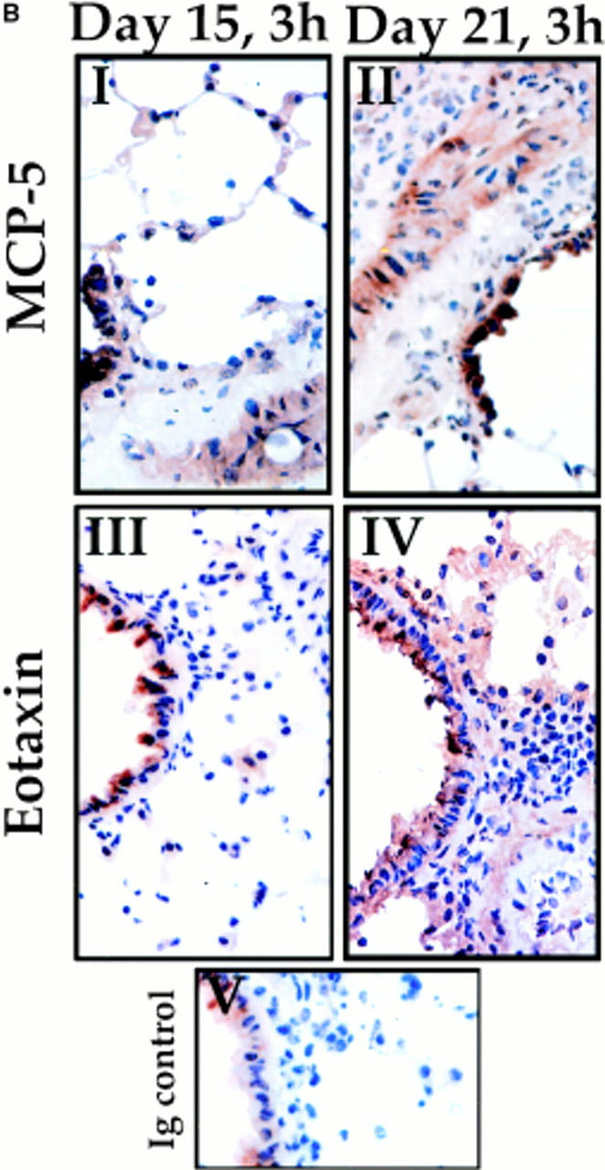
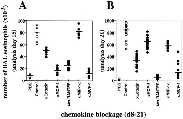
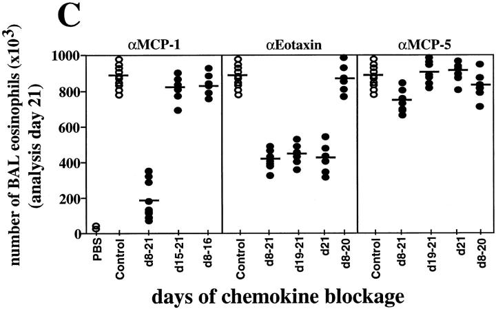
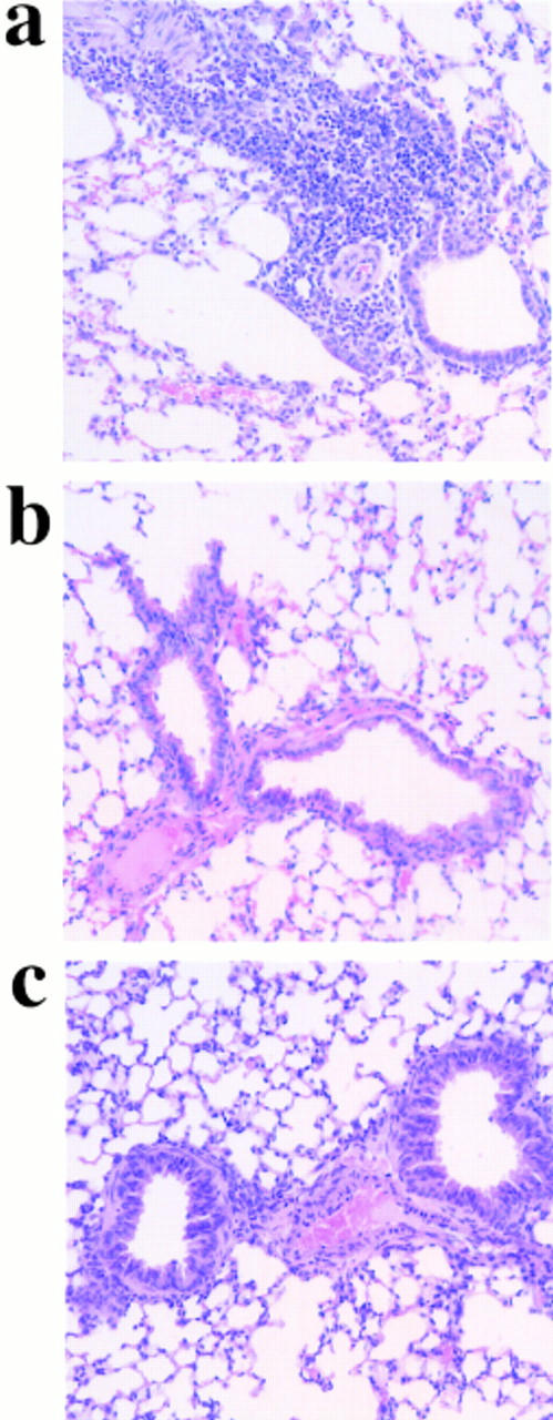
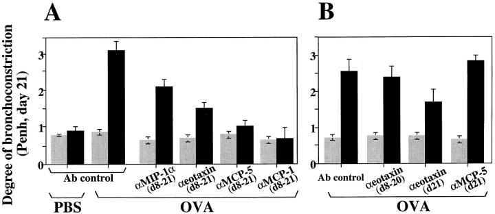
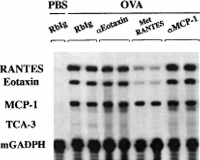
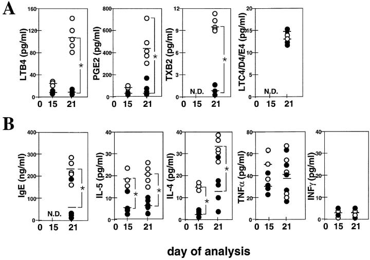
The complex pathophysiology of lung allergic inflammation and bronchial hyperresponsiveness (BHR) that characterize asthma is achieved by the regulated accumulation and activation of different leukocyte subsets in the lung. The development and maintenance of these processes correlate with the coordinated production of chemokines. Here, we have assessed the role that different chemokines play in lung allergic inflammation and BHR by blocking their activities in vivo. Our results show that blockage of each one of these chemokines reduces both lung leukocyte infiltration and BHR in a substantially different way. Thus, eotaxin neutralization reduces specifically BHR and lung eosinophilia transiently after each antigen exposure. Monocyte chemoattractant protein (MCP)-5 neutralization abolishes BHR not by affecting the accumulation of inflammatory leukocytes in the airways, but rather by altering the trafficking of the eosinophils and other leukocytes through the lung interstitium. Neutralization of RANTES (regulated upon activation, normal T cell expressed and secreted) receptor(s) with a receptor antagonist decreases significantly lymphocyte and eosinophil infiltration as well as mRNA expression of eotaxin and RANTES. In contrast, neutralization of one of the ligands for RANTES receptors, macrophage-inflammatory protein 1alpha, reduces only slightly lung eosinophilia and BHR. Finally, MCP-1 neutralization diminishes drastically BHR and inflammation, and this correlates with a pronounced decrease in monocyte- and lymphocyte-derived inflammatory mediators. These results suggest that different chemokines activate different cellular and molecular pathways that in a coordinated fashion contribute to the complex pathophysiology of asthma, and that their individual blockage results in intervention at different levels of these processes.


Figures

Figure 1
Figure 1
Chemokine expression and leukocyte infiltration in the lung of OVA-treated mice. (A) Eotaxin, RANTES, MCP-5, MCP-1, MIP-1α, and MIP-1β mRNA expression (gray bars) in the lung of each mouse were normalized to the 28S rRNA expression in the same organ of the same mice. The quantitation of total RNA was determined by hybridization of a 28S rRNAcDNA probe. Each bar represents data from five mice at the time points indicated (3 h after OVA challenge on days 15, 18, and 21) during treatment (bottom). Values are expressed as the mean ± SEM. The accumulation in the lung of BAL monocytes/macrophages (M/m), T lymphocytes (L), and eosinophils (E) during OVA treatment (3 h after antigen challenge on days 15, 18, and 21) is also shown. Although only BAL accumulation is presented here, there is correlation between these data and the accumulation in the interstitium (reference 23). Five data points at the different times indicated were collected per day as described previously (reference 23). Values are expressed as the mean ± SEM. i.n., intranasal. (B) Immunohistochemical staining of lung sections from OVA-treated mice on day 15 (3 h; I and III) or day 21 (3 h; II and IV) after OVA challenge. Sections were stained with anti–MCP-5 mAb (I and II; see Materials and Methods) or anti-Eot mAb (III and IV; see Materials and Methods). Note that positive staining (brown precipitate) is comparable between days 15 and 21 for MCP-5, but that there is clearly less eotaxin staining on day 15 compared with day 21. Ig-stained control is also shown (V).
Figure 1
Figure 1
Chemokine expression and leukocyte infiltration in the lung of OVA-treated mice. (A) Eotaxin, RANTES, MCP-5, MCP-1, MIP-1α, and MIP-1β mRNA expression (gray bars) in the lung of each mouse were normalized to the 28S rRNA expression in the same organ of the same mice. The quantitation of total RNA was determined by hybridization of a 28S rRNAcDNA probe. Each bar represents data from five mice at the time points indicated (3 h after OVA challenge on days 15, 18, and 21) during treatment (bottom). Values are expressed as the mean ± SEM. The accumulation in the lung of BAL monocytes/macrophages (M/m), T lymphocytes (L), and eosinophils (E) during OVA treatment (3 h after antigen challenge on days 15, 18, and 21) is also shown. Although only BAL accumulation is presented here, there is correlation between these data and the accumulation in the interstitium (reference 23). Five data points at the different times indicated were collected per day as described previously (reference 23). Values are expressed as the mean ± SEM. i.n., intranasal. (B) Immunohistochemical staining of lung sections from OVA-treated mice on day 15 (3 h; I and III) or day 21 (3 h; II and IV) after OVA challenge. Sections were stained with anti–MCP-5 mAb (I and II; see Materials and Methods) or anti-Eot mAb (III and IV; see Materials and Methods). Note that positive staining (brown precipitate) is comparable between days 15 and 21 for MCP-5, but that there is clearly less eotaxin staining on day 15 compared with day 21. Ig-stained control is also shown (V).
Figure 2
Figure 2
BAL eosinophilia after blockage of chemokines during OVA-induced lung allergic inflammation. Chemokine blockage was performed daily before OVA provocation on days (d) 8–21. BAL eosinophil accumulation was evaluated 3 h after OVA administration on day 15 (A) or on day 21 (B). MCP-1, eotaxin, or MCP-5 blockage was also performed after each OVA intranasal challenge for the periods of time indicated on the x-axis, and exclusively analyzed on day 21 (C). Each circle represents a single PBS- or OVA-treated control mouse (open circle) or a single test mouse (filled circle). Bars represent the mean of each group. Significant difference between control and test groups of mice was determined using the Student's t test (P <0.001). Quantitative data is shown in Table 1 for this and other leukocyte subsets.
Figure 2
Figure 2
BAL eosinophilia after blockage of chemokines during OVA-induced lung allergic inflammation. Chemokine blockage was performed daily before OVA provocation on days (d) 8–21. BAL eosinophil accumulation was evaluated 3 h after OVA administration on day 15 (A) or on day 21 (B). MCP-1, eotaxin, or MCP-5 blockage was also performed after each OVA intranasal challenge for the periods of time indicated on the x-axis, and exclusively analyzed on day 21 (C). Each circle represents a single PBS- or OVA-treated control mouse (open circle) or a single test mouse (filled circle). Bars represent the mean of each group. Significant difference between control and test groups of mice was determined using the Student's t test (P <0.001). Quantitative data is shown in Table 1 for this and other leukocyte subsets.
Figure 3
Figure 3
OVA-induced lung inflammation after chemokine blockage. On day 21, lung tissue was excised from OVA-treated mice injected with control (a), anti–MCP-1 (b), or anti–MCP-5 (c) antibodies. Chemokine blockage was performed daily before OVA provocation on days 8–21. One representative mouse out of ten is shown. Quantitative data corresponding to this set of experiments is shown in Table 2.
Figure 4
Figure 4
Inhibition of OVA-induced airway hyperresponsiveness after chemokine blockage. Results are shown as the mean ± SEM for Penh before (gray bars) and after (black bars) methacholine provocation (n = 10, two independent experiments). Mice were exposed to an aerosol of methacholine for 1 min, and airway constriction was evaluated for the next 5 min. PBS- or OVA plus irrelevant Ab–treated mice were used as control for OVA-treated littermates in which MIP-1α, eotaxin, MCP-5, or MCP-1 was blocked from days (d) 8 to 21 (A) or at the time points indicated (B).
Figure 5
Figure 5
Chemokine expression after chemokine blockage. On day 21, total RNA from lungs of OVA plus Ab control–, OVA plus anti-Eot–, OVA plus Met-RANTES–, or OVA plus anti–MCP-1–treated mice was extracted 3 h after antigen challenge. Chemokine blockage was performed daily before OVA provocation on days 8–21. In a control group, mice were treated with PBS (intranasally) and the irrelevant Ab (intravenously) instead of OVA (intranasally) and the chemokine-neutralizing Ab (intravenously), respectively. RANTES, eotaxin, and MCP-1 mRNA expression were determined by RPA. Both TCA and GAPDH mRNA expression were measured as negative and positive control, respectively. The intensity of the band for each chemokine was normalized to the intensity of the band of GAPDH in the control sample (not shown) and analyzed. Chemokine expression of two representative mice out of four is shown.
Figure 6
Figure 6
Production of OVA-induced mediators of inflammation in the BAL fluid after MCP-1 blockage. 1 h after OVA administration on day 15 or 21, inflammatory mediator release was measured in the BAL fluid of OVA plus irrelevant Ab–treated control mice (open circles) and OVA plus anti–MCP-1(days 8–21)–treated mice (filled circles). Anti– MCP-1 or control Abs were administered daily before OVA provocation on days 8–21. Each circle represents a single mouse. Bars represent the mean of each group. Significant difference between control and test groups of mice was determined using the Student's t test (*P <0.01).

References

    1. Gleich GJ, Flavahan NA, Fujisawa T, Vanhoutte PM. The eosinophil as a mediator of damage to respiratory epithelium: a model for bronchial hyperreactivity. J Allergy Clin Immunol. 1988;81:776–781. - PubMed
    1. Gleich GJ. The eosinophil and bronchial asthma. Current understanding. J Allergy Clin Immunol. 1990;85:422–436. - PubMed
    1. Eum S-Y, Hailé S, Lefort J, Huerre M, Vargaftig BB. Eosinophil recruitment into the respiratory epithelium following antigenic challenge in hyper-IgE mice is accompanied by interleukin 5-dependent bronchial hyperresponsiveness. Proc Natl Acad Sci USA. 1995;92:12290–12294. - PMC - PubMed
    1. Bradley BL, Azzawi M, Jacobson M, Assoufi B, Collins JV, Irani A-MA, Schwartz LB, Durham SR, Jeffrey PK, Kay AB. Eosinophils, T-lymphocytes, mast cells, neutrophils and macrophages in bronchial biopsy specimens from atopic subjects with asthma: comparison with biopsy specimens from atopic subjects without asthma and normal control subjects and relationship to bronchial hyperresponsiveness. J Allergy Clin Immunol. 1991;88:661–667. - PubMed
    1. Gonzalo JA, Lloyd CM, Kremer L, Finger E, Martinez-A C, Siegelman MH, Cybulski M, Gutierrez-Ramos JC. Eosinophil recruitment to the lung in a murine model of allergic inflammation. The role of T cells, chemokines, and endothelial adhesion receptors. J Clin Invest. 1996;98:2332–2345. - PMC - PubMed

Publication types

MeSH terms