. 1998 Jul 6;188(1):211-6.
doi: 10.1084/jem.188.1.211.

Nuclear factor (NF)-kappaB-regulated X-chromosome-linked iap gene expression protects endothelial cells from tumor necrosis factor alpha-induced apoptosis

Affiliations

Nuclear factor (NF)-kappaB-regulated X-chromosome-linked iap gene expression protects endothelial cells from tumor necrosis factor alpha-induced apoptosis

C Stehlik et al. J Exp Med. .

Abstract

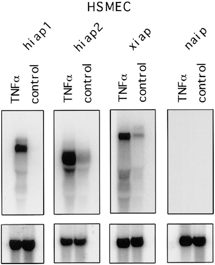
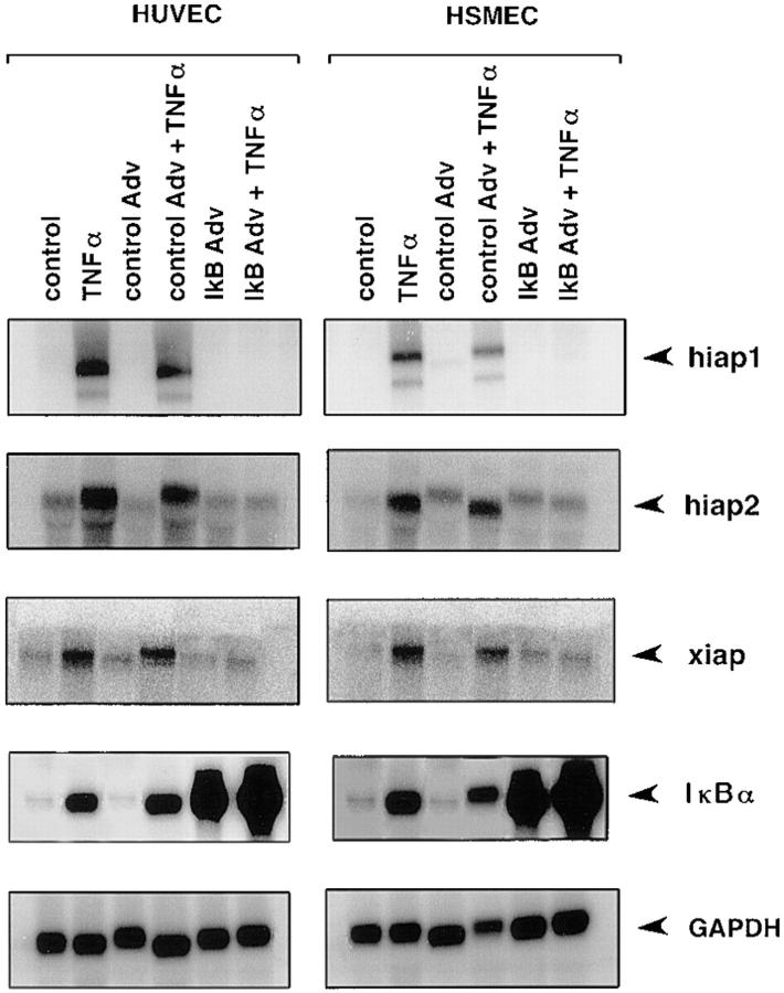
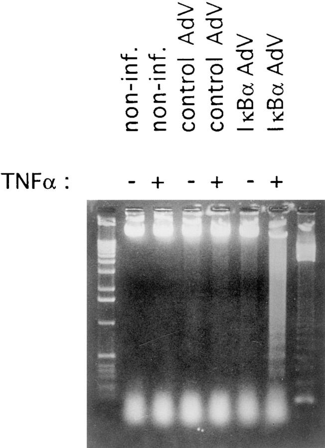
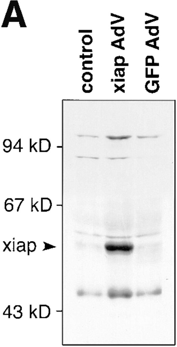
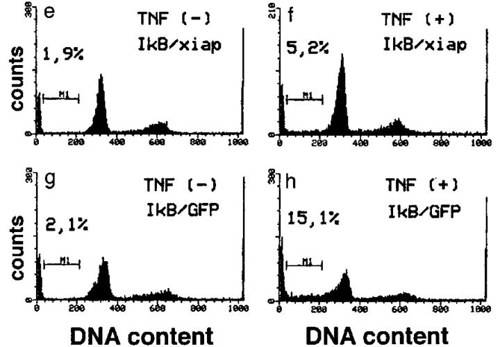
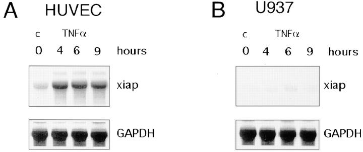
By differential screening of tumor necrosis factor alpha (TNF-alpha) and lipopolysaccharide (LPS)- activated endothelial cells (ECs), we have identified a cDNA clone that turned out to be a member of the inhibitor of apoptosis (iap) gene family. iap genes function to protect cells from undergoing apoptotic death in response to a variety of stimuli. These iap genes, hiap1, hiap2, and xiap were found to be strongly upregulated upon treatment of ECs with the inflammatory cytokines TNF-alpha, interleukin 1beta, and LPS, reagents that lead to activation of the nuclear transcription factor kappaB (NF-kappaB). Indeed, overexpression of IkappaBalpha, an inhibitor of NF-kappaB, suppresses the induced expression of iap genes and sensitizes ECs to TNF-alpha-induced apoptosis. Ectopic expression of one member of the human iap genes, human X-chromosome-linked iap (xiap), using recombinant adenovirus overrules the IkappaBalpha effect and protects ECs from TNF-alpha- induced apoptosis. We conclude that xiap represents one of the NF-kappaB-regulated genes that counteracts the apoptotic signals caused by TNF-alpha and thereby prevents ECs from undergoing apoptosis during inflammation.


Figures

Figure 1
Figure 1
Northern blot analysis of iap gene expression in HSMECs. 10 μg of total RNA from either nontreated or TNF-α–treated (500 U/ml) HSMECs was loaded in each lane and hybridized to oligonucleotides specific to hiap1, hiap2, xiap, and naip. The predicted transcript size corresponds to the published one (7) for hiap1 (6.5 kb), hiap2 (4.5 kb), and xiap (9 kb). To confirm the equal loading of RNA, membranes were stripped and reprobed with GAPDH.
Figure 2
Figure 2
Northern blot analysis of iap gene expression in adenovirus IκBα–infected ECs. HUVECs and HSMECs were not infected, were infected with a control adenovirus, or were infected with the recombinant adenovirus IκBα construct. Cells were either left untreated or treated with TNF-α (500 U/ ml) for 4 h. The membranes were probed with oligonucleotides specific to hiap1, hiap2, and xiap. Expression of IκBα was controlled by reprobing the membranes with an IκBα-cDNA. Equal loading was confirmed by hybridization with a GAPDH cDNA probe. Adv, adenovirus.
Figure 3
Figure 3
DNA fragmentation in adenovirus IκBα–infected and TNF-α–stimulated HUVECs. HUVECs were not infected, were infected, with a control adenovirus, or were infected with the recombinant adenovirus-IκBα construct. Noninfected cells and infected cells were left untreated or treated with TNF-α (500 U/ml) for 6 h. Appearance of fragmented genomic DNA was analysed by 1.3% agarose gel electrophoresis. Left lane: 1-kb ladder molecular weight standard; right lane: 123-bp ladder molecular weight standard. Non-inf.: noninfected cells; AdV: adenovirus.
Figure 4
Figure 4
Inhibition of apoptosis by ectopic xiap expression. (A) Lysates of noninfected or infected HUVECs were separated by SDS-PAGE, blotted onto nylon membranes, and stained for myc-tagged XIAP protein. AdV, adenovirus; GFP, green fluorescent protein. (B) HUVECs were infected with IκBα alone (c and d), together with xiap (e and f    ), or together with GFP (g and h) recombinant adenovirus. 48 h after infection cells were treated with TNF-α (500 U/ml) for 6 h or left untreated and analyzed by FACS® after propidium iodide staining. Cells with a DNA content <2 N appear in the sub-G1 region (M1). The percentage of cells found in the M1 region is indicated. The data show one out of three representative experiments.
Figure 4
Figure 4
Inhibition of apoptosis by ectopic xiap expression. (A) Lysates of noninfected or infected HUVECs were separated by SDS-PAGE, blotted onto nylon membranes, and stained for myc-tagged XIAP protein. AdV, adenovirus; GFP, green fluorescent protein. (B) HUVECs were infected with IκBα alone (c and d), together with xiap (e and f    ), or together with GFP (g and h) recombinant adenovirus. 48 h after infection cells were treated with TNF-α (500 U/ml) for 6 h or left untreated and analyzed by FACS® after propidium iodide staining. Cells with a DNA content <2 N appear in the sub-G1 region (M1). The percentage of cells found in the M1 region is indicated. The data show one out of three representative experiments.
Figure 5
Figure 5
Lack of TNF-α–inducible xiap gene expression correlates with apoptosis in U937 cells. Northern blot analysis of xiap gene expression in HUVECs (A) and U937 cells (C). HUVECs and U937 cells were treated for 4, 6, and 9 h with TNF-α. To confirm equal loading of RNA, membranes were stripped and reprobed with GAPDH. TNF-α–induced genomic DNA fragments from HUVECs (B) and U937 cells (D) were determined by colorimetric enzyme immunoassay. Columns represent the mean of three independent experiments. SD is indicated by error bars. TNF-α (500 U/ml); c, nontreated cells.
Figure 5
Figure 5
Lack of TNF-α–inducible xiap gene expression correlates with apoptosis in U937 cells. Northern blot analysis of xiap gene expression in HUVECs (A) and U937 cells (C). HUVECs and U937 cells were treated for 4, 6, and 9 h with TNF-α. To confirm equal loading of RNA, membranes were stripped and reprobed with GAPDH. TNF-α–induced genomic DNA fragments from HUVECs (B) and U937 cells (D) were determined by colorimetric enzyme immunoassay. Columns represent the mean of three independent experiments. SD is indicated by error bars. TNF-α (500 U/ml); c, nontreated cells.

References

    1. Hale AJ, Smith CA, Sutherland LC, Stoneman VEA, Longthorne VL, Culhane AC, Williams GT. Apoptosis: molecular regulation of cell death. Eur J Biochem. 1996;236:1–26. - PubMed
    1. Barinaga M. Forging a path to cell death. Science. 1996;273:735–737. - PubMed
    1. Wang CY, Mayo MW, Baldwin AS., Jr TNF- and cancer therapy–induced apoptosis: potentiation by inhibition of NF-κB. Science. 1996;274:784–787. - PubMed
    1. Van Antwerpen DJ, Martin SJ, Kafri T, Green DR, Verma IM. Suppression of TNF-α–induced apoptosis by NF-κB. Science. 1996;274:787–789. - PubMed
    1. Beg AA, Baltimore D. An essential role for NF-κB in preventing TNF-α–induced cell death. Science. 1996;274:782–784. - PubMed

Publication types

MeSH terms