

doi: 10.1073/pnas.95.14.7893.
Structural, functional, and evolutionary relationships between lambda-exonuclease and the type II restriction endonucleases
Affiliations
- PMID: 9653111
- PMCID: PMC20900
- DOI: 10.1073/pnas.95.14.7893
Item in Clipboard
Structural, functional, and evolutionary relationships between lambda-exonuclease and the type II restriction endonucleases
Proc Natl Acad Sci U S A.
.
Abstract
lambda-exonuclease participates in DNA recombination and repair. It binds a free end of double-stranded DNA and degrades one strand in the 5' to 3' direction. The primary sequence does not appear to be related to any other protein, but the crystal structure shows part of lambda-exonuclease to be similar to the type II restriction endonucleases PvuII and EcoRV. There is also a weaker correspondence with EcoRI, BamHI, and Cfr10I. The structure comparisons not only suggest that these enzymes all share a similar catalytic mechanism and a common structural ancestor but also provide strong evidence that the toroidal structure of lambda-exonuclease encircles its DNA substrate during hydrolysis.
Figures
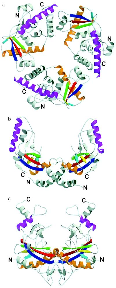
Ribbon diagrams of λ-exonuclease (a), PvuII (b), and EcoRV (c) with corresponding secondary structure elements between the three proteins colored similarly. Figure created with molscript (26) and rendered in raster 3d (27, 28).
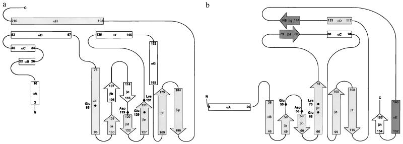
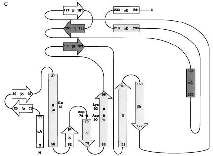
Schematic representations of the topology of the secondary structure for λ-exonuclease (a), PvuII (b), and EcoRV (c). Regions that structurally correspond in all three proteins are shaded in gray. Regions of structural similarity between PvuII and EcoRV that are not seen in λ-exonuclease are shaded in dark gray. b and c are modified from refs. –. Regions of secondary structure for λ-exonuclease were determined by using the program dssp (29). Structurally corresponding active site residues also are identified.
Schematic representations of the topology of the secondary structure for λ-exonuclease (a), PvuII (b), and EcoRV (c). Regions that structurally correspond in all three proteins are shaded in gray. Regions of structural similarity between PvuII and EcoRV that are not seen in λ-exonuclease are shaded in dark gray. b and c are modified from refs. –. Regions of secondary structure for λ-exonuclease were determined by using the program dssp (29). Structurally corresponding active site residues also are identified.

Stereoview overlay of Cα atoms for λ-exonuclease (yellow) and PvuII (cyan) protein fragments (a) and λ-exonuclease (yellow) and EcoRV (magenta) protein fragments (b). The segments shown include all of the regions of structural similarity identified by the Dali database server (Table 1). Figure prepared by using molscript (26) and rendered with raster 3d (27, 28).
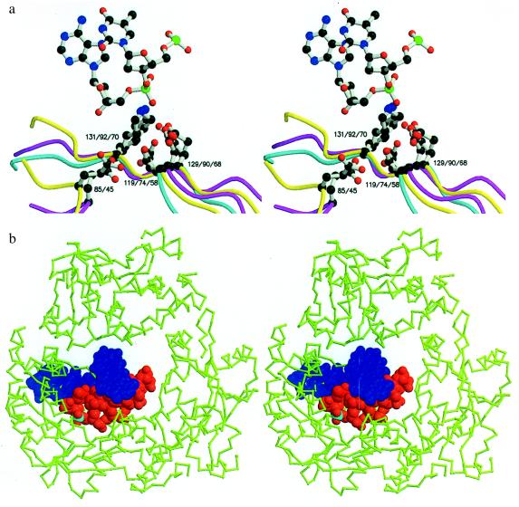
(A) Stereoview showing the superposition of the active site residues of λ-exonuclease (yellow), (Glu-85, Asp-119, Glu-129, and Lys-131), PvuII (cyan) (Asp-58, Glu-68, and Lys-70), and EcoRV (magenta) (Glu-45, Asp-74, Asp-90, and Lys-92). The figure includes the central nucleotide (TA) from the co-crystal structure of EcoRV bound to its cognate DNA sequence (17). (B) Superposition on the trimeric structure of λ-exonuclease of the cognate DNA decamer from the EcoRV:DNA complex (17). To make the DNA resemble the substrate of λ-exonucleases, five nucleotides were removed from the 5′ end of the strand that is hydrolyzed. The next phosphate that would be attacked is shown in cyan. The DNA was aligned based on the correspondence between the active site residues of EcoRV and λ-exonuclease (A). In the stereoview, the wider end of the central channel through the λ-exonuclease trimer, i.e., the end of the channel that is thought to accept the double-strand DNA substrate, is toward the viewer. Figure created by using molscript (26) and rendered with raster 3d (27, 28).
References
-
- Linn S M, Lloyd R S, Roberts R J, editors. Nucleases. Plainview, NY: Cold Spring Harbor Lab. Press; 1993.
-
- Radding C M. J Mol Biol. 1966;18:235–250. - PubMed
-
- Little J W. J Biol Chem. 1967;242:679–686. - PubMed
-
- Carter D M, Radding C M. J Biol Chem. 1971;246:2502–2512. - PubMed
-
- Cassuto E, Radding C M. Nat N Biol. 1971;229:13–16. - PubMed
Publication types
MeSH terms
Substances
Grants and funding
LinkOut - more resources
Full Text Sources
Other Literature Sources
Molecular Biology Databases
