. 1998 Jul 7;95(14):7987-92.
doi: 10.1073/pnas.95.14.7987.

Regulation of hypoxia-inducible factor 1alpha is mediated by an O2-dependent degradation domain via the ubiquitin-proteasome pathway

Affiliations

Regulation of hypoxia-inducible factor 1alpha is mediated by an O2-dependent degradation domain via the ubiquitin-proteasome pathway

L E Huang et al. Proc Natl Acad Sci U S A. .

Abstract

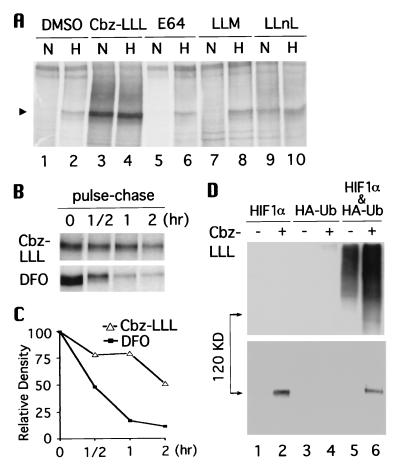
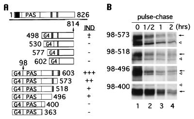
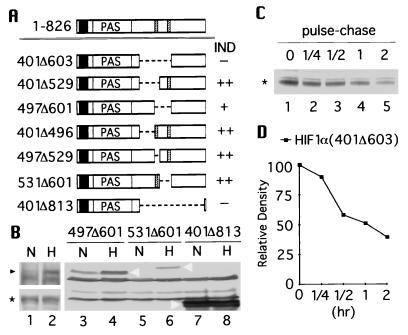
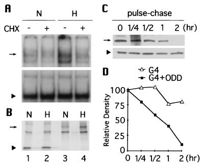
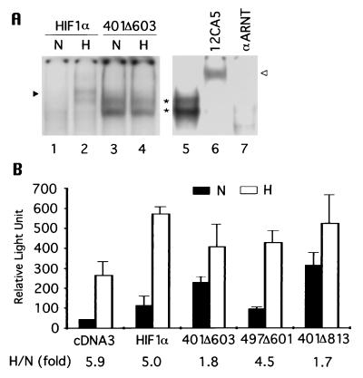
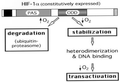
Hypoxia induces a group of physiologically important genes such as erythropoietin and vascular endothelial growth factor. These genes are transcriptionally up-regulated by hypoxia-inducible factor 1 (HIF-1), a global regulator that belongs to the basic helix-loop-helix PAS family. Although HIF-1 is a heterodimer composed of alpha and beta subunits, its activity is primarily determined by hypoxia-induced stabilization of HIF-1alpha, which is otherwise rapidly degraded in oxygenated cells. We report the identification of an oxygen-dependent degradation (ODD) domain within HIF-1alpha that controls its degradation by the ubiquitin-proteasome pathway. The ODD domain consists of approximately 200 amino acid residues, located in the central region of HIF-1alpha. Because portions of the domain independently confer degradation of HIF-1alpha, deletion of this entire region is required to give rise to a stable HIF-1alpha, capable of heterodimerization, DNA-binding, and transactivation in the absence of hypoxic signaling. Conversely, the ODD domain alone confers oxygen-dependent instability when fused to a stable protein, Gal4. Hence, the ODD domain plays a pivotal role for regulating HIF-1 activity and thereby may provide a means of controlling gene expression by changes in oxygen tension.


Figures

Figure 1
Figure 1
Degradation of HIF-1α is via the ubiquitin-proteasome pathway. (A) Hep3B cells were pretreated with dimethyl sulfoxide, 10 μM Cbz-LLL, and 50 μM trans-epoxysuccinyl-l-leucylamido-(4-guanidino) butane, N-acetyl-l-leucinyl-l-leucinyl-methioninal, and a N-acetyl-l-leucinyl-l-leucinyl-l-norleucinal, respectively, for 16 hr, and radiolabeled with [35S]methionine in the presence of these reversible (39) inhibitors under normoxia (N) or hypoxia (H) for another 4 hr. Arrow heads indicate HIF-1α. B. Hep3B cells were incubated under normoxia with 10 μM Cbz-LLL (Upper) or 130 μM desferrioxamine (DFO) for 4 hr in the presence of [35S]methionine, and then chased for 0.5, 1, and 2 hr. Cell lysates were immunoprecipitated with anti-HIF-1α antibodies. The intensity of HIF-1α signals in B was quantified and plotted in C. (D) 293 cells were transfected with HIF-1α expression vector, HA-tagged ubiquitin vector, or both, and were treated with Cbz-LLL (+) or dimethyl sulfoxide (−) for 16 hr before harvest. Expressed HIF-1α was immunoprecipitated with anti-HIF-1α, determined by Western blot analysis with anti-HA antibody (Upper), and reprobed with anti-HIF-1α antibodies (Lower).
Figure 2
Figure 2
Identification of the regions responsible for oxygen-induced degradation by means of Gal4 fusion proteins. (A) The full-length HIF-1α (aa 1–826) is depicted on top; bHLH domain is represented by a black box, PAS domains labeled as PAS, and two PEST-like sequences drawn as hatched boxes. The Gal4 DNA binding domain (G4) is fused to stretches of HIF-1α downstream of the PEST-like sequences (aa 498, 530, 577, and 602–814) and to stretches of HIF-1α upstream of the PEST-like sequences (aa 98–603, 573, 518, 496, 400, and 363). The hypoxia inducibility (IND) of each construct is summarized on the right. (B) 293 cells were transfected with Gal-HIF-1α (98 to 573, 518, 496, and 400), labeled with [35S]methionine at 1% O2, and chased at 21% O2 for 0.5, 1, and 2 hr. Immunoprecipitation was carried out with anti-Gal4 antibody. Gal4-HIF-1α fusions are denoted by an arrow, and nonspecific bands by open arrowheads.
Figure 3
Figure 3
Analyses of internal deletions of HIF-1α. (A) Schematic drawings of internal deletions of HIF-1α where aa 401–603, 401–529, 497–601, 401–496, 497–529, 531–601, and 401–813 were deleted. Hypoxic inducibility of each construct is summarized on the right. (B) HA-tagged full-length HIF-1α (Upper) and HIF-1α(401Δ603) (Lower) were cotransfected with pARNT into 293 cells and examined by Western blot analysis using anti-HIF-1α antibodies. Likewise, HA-tagged HIF-1α(497Δ601, 531Δ601, and 401Δ813) were cotransfected with pARNT and analyzed by Western blot using anti-HA antibody (lanes 3–8). (C) 293 cells were transfected with HA-tagged HIF-1α(401Δ603), labeled with [35S]methionine, and then chased for 0.25, 0.5, 1, and 2 hr under normoxia. Immunoprecipitation was done with anti-HA antibody. The intensity of each band was quantified and plotted in D. The full-length HIF-1α is marked with black arrow head, HIF-1α(401Δ603) with asterisk, and HIF-1α(497Δ601, 531Δ601, and 401Δ813) by white arrowhead.
Figure 4
Figure 4
The function of ODD domain is transportable. (A) Gal4-HIF-1α(401–603) (Upper) and Gal4 (Lower) expression vectors were transfected into 293 cells respectively and their DNA-binding activity was tested by electrophoretic mobility-shift assay. (B) [35S]methionine-labeling and immunoprecipitation were performed to quantify abundance of Gal4 (lanes 1 and 2) and Gal4-HIF-1α(401–603) (lanes 3 and 4) under normoxia and hypoxia. (C) Similarly, Gal4-HIF-1α(401–603) (Upper) and Gal4 (Lower) were pulse-labeled and chased for 0.25, 0.5, 1, and 2 hr at 21% O2. The intensity of each signal was plotted in D. Arrow indicates Gal4-HIF-1α(401–603) and arrowhead marks Gal4.
Figure 5
Figure 5
Autonomous heterodimerization, DNA binding, and transactivation of stable HIF-1α. (A) (HA)HIF-1α or (HA)HIF-1α(401Δ603) were cotransfected with pARNT into 293 cells and HIF-1 DNA binding was examined directly by electrophoretic mobility-shift assay (lanes 1–4), or in the presence of antibodies against HA or ARNT (21) (lanes 6 and 7). An arrowhead denotes HIF-1, an asterisk indicates HIF-1α(401Δ603), and an open arrowhead marks supershifted HIF-1α(401Δ603). (B) Hep3B cells were transfected with pEpoE-luc and pcDNA3 or with pEpoE-luc and pARNT plus HA-tagged HIF-1α, HIF-1α(401Δ603), HIF-1α(497–601), or HIF-1α(401Δ813). Luciferase activity was measured and plotted. The results are the mean of three independent transfections. The corresponding hypoxic induction fold is indicated at bottom.
Figure 6
Figure 6
A model for hypoxia-induced activation of HIF-1. A schematic drawing of HIF-1α is illustrated. The bHLH domain is marked by a black box and the C terminus transactivation domain by a hatched box. PAS and ODD domains are indicated. Steps regulated by changes of oxygen tension are boxed with bold lines.

References

    1. Bunn H F, Poyton R O. Physiol Rev. 1996;76:839–885. - PubMed
    1. Guillemin K, Krasnow M A. Cell. 1997;89:9–12. - PubMed
    1. Semenza G L, Wang G L. Mol Cell Biol. 1992;12:5447–5454. - PMC - PubMed
    1. Wang G L, Semenza G L. J Biol Chem. 1993;268:21513–21518. - PubMed
    1. Wang G L, Jiang B-H, Rue E A, Semenza G L. Proc Natl Acad Sci USA. 1995;92:5510–5514. - PMC - PubMed

Publication types

MeSH terms

LinkOut - more resources