. 1998 Jul 7;95(14):8187-92.
doi: 10.1073/pnas.95.14.8187.

HD mice: a novel mouse mutant with a specific defect in the generation of CD4(+) T cells

Affiliations

HD mice: a novel mouse mutant with a specific defect in the generation of CD4(+) T cells

V P Dave et al. Proc Natl Acad Sci U S A. .

Abstract

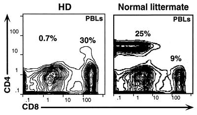
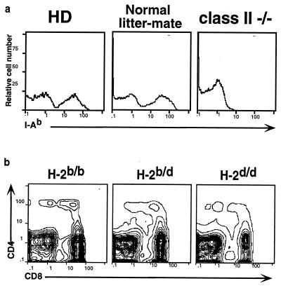
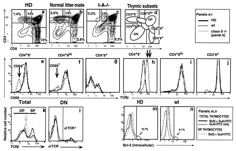
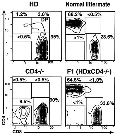
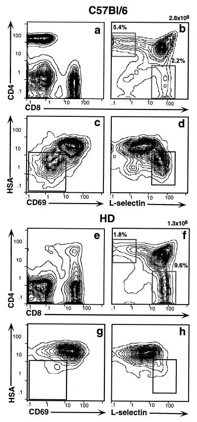
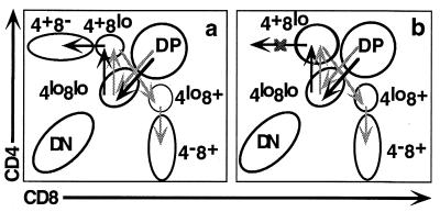
We have identified a spontaneous mutation in mice, which we term HD for "helper T cell deficient." This mouse is distinguished by the virtual absence of peripheral T cells of the CD4(+)8(-) major histocompatibility complex (MHC) class II-restricted T helper subset due to a specific block in thymic development. The developmental defect is selective for CD4(+)8(-) cells; the maturation of CD4(-)8(+) and gamma delta T cells is normal. The autosomal recessive mutation underlying the HD phenotype is unrelated to MHC class II, since it segregates independently of the MHC class II locus. Moreover, the HD phenotype is not caused by a defect of the CD4 gene. Bone marrow transfer experiments demonstrate that the defect is intrinsic to cells of the hematopoietic lineage, i.e., most likely to developing thymocytes themselves. The frequency of CD4(+)8(low) intermediate cells is markedly increased in HD mice, suggesting that class II-restricted thymocytes are arrested at this stage. This is the first genetic defect of its kind to be described in the mouse and may prove highly informative in understanding the molecular pathways underlying lineage commitment.


Figures

Figure 1
Figure 1
HD mice specifically lack peripheral CD4+ T lymphocytes. PBLs from a HD mouse or a normal littermate were stained with antibodies specific for CD4, CD8, and αβTCR and then analyzed by flow cytometry.
Figure 2
Figure 2
HD phenotype is not caused by the absence of MHC class II products and is genetically unlinked to the class II locus. (a) PBLs from HD mice, normal littermates, and MHC class II-deficient mice were stained with antibody specific for the MHC class II I-Ab product. (b) F2 progeny from the intercross between HD and BALB/c mice were stained with antibodies against CD4 and CD8. Data are shown for one mouse each of the HD phenotype that typed as H-2 b/b, b/d, or d/d.
Figure 3
Figure 3
Thymic development of CD4 lineage cells is abrogated in HD mice. Thymocytes from HD mice, normal littermates, or MHC class II-deficient controls were stained with antibodies specific for CD4, CD8, αβTCR, and CD69 and analyzed by flow cytometry. Histograms represent total thymocytes (a–c and k) or thymic subsets, as specified. In e–l, solid and dashed dark lines correspond to HD and wt mice, respectively, whereas the solid light line in k represents class II−/− mice. In m and n, solid and dashed dark lines correspond to total thymocytes stained either with anti-Bcl-2 and fluoresceinated goat anti-hamster fluorescein isothiocyanate (GαH–FITC) or only GαH–FITC, respectively, whereas the solid light lines represent DP thymocytes stained with anti-Bcl-2 and GαH-FITC. Bcl-2high populations correspond to SP thymocytes. Thymic subset designations are defined in d. Gates used to define CD4+8, CD4+8low, and CD48+ subsets are shown in a–c. Arrows point out key differences between HD mice and wt or class II−/− controls as follows: In e and f, arrows identify the mature CD69 population that is present in wt but absent in HD mice. In h, arrows highlight the 2-fold difference in TCR expression between CD4+8 cells from HD versus wt mice. In k, the arrow highlights the higher TCR expression levels seen on DP thymocytes from class II−/− but not HD or wt mice.
Figure 4
Figure 4
HD mice lack the αβTCR+ peripheral T cell subset found in CD4−/− mice. PBLs from a HD mouse, a wt littermate, or a CD4-deficient mouse were stained with antibodies against CD4, CD8, and αβTCR and analyzed by flow cytometry. Plots are gated to show CD4:CD8 expression profiles for αβTCR+ cells.
Figure 5
Figure 5
The HD defect maps to the hematopoietic compartment. Sublethally irradiated RAG2−/− recipients were reconstituted with T cell-depleted bone marrow from wt (a–d) or HD (e–h) mice. PBLs and thymocytes were collected after 4 wk and stained with antibodies against CD4, CD8, HSA, CD69, and l-selectin. Staining profiles are shown for total PBLs (a and e), total thymocytes (b and f), and SP CD4+ thymocytes (c, d, g, and h). Insets in c, d, g, and h indicate the position of the most mature SP CD4+ subpopulation which is present in wt but not in HD mice.
Figure 6
Figure 6
Absence of mature SP CD4+ thymocytes in HD mice reflects a blockade of development at the CD4+8low stage. (a) Developmental routes postulated to be followed by class I- (light arrows) and class II-restricted (dark arrows) thymocytes in wt mice. (b) Model of HD defect as a blockade in CD4 development at the CD4+8low stage.

Similar articles

Cited by

References

    1. Robey E A, Fowlkes B J, Gordon J W, Kioussis D, von Boehmer H, Ramsdell F, Axel R. Cell. 1991;64:99–107. - PubMed
    1. Chan S H, Cosgrove D, Waltzinger C, Benoist C, Mathis D. Cell. 1993;73:225–236. - PubMed
    1. Davis C B, Killeen N, Crooks M E C, Raulet D, Littman D R. Cell. 1993;73:237–247. - PubMed
    1. Suzuki H, Punt J A, Granger L G, Singer A. Immunity. 1995;2:413–425. - PubMed
    1. Matechak E O, Killeen N, Hedrick S M, Fowlkes B J. Immunity. 1996;4:337–347. - PubMed

Publication types

MeSH terms

Substances

LinkOut - more resources