. 1998 Jul 7;95(14):8199-204.
doi: 10.1073/pnas.95.14.8199.

Intrathymic selection of NK1.1(+)alpha/beta T cell antigen receptor (TCR)+ cells in transgenic mice bearing TCR specific for chicken ovalbumin and restricted to I-Ad

Affiliations

Intrathymic selection of NK1.1(+)alpha/beta T cell antigen receptor (TCR)+ cells in transgenic mice bearing TCR specific for chicken ovalbumin and restricted to I-Ad

C Iwabuchi et al. Proc Natl Acad Sci U S A. .

Abstract

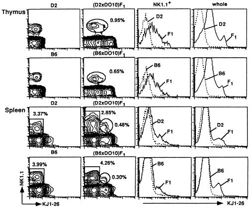
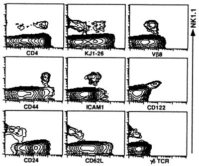
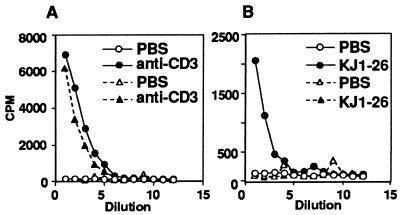
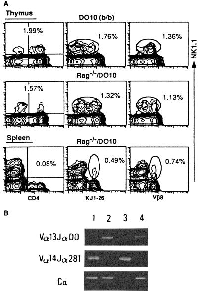
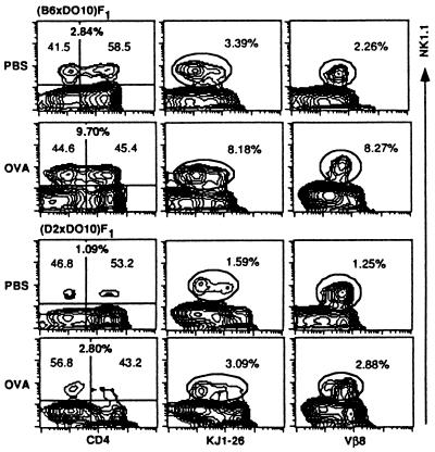
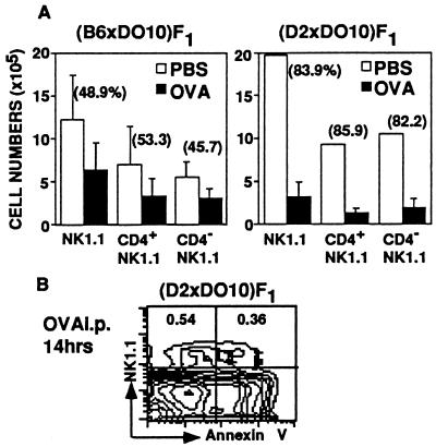
Generation and negative selection of NK1.1(+)alpha/beta T cell receptor (TCR)+ thymocytes were analyzed using TCR-transgenic (B10. D2 x DO10)F1 and (C57BL/6 x DO10)F1 mice and Rag-1(-/-)/DO10 mice, which had been established by breeding and backcrossing between Rag-1(-/-) and DO10 mice. Almost all T cells from these mice were shown to bear Valpha13/Vbeta8.2 that is specific for chicken ovalbumin (cOVA) and restricted to I-Ad. A normal proportion of the NK1.1(+) Valpha13/Vbeta8.2(+) thymocytes was generated in these mice. However, the actual cell number of both NK1.1(+) and NK1.1(-) thymocytes in I-Ad/d mice (positive selecting background) was larger than that in I-Ab/d mice (negative selecting background). Markedly low but significant proportions of NK1.1(+) Valpha13/Vbeta8.2(+) cells were detected in the spleens from I-Ad/d and I-Ab/d mice. It was shown that the splenic NK1.1(+) T cells of the I-Ab/d mice were anergized against stimulation through TCR. When (B10.D2 x DO10)F1 and (C57BL/6 x DO10)F1 mice were given cOVA, extensive or intermediate elimination of NK1.1(+)alpha/betaTCR+ thymocytes was induced in I-Ad/d or I-Ab/d mice, respectively. However, the clonal elimination was not as complete as that seen in the major NK1.1(-) thymocyte population. The present findings indicate that normal generation of NK1.1(+)alpha/betaTCR+ thymocytes occurs in the absence of Valpha14-Jalpha281 and that substantial negative selection operates on the NK1.1(+)alpha/betaTCR+ cells.


Figures

Figure 1
Figure 1
Analysis of NK1.1 expression on thymocytes and spleen cells from D2, B6, (D2 × DO10)F1, and (B6 × DO10)F1 mice. Thymocytes and splenocytes were stained with PE-anti-NK1.1 and biotin-KJ1-26 followed by Streptavidin-FITC. The proportions of NK1.1+KJ1-26+ cells (thymus) and NK1.1+KJ1-26 and NK1.1+ KJ1-26+ cells (spleen) of (D2 × DO10)F1 and (B6 × DO10)F1 mice are indicated (Middle Left). Fluorescence intensities of KJ1-26 among the NK1.1+ population in the thymus as well as in whole thymocyte and splenocyte populations are also shown in the histograms to compare those between D2 and (D2 × DO10)F1 or between B6 and (B6 × DO10)F1, respectively (Right).
Figure 2
Figure 2
Phenotypic analysis of thymocytes from (B6 × DO10)F1 mice. Thymocytes were stained either with combination of PE-anti-NK1.1 and biotin-Abs (KJ1-26, anti-Vβ8, CD44, ICAM-1, CD24, CD62L, and γ/δTCR) plus Streptavidin-FITC or PE-anti-CD122 and biotin-anti-NK1.1 plus Streptavidin-FITC. Expression of NK1.1 (Vertical) is indicated. Other markers on thymocytes (Horizontal) are indicated.
Figure 3
Figure 3
Induction of IL-4 by in vivo administration of anti-CD3 or KJ1-26. (D2 × DO10)F1 and (B6 × DO10)F1 mice were injected intravenously with 4 μg of anti-CD3 or KJ1-26, and spleens were removed after 90 min. Spleen cells (5 × 10⁶) were cultured in 96-well plates for 2 h. Supernatants were harvested, and IL-4 was measured using the CT.4S cells. (A) Induction of IL-4 in response to in vivo treatment with anti-CD3. (B) Induction of IL-4 in response to in vivo treatment with KJ1-26. (D2 × DO10)F1 mice were injected with PBS (○) or anti-CD3 or KJ1-26 (•). (B6 × DO10)F1 mice were injected with PBS (▵) or anit-CD3 or KJ1-26 (▴).
Figure 4
Figure 4
NK1.1 expression and Vα usage on thymocytes and splenocytes from DO10 (H-2b/b) and Rag-1−/−/DO10 mice. (A) FACS analysis of NK1.1 expression on thymocytes and splenocytes. Cells were stained with PE-anti-NK1.1 and FITC-anti-CD4, biotin-KJ1-26, or biotin-anti-Vβ8 followed by Streptavidin-FITC. The proportions of whole NK1.1+ cells are shown in the thymus, and NK1.1+CD4+, NK1.1+KJ1-26+, and NK1.1+Vβ8+ cells are shown in the spleen. (B) Analysis of TCRα chain usage in thymocytes and splenocytes using reverse transcription-PCR. Vα13-Jα DO (DO10 TCRα), Vα14-Jα281, and Cα transcripts were amplified with specific primers as described in Materials and Methods from B6 thymocytes (lane 1), Rag-1−/−/DO10 thymocytes (lane 2), B6 splenocytes (lane 3), and Rag-1−/−/DO10 splenocytes (lane 4), respectively.
Figure 5
Figure 5
Influence of cOVA administration on NK1.1+ thymocytes in (B6 × DO10)F1 and (D2 × DO10)F1 mice. Thymocytes from cOVA- or PBS-treated mice were isolated and stained with PE-anti-NK1.1, FITC-anti-CD4, and biotin-KJ1-26 or biotin-anti-Vβ8 followed by Streptavidin-FITC. A representative profile from three separate experiments is shown. The proportions of NK1.1+, NK1.1+ CD4, and NK1.1+ CD4+ cells (Left), NK1.1+KJ1-26+ (NK1.1+KJ1-26low and NK1.1+KJ1-26intermediate) (Middle), and NK1.1+Vβ8+ (Right) cells are indicated.
Figure 6
Figure 6
Clonal deletion of NK1.1+ thymocytes following cOVA administration. (A) The total number of NK1.1+ thymocytes from cOVA- or PBS-injected (B6 × DO10)F1 and (D2 × DO10)F1 mice. Absolute cell numbers of NK1.1+, NK1.1+CD4+, and NK1.1+CD4 thymocytes in mice given cOVA (closed bar) or PBS (open bar) are shown. Results represent means and SD of the calculated cell numbers (n = 3–5). Percent reduction is also indicated. (B) Expression of an early apoptotic marker, Annexin V, on NK1.1+ thymocytes obtained from (D2 × DO10)F1 mice given cOVA. Thymocytes were stained with Annexin V-FITC and NK1.1-PE, and the proportions of NK1.1+ Annexin V and NK1.1+ Annexin V+ cells are indicated.

References

    1. Bendelac A. Curr Opin Immunol. 1995;7:367–374. - PubMed
    1. MacDonald H R. J Exp Med. 1995;182:633–638. - PMC - PubMed
    1. Arase H, Arase N, Ogasawara K, Good R A, Onoé K. Proc Natl Acad Sci USA. 1992;89:6506–6510. - PMC - PubMed
    1. Adachi Y, Koseki H, Zijlstra M, Taniguchi M. Proc Natl Acad Sci USA. 1995;92:1200–1204. - PMC - PubMed
    1. Abo T, Ohteki T, Seki S, Koyamada N, Yoshikai Y, Masuda T, Rikiishi H, Kumagai K. J Exp Med. 1991;174:417–424. - PMC - PubMed

Publication types

MeSH terms

Substances

LinkOut - more resources